What are arachnoid granulations?
Arachnoid granulations represent growths of the arachnoid membrane into dural venous sinuses, through which CSF enters the venous system from subarachnoid space They are usually idiopathic and are identified as incidental osteolytic findings.
Are there arachnoid granulations in the dural sinuses?
Arachnoid granulations were found in 19 (66%) of the cadaveric specimens, in a similar distribution as that seen on the imaging studies. Conclusion: Discrete filling defects, consistent with arachnoid granulations, may be seen in the dural sinuses on 24% of contrast-enhanced CT scans and on 13% of MR studies.
Can MRI detect arachnoid granulations?
The MRI can be used for the identification of these small protrusions, with general signal characteristics for the cerebrospinal fluid. Arachnoid granulations, also known as a Pacchionian granulation, are projections of the arachnoid membrane (villi) into the dural sinuses that allow CSF entrance from the subarachnoid space into the venous system.
What is the difference between arachnoid granulation and dural venous thrombosis?
The arachnoid granulations often have a parasagittal location, leading to specific changes on the X-rays of the skull, such as the osteolytic lucency or the filling defect that appears at the level of the dural venous sinuses. Because of the latter modification, it is possible to mistake the arachnoid granulation for dural venous thrombosis.

What causes arachnoid granulation?
Arachnoid granulations increase in numbers and enlarge with age in response to increased CSF pressure from the subarachnoid space and are usually quite apparent by 4 years of age.
Why is arachnoid granulations important?
Function. They allow cerebrospinal fluid (CSF) to exit the subarachnoid space and enter the blood stream. The diffusion across the arachnoid granulations into the superior sagittal sinus returns CSF to the venous circulation. They act as one-way valves.
What does arachnoid granulation mean?
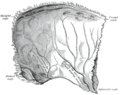
Arachnoid granulations (AGs) are tufts of arachnoid membrane invaginated into the dural sinuses through which cerebrospinal fluid (CSF) enters the venous system. The lesions are primarily located in the parasagittal region along the superior sagittal sinus[1], which is occasionally seen at the transverse sinus.
Can arachnoid granulations cause headaches?
Giant arachnoid granulations have been reported to be associated with headaches, which can be acute or chronic in presentation. In some cases, idiopathic intracranial hypertension, previously called pseudotumor cerebri, may occur.
How are arachnoid granulations treated?
As was the case in our patient, asymptomatic arachnoid granulations incidentally found do not require treatment. However, arachnoid granulations can grow to fill and dilate the dural sinuses, sometimes causing symptoms of increased intracranial pressure from venous hypertension secondary to partial sinus occlusion.
Do arachnoid granulations enhance?
The key MRI features of giant arachnoid granulations are non-enhancing granules with central linear enhancement and surrounding enhancing flowing blood on contrast-enhanced MR venography3). Intrasinus thrombus may show contrast enhancement and occlude venous flow.
What is large arachnoid granulations?
Arachnoid granulations (AG) are extensions of the arachnoid membrane into the dural venous sinuses, serving to drain the cerebrospinal fluid (CSF) from the subarachnoid space into the venous system. In some people, these extensions grow to become “giant” AG 1-4.
Do arachnoid cysts go away?
Most arachnoid cysts never cause symptoms, but on the rare occasions that they do, treatment for arachnoid cysts usually relieves symptoms. But cysts can grow back or fill with fluid after treatment. If that happens, you may need another procedure to drain the fluid or remove the cyst.
Do arachnoid granulations produce or drain CSF?
The arachnoid granulations (AGs) are herniations of the arachnoid membrane that protrude through the dura mater and into the lateral lacunae and venous sinuses on the surface of the brain. They are associated primarily with drainage of cerebrospinal fluid (CSF) into the venous sinuses [1].
What is venous sinus stenosis?
Venous sinus stenosis (VSS) is a kind of cerebral venous system disease that obstructs venous blood outflow. Some studies have shown that it may cause increased intravenous pressure, decreased regional blood flow, destruction of the blood-brain barrier, and intracranial hypertension [4].
What is the function of an arachnoid granulation quizlet?
Arachnoid granulations act as one-way valves. Normally the pressure of the CSF is higher than that of the venous system, so CSF flows through the villi and granulations into the blood. If the pressure is reversed for some reason, fluid will pass back into the subarachnoid space (of the brain).
What is dural sinus thrombosis?
What is Cerebral Venous Sinus Thrombosis? Cerebral venous sinus thrombosis is a rare condition when a large blood clot forms in a large vein in the brain called a dural venous sinus. The clot blocks the dural sinus and prevents the blood flow draining from the brain (Figure 1).
What happens if the arachnoid granulations are blocked?
Anytime there is a blockage in one of the channels of the brain or the arachnoid granulations, the plumbing system can get backed up. That backup can cause increased pressure in the brain because CSF is still produced in spite of the blockage. This condition is called hydrocephalus.
What is the function of the arachnoid granulations quizlet?
Arachnoid granulations act as one-way valves. Normally the pressure of the CSF is higher than that of the venous system, so CSF flows through the villi and granulations into the blood. If the pressure is reversed for some reason, fluid will pass back into the subarachnoid space (of the brain).
Do arachnoid granulations allow for reabsorption of cerebrospinal fluid?
After circulating over the convexities of the brain, CSF gets resorbed through the small arachnoid villi and the larger arachnoid granulations.
Do arachnoid granulations produce or drain CSF?
The arachnoid granulations (AGs) are herniations of the arachnoid membrane that protrude through the dura mater and into the lateral lacunae and venous sinuses on the surface of the brain. They are associated primarily with drainage of cerebrospinal fluid (CSF) into the venous sinuses [1].
Who is the arachnoid granulation?
Arachnoid granulations are named after Antonio Pacchioni (1665-1726), an Italian physician , who wrote extensively on the anatomy of the dura mater, and provided the first written description of his eponymous granulations in 1705 in the Dissertatio Epistolaris de Glandulis Conglobatis Durae Meningis Humanae, one of his monographs 1.
What is the granulation of the arachnoid membrane?
Arachnoid granulations , also known as Pacchionian granulations, are projections of the arachnoid membrane (villi) into the dural sinuses that allow CSF to pass from the subarachnoid space into the venous system.
Where do granulations occur?
The granulations typically occur next to the entrance of a superficial draining cortical vein into a sinus (similar to colonic diverticula occurring next to penetrating vessels).
What is granulation in a sinus?
The granulations are typically of CSF density and protrude into the calvaria or a dural venous sinus causing a filling defect. They may simulate a dural venous sinus thrombosis but are usually easy differentiated given their round well-defined shape and classic location.
Petrous Apex
Arachnoid granulations Arachnoid granulations are small outpouchings of the pia-arachnoid that typically protrude into the venous sinuses or inner table of the skull along the high convexities.22 They can, however, affect any part of the skull or skull base and mimic an epidermoid cyst or metastasis.
Idiopathic intracranial hypertension (idiopathic pseudotumor cerebri)
The AGs, described by Pacchioni in 1705 as “peculiar wartlike excrescences” were functionally investigated by Key and Retzius in 1876.
Nervous System
These are thought to arise from arachnoid granulations and so are found most commonly adjacent to venous sinuses. They account for 15–20% of intracranial tumours. They are slow growing and essentially ‘benign’. A few more aggressive tumours may metastasise.
HYDROCEPHALUS, INCLUDING NORMAL-PRESSURE HYDROCEPHALUS
Noojan J. Kazemi, ... Elsdon Storey, in Neurology and Clinical Neuroscience, 2007
Adult communicating hydrocephalus
Incomplete leptomeningeal fibrosis with or without changes in the arachnoid granulations is the most common finding, combined with ventricular ependymal disruption, subependymal glial reaction, and periventricular loss of myelin staining (see Di Rocco et al, 1977, for a review).
Idiopathic Intracranial Hypertension
Jonathan Baird-Gunning, Christian J. Lueck, in Reference Module in Neuroscience and Biobehavioral Psychology, 2018
Meningiomas
Meningiomas are believed to derive from the arachnoid cap cells around arachnoid granulations near venous sinuses, cisterns, ventricles, and brain. They can be found anywhere there is known pia, arachnoid, or dura. These tumors exhibit a wide variety of behaviors from benign to extremely aggressive.
What is the function of the arachnoid granulations?
Function. They allow cerebrospinal fluid (CSF) to exit the subarachnoid space and enter the blood stream. The diffusion across the arachnoid granulations into the superior sagittal sinus returns CSF to the venous circulation. They act as one-way valves.
Where do granulations occur?
The granulations typically occur next to the entrance of a superficial draining cortical vein into a sinus (similiar to colonic diverticuli occuring next to penetrating vessels).
What is granulation in a sinus?
The granulations are typically of CSF density and protrude into the calvaria or a dural venous sinus causing a filling defect. They may simulate a dural venous sinus thrombosis but are usually easy differentiated given their round well-defined shape and classic location.
Can arachnoid granulations cause venous hypertension?
Although usually incidental, giant arachnoid granulations that are of sufficient size to fill the lumen of a dural sinus and cause local dilation or filling defects can rarely cause symptoms due to sinus obstruct ion leading to venous hypertension.
Where are arachnoid granulations found?
Aberrant arachnoid granulations can be seen at the floor of the anterior and middle cranial fossa and, less frequently, at the posterior temporal bone wall. They contain cerebrospinal fluid but do not communicate with the dural venous sinuses.
What is an aberrant arachnoid pit?
Aberrant arachnoid granulations, also known as arachnoid pits, are arachnoid granulations that penetrated the dura but failed to migrate normally in the venous sinus.