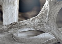
These three are part of an osteon, which is a functional unit of compact bone matter. Bones have two types of tissues: the hard, strong exterior and the spongy interior marrow. Osteocytes
Osteocyte
An osteocyte, a star-shaped type of bone cell, is the most commonly found cell in mature bone tissue, and can live as long as the organism itself. The adult human body has about 42 billion of them. Osteocytes do not divide and have an average half life of 25 years. They are derived from osteoprogenitor cells, some of which differentiate into active osteoblasts. Osteoblasts/osteocytes develop in mesenchyme.
Osteoblast
Osteoblasts are cells with a single nucleus that synthesize bone. However, in the process of bone formation, osteoblasts function in groups of connected cells. Individual cells cannot make bone. A group of organized osteoblasts together with the bone made by a unit of cells is usually called the osteon.
Osteoclast
An osteoclast is a type of bone cell that breaks down bone tissue. This function is critical in the maintenance, repair, and remodelling of bones of the vertebral skeleton. The osteoclast disassembles and digests the composite of hydrated protein and mineral at a molecular level by secreting acid and a collagenase, a process known as bone resorption. This process also helps regulate the level of blood calciu…
What is the difference between osteocytes and osteoclast?
Doesn't contain osteons osteocyte mature bone cells osteoclast huge cells from fusion of as many as 50 monocytes or white blood cells spongy bone red bone marrow is located here osteocyte main cells in bone tissue osteoclast concentrated in endosteum spongy bone osteocytes in lacuna osteocyte doesn't undergo cell division osteoclast
What is the function of osteoclast?
An osteoclast (from Ancient Greek ὀστέον (osteon) 'bone', and κλαστός (clastos) 'broken') is a type of bone cell that breaks down bone tissue. This function is critical in the maintenance, repair, and remodelling of bones of the vertebral skeleton.
Where is the nucleus of osteoclasts located?
Since osteoclasts are made up of two or more cells that fuse, they normally have more than one nucleus. They're located near the dissolving bone on the surface of the bone mineral. The cells that shape new bones are known as OSTEOBLASTS.
How many osteoclasts are there in bone?
Osteoclasts showed completely different behavior as compared to other types of bone cells. The occurrence of osteoclasts is quite scarce in the bony tissue. It is estimated that in an area of 1mm of the bony tissue, almost 2 to 3 osteoclasts are found. The structure of osteoclasts is related to their function.
See more

Where are osteoclasts found in bone?
Howship lacunaeOsteoclasts occupy small depressions on the bone's surface, called Howship lacunae; the lacunae are thought to be caused by erosion of the bone by the osteoclasts' enzymes. Osteoclasts are formed by the fusion of many cells derived from circulating monocytes in the blood. These in turn are derived from the bone marrow.
Are osteoblasts in compact bone?
The increase in diameter is called appositional growth. Osteoblasts in the periosteum form compact bone around the external bone surface. At the same time, osteoclasts in the endosteum break down bone on the internal bone surface, around the medullary cavity.
What is not found in compact bone?
Immature compact bone does not contain osteons and has a woven structure. It forms around a framework of collagen fibres and is eventually replaced by mature bone in a remodeling process of bone resorption and new bone formation that creates the osteons.
What is found in compact bone?
Compact bone consists of closely packed osteons or haversian systems. The osteon consists of a central canal called the osteonic (haversian) canal, which is surrounded by concentric rings (lamellae) of matrix. Between the rings of matrix, the bone cells (osteocytes) are located in spaces called lacunae.
Where are osteoclasts and osteoblasts found?
Osteoblasts, bone lining cells and osteoclasts are present on bone surfaces and are derived from local mesenchymal cells called progenitor cells. Osteocytes permeate the interior of the bone and are produced from the fusion of mononuclear blood-borne precursor cells.
What is are found in compact bone but not spongy bone?
Whereas compact bone tissue forms the outer layer of all bones, spongy bone or cancellous bone forms the inner layer of all bones. Spongy bone tissue does not contain osteons that constitute compact bone tissue. Instead, it consists of trabeculae, which are lamellae that are arranged as rods or plates.
What is found in compact bone and cancellous bone?
Cancellous Bone – The softer, less dense tissue that makes up the ends of bones and creates blood cells. Osteons – Functional units of compact bone, created by a network of bone cells and blood vessels. Osteocyte – A cell which function to maintain and repair bone tissue.
What are the 4 main parts of compact bone?
To wrap it all up, osteons are structural and functional units of compact bone. Every osteon is made up of a central canal (which contains nerves and blood vessels), perforating canals, lamellae (configurations of bone matrix), and osteocyte-containing lacunae (holes).
What is the major difference between compact and spongy bone?
Compact bone is a robust and heavy bony structure that forms the diaphysis of long bones, whereas spongy bone is a soft and smooth bony structure that forms the epiphysis of long bones.
What is the difference between compact bone and spongy bone quizlet?
Compact bone has more bone matrix and less space due to osteons. Spongy bones have less bone matrix and more space due to trabeculae.
What makes up compact and spongy bone?
Compact bone is dense and composed of osteons, while spongy bone is less dense and made up of trabeculae. Blood vessels and nerves enter the bone through the nutrient foramina to nourish and innervate bones.
Where are the osteoblast cells located?
bone surface2.1. Osteoblasts. Osteoblasts are cuboidal cells that are located along the bone surface comprising 4–6% of the total resident bone cells and are largely known for their bone forming function [22].
Are osteoblasts found in spongy bone?
Spongy bone tissue is composed of trabeculae and forms the inner part of all bones. Four types of cells compose bony tissue: osteocytes, osteoclasts, osteoprogenitor cells, and osteoblasts.
What are the 4 main parts of compact bone?
To wrap it all up, osteons are structural and functional units of compact bone. Every osteon is made up of a central canal (which contains nerves and blood vessels), perforating canals, lamellae (configurations of bone matrix), and osteocyte-containing lacunae (holes).
Are osteoblasts in the osteon?
However, in the process of bone formation, osteoblasts function in groups of connected cells. Individual cells cannot make bone. A group of organized osteoblasts together with the bone made by a unit of cells is usually called the osteon.
What is the secretion of cathepsin K?
Upon polarization of the osteoclast over the site of resorption, cathepsin K is secreted from the ruffled border into the resorptive pit. Cathepsin K transmigrates across the ruffled border by intercellular vesicles and is then released by the functional secretory domain. Within these intercellular vesicles, cathepsin K, along with reactive oxygen species generated by TRAP, further degrades the bone extracellular matrix.
How do osteoclasts move?
Once activated, osteoclasts move to areas of microfracture in the bone by chemotaxis. Osteoclasts lie in small cavities called Howship's lacunae, formed from the digestion of the underlying bone. The sealing zone is the attachment of the osteoclast's plasma membrane to the underlying bone. Sealing zones are bounded by belts of specialized adhesion structures called podosomes. Attachment to the bone matrix is facilitated by integrin receptors, such as αvβ3, via the specific amino acid motif Arg-Gly-Asp in bone matrix proteins, such as osteopontin. The osteoclast releases hydrogen ions through the action of carbonic anhydrase ( H 2 O + CO 2 → HCO 3− + H +) through the ruffled border into the resorptive cavity, acidifying and aiding dissolution of the mineralized bone matrix into Ca 2+, H 3 PO 4, H 2 CO 3, water and other substances. Dysfunction of the carbonic anhydrase has been documented to cause some forms of osteopetrosis. Hydrogen ions are pumped against a high concentration gradient by proton pumps, specifically a unique vacuolar-ATPase. This enzyme has been targeted in the prevention of osteoporosis. In addition, several hydrolytic enzymes, such as members of the cathepsin and matrix metalloprotease (MMP) groups, are released to digest the organic components of the matrix. These enzymes are released into the compartment by lysosomes. Of these hydrolytic enzymes, cathepsin K is of most importance.
What is the resorption bay?
The resorption bays are created by erosive action of osteoclasts on the underlying bone. The border of the lower part of an osteoclast exhibits finger-like processes due to presence of deep infoldings of the cell membrane; this border is called ruffled border.
How does osteoclast resorption work?
Resorption of bone matrix by the osteoclasts involves two steps: (1) dissolution of inorganic components (minerals), and (2) digestion of organic component of the bone matrix. The osteoclasts pump hydrogen ions into subosteoclastic compartment and thus create an acidic microenvironment, which increases solubility of bone mineral, ...
What is the ruffled border of the osteoclast?
At a site of active bone resorption, the osteoclast forms a specialized cell membrane, the "ruffled border", that opposes the surface of the bone tissue. This extensively folded or ruffled border facilitates bone removal by dramatically increasing the cell surface for secretion and uptake of the resorption compartment contents and is a morphologic characteristic of an osteoclast that is actively resorbing bone.
What is an odontoclast?
An odontoclast (/odon·to·clast/; o-don´to-klast) is an osteoclast associated with absorption of the roots of deciduous teeth.
How many nuclei are in osteoclasts?
An osteoclast is a large multinucleated cell and human osteoclasts on bone typically have five nuclei and are 150–200 µm in diameter. When osteoclast-inducing cytokines are used to convert macrophages to osteoclasts, very large cells that may reach 100 µm in diameter occur.
Why is calcium important for osteoclasts?
Calcium is needed for several vital processes including nervous transmission, blood clotting, and muscle contraction. When levels of calcium in the blood become critically low, osteoclasts are stimulated to increase their workload. They reabsorb bone at a faster pace, which releases the stored calcium into the blood.
Why are osteoblasts important?
Osteoblasts and osteoclasts are both necessary for healthy bones, but it is the osteoclasts that enable bones to change once formed. Osteoclasts release enzymes that degrade bone material, take up the material for further degradation, and then recycle the collagen and mineral components. This releases calcium from the bone for use throughout the body, like several vital processes including nervous transmission, blood clotting, and muscle contraction. Osteoclasts are formed in the bone marrow from the same stem cells that form all blood cells. Osteoclast formation and activity increase in response to inactivity and low calcium blood levels, which causes bones to become thinner and weaker.
How do monocytes become osteoclasts?
To become an osteoclast (step 5), several monocytes fuse together and become a multinucleated cell, which develops a ruffled border and many lysosomes in order to degrade and reabsorb the bone matrix. The timing and location of this osteoclastogenesis is complex and under the control of many signaling molecules.
How are osteoblasts different from osteoblasts?
Osteoclasts are quite different than osteoblasts, both in the way they look and where they come from. This makes sense because osteoblasts and osteoclasts do very different things. Osteoclasts are multinucleated, meaning that they are cells that have more than one nuclei and have a foamy-looking cytoplasm due to large numbers of lysosomes and enzyme-filled vesicles. In addition, the cell membrane closest to the bone tissue is ruffled, which increases the surface area for secretion of digestive enzymes and absorption of digested bone tissue.
What are the two types of cells that work together to alter your bones in response to many environmental factors?
There are two types of cells that work together to alter your bones in response to many environmental factors: osteoblasts and osteoclasts. Bone is a hardened matrix composed mainly of the mineral calcium phosphate and the protein collagen.
What is the function of the ruffled cell membrane?
In addition, the cell membrane closest to the bone tissue is ruffled, which increases the surface area for secretion of digestive enzymes and absorption of digested bone tissue. Osteoclasts are derived from the same stem cells that make blood cells (red blood cells, various white blood cells, platelets, etc).
Where are osteoclasts formed?
Osteoclasts are formed in the bone marrow from the same stem cells that form all blood cells. Osteoclast formation and activity increase in response to inactivity and low calcium blood levels, which causes bones to become thinner and weaker. Things to Remember.
What is the process of bone remodeling?
bone remodeling. process begins with the removal of mature, mineralized bone tissue by osteoclasts. Their degradarive abilities allow osteoblasts to enter and secrete osteoid. Upon the osteoblasts becoming trapped in their own osteoid, new osteocytes are formed. osteoclasts.
How to dissolve bone in ossified matrix?
inject hydrochloric acid into ossified matrix, lowering the pH to about 4.5 , effectively dissolving the bone.
Why does exercise stress the bone?
exercising on a hard surface mechanically stresses the bone because muscles pull against them in order to contract. In response and with time, the bone becomes reinforced as - trigger the proliferation and activity of -. osteocytes osteoclasts. the process of reabsorption occurs when - activate -. osteoclasts.
How do bones change axis?
bones may widen or change axis by addition or removal of bone tissue to or from appropriate areas and surfaces. -requires coordination between osteoblasts/cytes/clasts and is regulated by the endocrine system. bone remodeling. process begins with the removal of mature, mineralized bone tissue by osteoclasts.
What is the name of the cell that is found in compact and spongy bones?
osteoblasts. -derived from mesenchymal stem cells. -found in compact and spongy bone of all bones. osteoclasts. bone degrading cells. balanced. osteoblasts activity is carefully - with the activity of osteoclasts. -many diseases can occur when this - is off.
What are the stages of osteoblasts?
osteoblasts. stages: proliferation, maturation and extra-cellular matrix synthesis, and matrix mineralization. matrix mineralization osteocytes. last stage where osteoblasts become stuck in their own products and become stuck. When this happens, they get a new identity and become -. ostoblasts.
What is the process of reabsorption?
the process of reabsorption occurs when - activate -. osteoclasts. derive from macrophages. osteoclasts. purpose is to degrade dead or dying chondrocytes in juveniles as well as degrade mature, mineralized bone in bone juveniles and adults. wolfe's law.
What are the repeating units of the skeletal system called?
arranged in repeating units called osteons and haversian systems
What is the role of the miniature canal system in bone?
miniature canal system throughout the bone provide routes for nutrients and oxygen to reach osteocytes and for waste to diffuse away
How many monocytes are in fusion?
huge cells from fusion of as many as 50 monocytes or white blood cells
What does light, reducing weight of bones do?
light, reducing weight of bones so it moves readily
How do osteoclasts affect bone structure?
Therefore, the number and amount of osteoclasts in the bone controls the bone structural integrity . Osteoclast resorbs bones by creating sealed compartments adjacent to the bone surface. Then, osteoclasts secrete acid phosphatases. These enzymes are acidic that functions to degrade the bone.
Why are osteocytes defined?
Initially, osteocytes were defined according to their morphology rather than their function. This was because their function remained unknown for decades. Later, it was recognized that they play many different yet important roles in bone development and maintenance.
What is the most abundant bone cell?
History of Osteocytes. Osteocytes are the most abundant and long-lived bone cells with speculation of living for about 25 years. During the 1950s, Gordan and Ham extensively studied osteocytes. In prior days of osteocyte discovery, it was thought that osteocytes are dormant cells and do not perform any function.
What is the role of osteocytes in mechanical endurance?
The shape and arrangement of osteocytes help in the mechanical functioning of bones. The sensing ability and signal transportation characteristics also play a crucial role in this particular function. This phenomenon of osteocyte mechanical endurance occurs through piezoelectric effect.
How many osteoclasts are there in the bone?
The occurrence of osteoclasts is quite scarce in the bony tissue. It is estimated that in an area of 1mm of the bony tissue, almost 2 to 3 osteoclasts are found. The structure of osteoclasts is related to their function.
How do osteocytes differ from each other?
It was also observed that osteocyte might differ in morphology from one another, by the bone type in which they are present.
When were osteoclasts discovered?
History of Osteoclasts. Albert von Kolliker (Source: Wikimedia) Osteoclasts are distinct bone cells doing bone demolishing work. They were discovered in 1873 by Albert von Kolliker. The resorption properties of osteoclasts, inside the bony tissue, were established in the early days of their discovery.
