
EKG may be diagnostic in acute pericarditis, changes might occur within a few hours of the onset of symptoms, but changes are not always present. Serial electrocardiograms are helpful in patients with acute pericarditis because it causes characteristic 12-lead EKG changes that have typically evolved sequentially through 4 stages 1 2.
What ECG changes are characteristic of pericarditis?
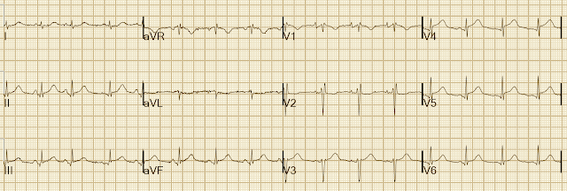
ST- and PR-segment changes are relative to the baseline formed by the T-P segment. The degree of ST elevation is typically modest (0.5 – 1mm). Pericarditis is classically associated with ECG changes that evolve through four stages. Stage 1 – widespread STE and PR depression with reciprocal changes in aVR (occurs during the first two weeks)
Is there a negative T wave on ECG with pericarditis?
There are no reciprocal ST segment depressions and no simultaneous T-wave inversions (negative T-waves). The ECG is used to diagnose acute pericarditis. One must always rule out the most serious differential diagnosis, which is ST elevation myocardial infarction (STEM).
Are serial EKG electrocardiograms helpful in acute pericarditis?
Serial electrocardiograms are helpful in patients with acute pericarditis because it causes characteristic 12-lead EKG changes that have typically evolved sequentially through 4 stages 1 2. Concave ST-elevation, PR segment depression, positive T wave. Changes occur within a few hours of the onset of symptoms.
What is the difference between acute and chronic pericarditis?
Recurrent pericarditis: recurrence of pericarditis after a documented first episode of acute pericarditis and a symptom-free interval of 4 – 6 weeks or longer. Chronic pericarditis: pericarditis lasting for >3 months.
Does pericarditis always show on EKG?
In pericarditis, there are hallmark changes that are seen and can help make the diagnosis. While an abnormal EKG is helpful in making the diagnosis, in the early stages of inflammation, the EKG may be normal. In most cases of uncomplicated pericarditis, a chest X-ray is usually normal.
Can pericarditis be missed on an ECG?
] suggested that PR-segment deviation is the earliest ECG change in patients with acute pericarditis. Therefore, PR-segment deviation seen in ECG must be important in terms of acute pericarditis diagnosis. We have called this type of pericarditis as atypical pericarditis, for acute pericarditis diagnosis may be missed.
What can mimic pericarditis?
In addition to these conditions, chest pain that can mimic pericarditis is seen in a wide range of conditions including gastric inflammation (gastritis) or ulcers, esophageal inflammation (esophagitis) and gastroesophageal reflux disease (GERD), clots in the arteries of the lung (pulmonary embolism), inflammation of ...
Does pericarditis show on echocardiogram?
Echocardiogram (echo) to see how well your heart is working and check for fluid or pericardial effusion around the heart. An echo will show the classic signs of constrictive pericarditis, including a stiff or thick pericardium that constricts the heart's normal movement.
Can doctors miss pericarditis?
It is often challenging to make a definitive diagnosis of constrictive pericarditis, as no gold standard diagnostic test exists. As a result, the diagnosis of constrictive pericarditis is commonly missed.
What is the best test for pericarditis?
The diagnostic test of choice for large effusions, cardiac tamponade, and constrictive pericarditis is two-dimensional Doppler echocardiography.
Will chest xray show pericarditis?
Cardiac CT scans use X-rays to create images of the heart and chest. The test can be used to look for heart thickening that may be a sign of constrictive pericarditis.
Can you have pericarditis without pericardial effusion?
The shadow of the heart may appear enlarged if there is a large accumulation of fluid (pericardial effusion) in the pericardial sac. However, most people with sudden onset (acute) pericarditis have a normal chest x-ray since there is frequently only a small or no pericardial effusion.
Can you have pericarditis without fever?
Sometimes acute pericarditis does not cause any symptoms. Pericarditis due to tuberculosis begins insidiously, sometimes without obvious symptoms of infection. It may cause fever and symptoms of heart failure, such as weakness, fatigue, and difficulty breathing.
What are two classic findings of pericarditis?
Characteristic clinical findings in pericarditis include pleuritic chest pain and pericardial friction rub on auscultation of the left lower sternal border. Electrocardiography may reveal diffuse PR-segment depressions and diffuse ST-segment elevations with upward concavity.
Is pericarditis hard to diagnose?
The diagnosis of constrictive pericarditis is difficult both for its rarity and because it is often obscured by other, more common, diagnoses.
Is pericarditis pain intermittent?
Incessant pericarditis lasts about four to six weeks but less than three months. The symptoms are continuous. Chronic constrictive pericarditis usually develops slowly and lasts longer than three months.
What is pericardial inflammation?
Definition: Diffuse inflammation of the pericardial lining surrounding the heart and characterized by sharp pleuritic, retrosternal chest pain worsened with recumbency and relieved by leaning forwards.
How long does it take for an EKG to evolve?
The duration for evolution through each of the 4 ECG stages is highly variable ranging from hours to weeks. Practically, Stage I is the only diagnostic phase because Stage II looks normal and Stage III mimics ischemia.
What are the two forms of pericarditis?
There are two forms of pericarditis: acute and chronic. This article will focus on the former, as it has implications for all clinicians and the ECG. Acute pericarditis causes chest pain, which may be very difficult to discern from pain caused by acute myocardial infarction.
What causes acute pericarditis?
Causes of acute pericarditis/myocarditis. The most frequent cause of pericarditis is infections, in particular viral infections. This explains why pericarditis may affect individuals of all ages. However, a wide range of local and systemic conditions may cause pericarditis. The most common causes are as follows:
What is the ECG used for?
The ECG is used to diagnose acute pericarditis. One must always rule out the most serious differential diagnosis, which is ST elevation myocardial infarction (STEM). In order to provide the reader with knowledge on this matter, we will now discuss the characteristics of all ECG changes seen in acute pericarditis, and contrast them to ECG changes seen in STEMI.
What is the pericardium?
The pericardium is a double-walled sac in which the heart and the roots of the great vessels are contained (Figure 1). The pericardial sac encloses the pericardial cavity which contains pericardial fluid. Numerous conditions may cause inflammation in the pericardium, ...
Is pericarditis a myocardial sac?
Note that pericarditis (inflammation of the pericardial sac) is difficult to discern from myocarditis (inflammation of the myocardial tissue) and because they tend to accompany each other, the term perimyocarditis is often used. Image by Bruce Blausen, Blausen Gallery 2014.
Is perimyocarditis a clinical term?
Therefore, the term perimyocarditis is often used in clinical practice (this article will use all three terms interchangeably). The etiology, clinical characteristics and ECG features of pericarditis will be discussed here.
Is myocarditis the same as pericarditis?
Pericard itis refers to inflammation of the pericardium, and myocarditis refers to inflammation of the myocardial ( muscle) tissue. However, it is often difficult to differentiate pericarditis and myocarditis, and they tend to accompany each other.
What are the criteria for acute pericarditis?
A. A. Currently, the diagnosis of acute pericarditis is based on demonstrating at least two of the following four criteria: 1. Non-ischemic chest pain, 2. ECG evidence of PR depression or ST segment deviation, 3. Detection of a pericardial rub on auscultation and 4.
What is the pain in the chest from pericarditis?
The chest pain of pericarditis can vary from severe substernal discomfort to a vague "ache". The chest pain is usually positional, not related to exertion and often radiates to the neck, ridge of the trapezius muscle or shoulder.
Can pericarditis be detected with MRI?
Many of these patients, on the verge of being labeled with a psychogenic etiology, are extremely grateful when their pericarditis is detected with cardiac MRI, opening the door for effective therapy and most importantly, relief of their persistent symptoms.
What is the most common symptom of pericarditis?
Chest pain is the most common symptom of pericarditis, it is most often sharp and pleuritic in nature. Usually it is precordial or retrosternal, with exacerbation by inspiration or coughing, and it decreases in intensity when the patient sits up and leans forward.
How long does it take for pericarditis to recur?
Recurrent pericarditis: Recurrent pericarditis is diagnosed with a documented first episode of acute pericarditis, a symptom-free interval of 4 – 6 weeks or longer and evidence of subsequent recurrence of pericarditis.
What is acute pericarditis?
Acute pericarditis is an inflammation of the pericardium. This inflammation causes EKG changes that have typically evolved sequentially through 4 stages 1. Acute pericarditis can be difficult to distinguish from ST-segment elevation myocardial infarction.
What is pericardial friction rub?
The presence of a pericardial friction rub on physical examination is pathognomonic for acute pericarditis. It is generated by friction between the two inflamed layers of the pericardium.
When should an electrocardiogram be performed?
An electrocardiogram should be performed when there is suspicion of acute pericarditis. EKG may be diagnostic in acute pericarditis, changes might occur within a few hours of the onset of symptoms, but changes are not always present.
Can corticosteroids cause pericarditis?
Low-dose corticosteroids should be considered for acute pericarditis in cases of contraindication or failure of aspirin/NSAIDs and colchicine, and when an infectious cause has been excluded, or when there is a specific indication such as autoimmune disease 3. Corticosteroids are not recommended as first-line therapy 3.
Can a PR segment depression be seen in EKG?
This electrocardiographic sign is more specific for acute pericarditis, although it is less sensitive. At this stage, T wa ve remains positive in most of the EKG leads.
What are the stages of acute pericarditis?
Typical ECG findings in acute pericarditis are present in no more than 60% of cases and evolve in four stages (Figure 1). In particular, ECG initially (within hours to days) depicts diffuse concave ST-segment elevation (in all leads, except aVR and often V1) with concomitant PR depression (first stage), followed by (within the first week) return to baseline of the latter deviations and T-wave flattening (second stage), diffuse T-wave inversion after the ST-segment has become isoelectric (third stage) and, finally, ECG normalisation or persistence of T-wave inversions (fourth stage). [ 6-8] Sustained atrial or ventricular arrhythmias are not frequent in acute pericarditis, and their presence denotes concomitant myocarditis or another not-relevant cardiac disease. [ 9]
What is the most common cause of pericarditis in developed countries?
In particular, the most common cause of pericarditis in developed countries is viruses, whereas tuberculosis is the most frequent cause in developing countries. With regard to endemic regions, tuberculosis often coexists with human immunodeficiency virus (HIV).
What is the most common form of pericardial disease?
Inflammation of the pericardial sac is called pericarditis. The main pericardial syndromes encompass pericarditis (acute, subacute, chronic, recurrent), pericardial effusion, cardiac tamponade and pericardial masses. Pericarditis is the most common form of pericardial disease worldwide and is typically encountered in young and middle-aged people.
How many phases does pericardial rub have?
It typically consists of three phases corresponding to movement of the heart during atrial systole (absent in atrial fibrillation), ventricular systole, and the rapid filling phase of early ventricular diastole. [ 2, 6] Sometimes pericardial rub has only two components or even one component.
What is the pericardial rub?
The medical history often reveals symptoms suggestive of viral infection. Auscultation exposes a pericardial friction rub (pathognomonic sign for pericarditis) due to increased friction of inflamed pericardial layers in about one-third of patients with acute pericarditis.
What are the most commonly used markers for pericarditis?
Among them, the most widely used are the white blood cells (WBCs), erythrocyte sedimentation rate (ESR) and C-reactive protein (CRP).
Is pericarditis a myocardial ischaemia?
Acute pericarditis is usually distinguished from myocardial ischaemia or infarction (based on the clinical findings, ECG, markers of myocardial necrosis and imaging modalities such as echocardiography), but coronary angiography is sometimes required to resolve the issue. Finally, not infrequen tly, a cute pericarditis is a manifestation of a presenting, silent MI.