
Which MRI findings are characteristic of adhesive capsulitis?
The MRI findings that suggest adhesive capsulitis include soft tissue thickening in the rotator interval, which may encase the coracohumeral and superior glenohumeral ligaments, and soft tissue thickening adjacent to the biceps anchor (2a, 6a). A thickened inferior glenohumeral ligament greater than 4 mm is often seen in the axillary pouch (2b).
Can you have adhesive capsulitis in one shoulder?
You can have adhesive capsulitis in one or both shoulders. The condition has 3 stages: Stage 1 is called the freezing or painful stage and may last 2 to 9 months. Stage 2 is called the adhesive stage and may last 4 to 12 months. You may have less pain. You may still have pain when you move your arm to reach.
Which tests are used to diagnose adhesive capsulitis (AC)?
In the early stages of disease, MRI is often able to identify and differentiate adhesive capsulitis from other shoulder pathologies, affording an earlier diagnosis and a more clinically effective treatment plan. Chi AS, Kim J, Long SS, Morrison WB, Zoga AC.
How long does adhesive capsulitis last?
Adhesive capsulitis may last from several months to years before it gets better on its own. You can have adhesive capsulitis in one or both shoulders. The condition has 3 stages: Stage 1 is called the freezing or painful stage and may last 2 to 9 months. Stage 2 is called the adhesive stage and may last 4 to 12 months. You may have less pain.
Can MRI detect frozen shoulder?
Medical imaging. A doctor may order: X-rays of the shoulder to identify any bone-related issues, such as bone spurs. Magnetic resonance imaging (MRI) to identify any damage to soft tissues, such as a rotator cuff tear. While an MRI can potentially show inflammation, it cannot definitively diagnose frozen shoulder.
What does adhesive capsulitis look like on MRI?
The MRI findings that suggest adhesive capsulitis include soft tissue thickening in the rotator interval, which may encase the coracohumeral and superior glenohumeral ligaments, and soft tissue thickening adjacent to the biceps anchor (2a, 6a).
How do you test for adhesive capsulitis?
A 2017 study concluded that adhesive capsulitis can be accurately and consistently diagnosed with noncontrast magnetic resonance imaging (MRI) of the shoulder in conjunction with appropriate clinical criteria.
What does frozen shoulder look like on an MRI?
0:4210:16How Frozen shoulder looks on MRI - YouTubeYouTubeStart of suggested clipEnd of suggested clipAnd you can see the ligament it's not really thickened it's how it's supposed to be in theMoreAnd you can see the ligament it's not really thickened it's how it's supposed to be in the literature some have a cut-off value of 3 or 4 millimeters depending on how you measure. So this is normal.
What are the stages of adhesive capsulitis?
Frozen shoulder, also known as adhesive capsulitis, is a common presentation in the primary care setting and can be significantly painful and disabling. The condition progresses in three stages: freezing (painful), frozen (adhesive) and thawing, and is often self-.
How is frozen shoulder syndrome treated?
Treatment for frozen shoulder involves range-of-motion exercises. Sometimes treatment involves corticosteroids and numbing medications injected into the joint. Rarely, arthroscopic surgery is needed to loosen the joint capsule so that it can move more freely.
What is the hallmark of adhesive capsulitis?
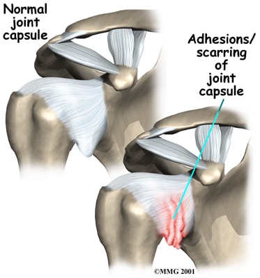
Contracture of the glenohumeral capsule is the hallmark of adhesive capsulitis. Findings include loss of the synovial layer of the capsule, adhesions of the axillary to itself and to the anatomical neck of the humerus, and overall decreased capsular volume.
Is there a special test for adhesive capsulitis?
pathologies that may be limiting shoulder ROM and causing pain. These tests include, but are not limited to the empty can test, Speed's test, drop arm test, and Neer and Hawkin's impingement tests. There is no one specific special test that confirms the diagnosis of adhesive capsulitis.
How can you tell the difference between adhesive capsulitis and rotator cuff tear?
One key finding that helps differentiate a frozen shoulder from a rotator cuff tear is how the shoulder moves. With frozen shoulder, the shoulder motion is the same whether the patient or the doctor tries to move the arm. With a rotator cuff tear, the patient may have difficulty moving the arm.
What can an MRI of the shoulder reveal?
MRI gives clear views of rotator cuff tears, injuries to the biceps tendon and damage to the glenoid labrum, the soft fibrous tissue rim that helps stabilize the joint. MR imaging of the shoulder is typically performed to diagnose or evaluate: degenerative joint disorders such as arthritis and labral tears.
Who can diagnose frozen shoulder?
To diagnose frozen shoulder, a Penn orthopaedic specialist will evaluate your symptoms and examine your shoulder and arm to assess your range of motion and pain levels. Musculoskeletal radiologists carefully review imaging to determine the severity of your condition so you can receive the best possible treatment.
Does frozen shoulder hurt in the front or back of the shoulder?
Signs Of Frozen Shoulder The pain associated with this condition can be both dull or aching and usually located over the front of the shoulder and sometimes the upper arm. Pain tends to be worse early in the course of the disease, during the freezing stage, and with movement of the arm.
What is primary adhesive capsulitis?
Idiopathic (“primary”) adhesive capsulitis occurs spontaneously without a specific precipitating event. Primary adhesive capsulitis results from a chronic inflammatory response with fibroblastic proliferation, which may actually be an abnormal response from the immune system.
What causes frozen shoulder symptoms?
What causes frozen shoulder? Although many shoulder diseases involve pain and loss of motion, frozen shoulder is most often caused by inflammation (swelling, pain and irritation) of the tissues surrounding the joint. The tissue that envelops the joint and holds it together is called the capsule.
What is a subacromial bursitis?
Subacromial bursitis is a common etiology of shoulder pain. It results from inflammation of the bursa, a sac of tissue present under the acromion process of the shoulder. It is usually brought about by repetitive overhead activities or trauma.
What is a Type 2 acromion of the shoulder?
A type II acromion is considered to be one that in which the acromion has a down-sloping character. A type III acromion is considered to be present when there is significant downward hooking of the acromion consequently greatly restricting the caliber of the subacromial arch area.
What is adhesive capsulitis?
Adhesive capsulitis of the shoulder (frozen shoulder) is a common cause of pain and limitation of motion with an incompletely understood and complex pathogenesis. The diagnosis is commonly made through a combination of clinical history and physical examination findings, but early signs of adhesive capsulitis can be nonspecific and overlap with other causes of shoulder stiffness. 1, 2 Recognizing the characteristic MRI findings of adhesive capsulitis enables the radiologist to make an earlier diagnosis of adhesive capsulitis in clinically confusing cases.
How long does adhesive capsulitis last?
While idiopathic adhesive capsulitis is generally considered a self-limited disease, resolving between 1 and 3 years, several studies have shown that a significant percentage of patients can have persistent symptoms with a more protracted and disabling course. 36,5,16, 37, 38,20
Which ligaments are affected by adhesive capsulitis?
The pathologic changes of adhesive capsulitis can affect the capsule diffusely, but the rotator interval, coracohumeral ligament, axillary joint capsule, and the inferior glenohumeral ligament have been most closely investigated because involvement in these locations is believed to be most closely associated with the early clinical findings of pain and limitation in motion. 23, 24 ,23, 25 ,19, 26
What is the arthroscopic view of the inferior capsule?
Arthroscopic view of the inferior capsule demonstrates a thickened, hyperemic synovium compatible with stage 2 adhesive capsulitis. (Image courtesy of David Moore, MD, Elite Sports Medicine and Orthopedics, Nashville, TN.)
Does gadolinium enhance rotator axillary joint?
Enhancement of the rotator interval and of the axillary joint capsule following intravenous gadolinium contrast administration shows the highest sensitivity (90%) for adhesive capsulitis with a specificity of >80%. 25 Enhancement is often associated with increased T2 hyperintensity and thickening of these structures (Figure 11).
Can a MRI show a rotator cuff injury?
Calcific tendinitis, rotator cuff injury, biceps tendon pathology, and glenohumeral and acromioclavicular arthritis can all present with similar clinical findings of shoulder stiffness and pain. The MRI features of synovial and capsular thickening can be observed following severe shoulder trauma, such as with a shoulder dislocation. A careful clinical history in conjunction with familiarity with the MRI findings help to effectively identify and differentiate adhesive capsulitis from these other conditions.
Is adhesive capsulitis a clinical diagnosis?
Adhesive capsulitis is a clinical diagnosis, but the physical exam findings can be confusing, especially in the early stages or with concomi tant shoulder pathologies such as rotator cuff impingement, bursitis, and labral pathology which may present with overlapping clinical features. 23,5,26 With its widespread availability, multiplanar capability, anatomic detail, and soft tissue differentiation, MRI is the favored imaging modality for the evaluation of shoulder pathology. The findings associated with adhesive capsulitis are depicted by routine MRI, MRI with intravenous contrast, and MR arthrography. In our practice, identification and evaluation of adhesive capsulitis is most often by routine MRI, followed by intravenous contrast-enhanced MRI. MR arthrography appears to improve diagnostic accuracy but it is unlikely to be necessary to confirm the diagnosis when clinical history and less invasive imaging procedures are considered. 20
What are the signs and symptoms of adhesive capsulitis?
Adhesive capsulitis may last from several months to years before it gets better on its own. You can have adhesive capsulitis in one or both shoulders. The condition has 3 stages:
How is adhesive capsulitis diagnosed?
Your healthcare provider will do an exam. He or she will check your neck and shoulder. He or she will check how your shoulder moves and how strong it is. Your provider may move your arm in different positions while you stand or lie down. You may also need the following:
What is the goal of adhesive capsulitis treatment?
The goal of treatment is to help you regain as much shoulder movement as possible. Treatment will depend on what stage you are in. Ask your healthcare provider about these and other treatments for adhesive capsulitis:
Why is my shoulder frozen?
The condition is often called frozen shoulder because the swollen tissues cause pain and decrease your shoulder movement.
How to get your shoulder to move?
Heat helps relax muscles and may help improve shoulder movement. Use a heat pack, or soak a small towel in warm water. Wring out the extra water before you apply the towel to your shoulder. Apply heat for 20 to 30 minutes every hour, or as directed.
How to help a swollen shoulder after stretching?
Apply ice to help ease pain after stretching. Use an ice pack, or put crushed ice in a plastic bag. Cover it with a towel before you apply it to your shoulder. Apply ice for 15 to 20 minutes every hour , or as directed. Apply heat as directed. Heat helps relax muscles and may help improve shoulder movement.
How long does a swollen shoulder last?
The condition has 3 stages: Stage 1 is called the freezing or painful stage and may last 2 to 9 months. Stage 2 is called the adhesive stage and may last 4 to 12 months. You may have less pain. You may still have pain when you move your arm to reach. Your shoulder may still be stiff, and you may not be able to move your shoulder much.
What is the clinical diagnosis of adhesive capsulitis?
The clinical diagnosis of idiopathic adhesive capsulitis relies on the detection of a global decreased range of motion at the glenohumeral joint, absence of previous major trauma, and a normal joint space on plain radiographs. 2 However, these diagnostic criteria are nonspecific, as the clinical features of rotator cuff pathology and impingement often mimic those of adhesive capsulitis.
How long does adhesive capsulitis last?
The natural history of idiopathic or secondary forms of adhesive capsulitis is quite variable with residual pain and stiffness persisting in some studies up to 7 years. 6 Treatment regimens vary greatly, reflecting the complexity of this condition. Supportive treatment, oral and injected medications, physical therapy, the brisement procedure, manipulative therapy, and surgical release are all used in addressing this disorder, depending on the severity and duration of symptoms.
What is frozen shoulder?
Adhesive capsulitis or “frozen shoulder” is an inflammatory condition of the glenohumeral joint synovium and capsule leading to a restricted range of motion. It is most commonly encountered in female patients who are 40 to 60 years of age. Primary or idiopathic adhesive capsulitis is encountered in the absence of preceding trauma. Secondary adhesive capsulitis or post-traumatic arthritis results from antecedent injury, low-level repetitive trauma, surgery, or rheumatologic conditions. Although poorly understood, adhesive capsulitis is felt to begin as an inflammatory hypervascular synovitis, which prompts a progressive fibroblastic response in the adjacent capsule. Capsular thickening and contraction ensue. 1 At arthroscopy, synovial inflammation or capsular thickening may be seen. The abnormalities most commonly involve the rotator interval capsule, the biceps tendon root, and the inferior and posterior capsule.
Epidemiology
Clinical Presentation
- Adhesive capsulitis presentation can be broken into three distinct stages: 1. freezing: painful stage 1.1. patients may not present during this stage because they think that eventually, the pain will resolve if self-treated 1.2. as the symptoms progress, pain worsens and both active and passive range of motion (ROM) becomes more restricted 1.3. thi...
Pathology
- Adhesive capsulitis is divided into two main types: 1. primary or idiopathic 1.1. absence of preceding trauma 2. secondary 2.1. major or minor repetitive trauma 2.2. shoulder or thoracic surgery 2.3. endocrine, e.g. diabetes, hyperthyroidism 12 2.4. rheumatological conditions
Radiographic Features
- Described features on fluoroscopic arthrography include: 1. limited injectable fluid capacity of the glenohumeral joint 2. small dependent axillary fold 3. small subscapularis bursa 4. irregularity of the anterior capsular insertion at the anatomic neck of the humerus 5. lymphatic filling may be present 1. limitation of movement of the supraspinatus is considered a sensitive feature 7 2. lim…
Treatment and Prognosis
- Adhesive capsulitis is typically a self-limiting disease that improves over 1-2 years. Treatment options include: 1. physiotherapy 2. corticosteroid injections 3. glenohumeral hydrodilatation 4. closed manipulation under anesthesia 5. arthroscopic capsular release with lysis of adhesions