How are blood vessels innervated by neurons?
Blood vessels can receive innervation from three main classes of neurons: sympathetic vasoconstrictor neurons, sympathetic or parasympathetic vasodilator neurons, and sensory neurons that mediate vasodilation.
What is the difference between parasympathetic and vasodilation?
Parasympathetic stimulation also relaxes the smooth musculature of the peripheral blood vessels, which results in the vasodilation of peripheral vasculature. In contrast to this, acting on the smooth muscles of the coronary vessels, the parasympathetic nerves cause their vasoconstriction in response to reduced demand for oxygen.
Where is the parasympathetic nerve found in the body?
An estimated 75 percent of all parasympathetic nerve fibers in the body come from this nerve. This nerve has branches in many key organs, including the stomach, kidneys, liver, pancreas, gallbladder, bladder, anal sphincter, vagina, and penis.
How do the sympathetic and parasympathetic nerves work together?
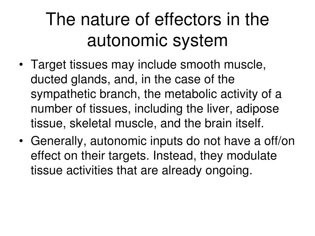
The sympathetic and parasympathetic nerves work together to balance the functions of autonomic effector organs. The neurotransmitters released from the varicosities in the ANS can regulate the vascular tone.
See more

Do blood vessels have sympathetic and parasympathetic innervation?
Most arteries and veins in the body are innervated by sympathetic adrenergic nerves, which release norepinephrine (NE) as a neurotransmitter. Some blood vessels are innervated by parasympathetic cholinergic or sympathetic cholinergic nerves, both of which release acetylcholine (ACh) as their primary neurotransmitter.
Does parasympathetic control blood vessels?
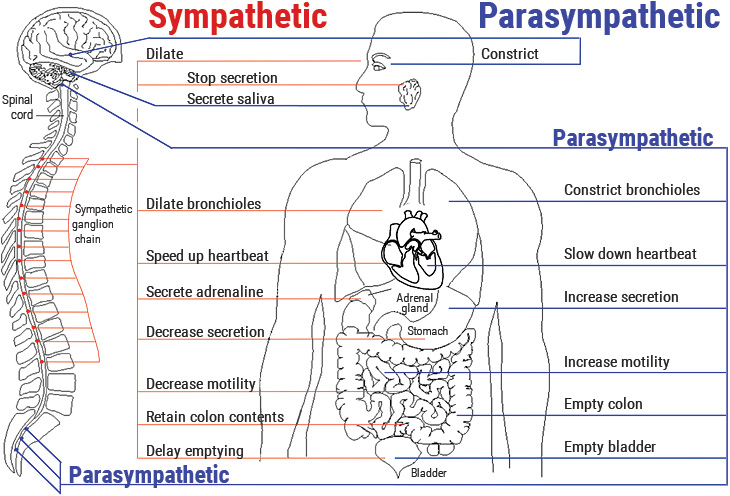
The parasympathetic division directs the body toward a “rest or digest” mode, generally decreasing heart rate and blood pressure. Under parasympathetic stimulation, blood vessels normally dilate, increasing blood flow but lowering pressure.
Do blood vessels have only sympathetic innervation?
Although most organs are innervated by both sympathetic and parasympathetic nerves, some-including the adrenal medulla, arrector pili muscles, sweat glands, and most blood vessels-receive only sympathetic innervation.
Is vasoconstriction of blood vessels sympathetic or parasympathetic?
Cutaneous vasoconstriction is predominantly controlled through the sympathetic part of the autonomic nervous system. Most sympathetic activation promotes vasoconstriction.
Does vagus nerve innervate blood vessels?
The right vagus nerve primarily innervates the SA node, whereas the left vagus innervates the AV node; however, there can be significant overlap in the anatomical distribution. Atrial muscle is also innervated by vagal efferents, whereas the ventricular myocardium is only sparsely innervated by vagal efferents.
What does the parasympathetic vagus nerve do to blood vessels?
The parasympathetic side, which the vagus nerve is heavily involved in, decreases alertness, blood pressure, and heart rate, and helps with calmness, relaxation, and digestion. As a result, the vagus nerve also helps with defecation, urination, and sexual arousal.
Which organ is not innervated by the parasympathetic division?
The answer is (a) adipose tissue. Adipose tissue is innervated by the sympathetic nervous system, but not the parasympathetic nervous system. The stomach, gallbladder, and large intestine are all involved in digestion and therefore receive innervation from the parasympathetic nervous system.
Which organ is innervated only by parasympathetic nerves?
The glossopharyngeal and vagus parasympathetic nerves innervate glands of the upper tract; these include the salivary glands, esophageal glands, gastric glands, pancreas, and Brunner's glands in the duodenum. Glands in the large intestine also receive parasympathetic innervation.
Is increased blood pressure sympathetic or parasympathetic?
Sympathetic activity is increased in hypertension and heart failure, and is responsible for initiation and development of the diseases [7, 8, 31].
How does the parasympathetic nervous system affect blood pressure?
The parasympathetic nervous system decreases respiration and heart rate and increases digestion. Stimulation of the parasympathetic nervous system results in: Construction of pupils. Decreased heart rate and blood pressure.
What happens to blood vessels in fight or flight?
As nitric oxide relaxes blood vessels, a reduction in its levels causes blood vessels to contract and can increase the risk of a heart attack.
What happens to blood vessels during flight and fight response?
In general, activation of alpha-adrenergic receptors results in the constriction of blood vessels, contraction of uterine muscles, relaxation of intestinal muscles, and dilation of the pupils.
Does the sympathetic nervous system constrict blood vessels?
In blood vessels, sympathetic activation constricts arteries and arterioles (resistance vessels), which increases vascular resistance and decreases distal blood flow. When this occurs throught the body, the increased vascular resistance causes arterial pressure to increase.
How does the parasympathetic nervous system affect blood pressure?
The parasympathetic nervous system decreases respiration and heart rate and increases digestion. Stimulation of the parasympathetic nervous system results in: Construction of pupils. Decreased heart rate and blood pressure.
Does parasympathetic increase blood pressure?
This study indicates the importance of behavioural modification in hypertension and that increased parasympathetic function is associated with success in reduction of BP.
Is increased blood pressure sympathetic or parasympathetic?
Sympathetic activity is increased in hypertension and heart failure, and is responsible for initiation and development of the diseases [7, 8, 31].
Where do parasympathetic nerves come from?
Trusted Source. of all parasympathetic nerve fibers in the body come from this nerve. This nerve has branches in many key organs, including the stomach, kidneys, liver, pancreas, gallbladder, bladder, anal sphincter, vagina, and penis.
Where does the parasympathetic nervous system start?
Parasympathetic nervous system function. Your PSNS starts in your brain and extends out via long fibers that connect with special neurons near the organ they intend to act on. Once PSNS signals hit these neurons, they have a short distance to travel to their respective organs.
What is the acronym for parasympathetic response?
Examples of parasympathetic responses. An easy acronym to remember how and where the PSNS works is SLUDD. This stands for: Salivation: As part of its rest-and-digest function, the PSNS stimulates production of saliva, which contains enzymes to help your food digest.
What happens if your parasympathetic nervous system doesn't work?
Your PSNS is a vital part of your body’s key functions. When it doesn’t work properly, you can face a number of bodily dysfunctions that affect your health. If you think you may be having trouble with one of your body’s parasympathetic nervous system functions, talk to your doctor to find out how you can get help.
What is the nervous system?
Your nervous system is a wild and wonderful network of nerves that act in different key functions to keep your body moving, responding, sensing, and more. This article is going to examine the parasympathetic nervous system, one of two majors divisions of the larger autonomic system. In the simplest terms, the parasympathetic ...
What are the receptors in the heart?
Parasympathetic nervous system and your heart. There are a number of special receptors for the PSNS in your heart called muscarinic receptors. These receptors inhibit sympathetic nervous system action. This means they’re responsible for helping you maintain your resting heart rate.
What is the difference between resting heart rate and sympathetic nervous system?
This means they’re responsible for helping you maintain your resting heart rate. For most people, the resting heart rate is between 60 and 100 beats per minute. On the other hand, the sympathetic nervous system (SNS) increases heart rate. A faster heart rate (usually) pumps more oxygen-rich blood to the brain and lungs.
Which nerves innervate the muscular and endothelial layers of blood vessels?
The cholinergic nerve endings innervate the muscular and endothelial layers of blood vessels [49]. It has been shown that the M3 AChRs on the endothelium mediate vasorelaxation by regulating the release of NO in arteries. In contrast, the activation of M2 and M3 receptors on smooth muscle cells by acetylcholine released from cholinergic nerve induces vascular contraction by inhibiting the NO production [50]. Another study showed that when M3 AChRs are inhibited by atropine on the endothelial cells, activation of nicotine receptors by Ach contributes to the endothelium-dependent relaxation through the PI3K/AKT pathway in the aorta of hypertensive animals [51]. Activation of the primary PSNS neurotransmitter acetylcholine receptors on endothelial cells induces vascular relaxation in human coronary arteries [52]. The cholinergic nerve innervation of vascular tone is a complicated process and needs to be further studied.
What is the role of the sympathetic nerve in the heart?
The sympathetic nerve enables organisms to respond in a proper manner to destabilizations in either internal or external circumstances. Sympathetic regulation is a critical component of vascular tone and blood pressure, and plays a pivotal role in maintaining cardiovascular homeostasis and regular physiological functions. Extrinsic stimuli, such as stress, trauma, hemorrhage and pain, can elevate the sympathetic nerve activity, which can directly increase vascular resistance. Major arteries and precapillary arterioles are innervated by sympathetic nerves, but other vessels, such as venules, capillaries and collecting veins are rarely innervated [5].
How does the endothelium regulate vessel tone?
In a healthy state, the endothelium and ANS work together to regulate vessel tone. The vasodilator and vasoconstrictor substances released from endothelial cells can maintain the vascular tone properly via affecting the activity of ANS. An earlier study reported that the release of NO by endothelium can blunt vasoconstrictive responses induced by α2-adrenergic nerve [31]. It has been shown that the endothelium inhibits neurogenic NE induced vasoconstriction in the presence of shear stress in isolated perfused rabbit carotid arteries, but the inhibition decreases by a guanylate cyclase inhibitor [76]. That suggested that endothelium under shear stress can modulate adrenergic vasoconstriction likely due to the production of NO [76]. A subsequent study reported that neurogenic vasoconstriction by NE is modulated by endothelial factor NO in the rat tail artery [77]. The endothelium may also control the metabolism of NE and provide a physical barrier of neurotransmitter diffusion into the blood vessel lumen [78]. In addition, NO not only inhibits the central but also the peripheral cardiac and vascular sympathetic activity [79]. ANS activity can also be influenced by endothelial constriction factors. The levels of endothelin can evoke different responses. Higher levels of endothelin increase the sensitivity of smooth muscle cells to NE and induce higher arterial pressure through enhanced vasoconstriction, while lower endothelin inhibits the sympathetic activity and results in slight decreases in arterial pressure through a suppression in vasoconstriction [80,81].
What is the function of the vascular system?
The function of the blood vessel is to nourish the living body and maintain homeostasis. The functional integrity of the endothelial cells is a vital factor in vascular homeostasis. The dysfunction of endothelium is a fundamental element in the progression of atherosclerosis [1]. Risk factors such as hypertension, heart failure and diabetes mellitus impair endothelial function. In addition, the factors outside of vessels can also influence the vascular system. The autonomic nervous system (ANS) is involved in mediating the behavior of the endothelial function. In the anatomical view of ANS, endothelial cells (ECs) do not receive direct SNS innervation due to the long distances [2]. However, Neurotransmitters released from varicosities in the perivascular plexus via autonomic neuroeffector junctions can reach endothelial receptors and regulate endothelial function [3]. In the development of vascular diseases, the co-existence of ANS abnormality and endothelial dysfunction suggests the complex interactions between them.
What is NPY in the nervous system?
NPY is a 36 amino-acid neuropeptide widely distributed in the central nerves and the peripheral nerves . It is a sympathetic cotransmitter colocalized and released with NE in the SNS innervating the cardiovascular system [67,68]. NPY was initially shown to have a mild direct constriction effect on smooth muscle cells in the cerebral vasculature [69] but potentiated the NE-induced vasoconstriction powerfully in the isolated rabbit blood vessels [67]. The release of NPY results in endothelium-dependent constriction and proliferation of smooth muscle cells. NPY interacts with its receptors expressed on the endothelium and regulates angiogenesis by an autocrine signaling mechanism during tissue development [70]. NPY also plays a potential role in increasing leukocyte adhesion to endothelial cells [71]. The NPY antagonist PP56 has been shown to have an anti-inflammatory effects on both acute and chronic arthritis [72].
What is the function of ATP in the body?
It is well established that ATP is a major intracellular energy source. As an excitatory cotransmitter in autonomic nerves, however, ATP acts at the purinergi c receptor (P2X and P2Y) to medicate the extracellular actions . The subtypes of purinergic receptors are P1 and P2 receptors. There are four P1 receptors (A1, A2A, A2B and A3) all binding with G proteins, and they are expressed on vascular endothelium and smooth muscle to mediate the vascular tone and smooth muscle proliferation. P2 receptors, based on signal transduction mechanisms and different molecular structure, are divided into ionotropic P2X receptors and G protein-coupled P2Y receptors [37]. To date, there are seven P2X receptor proteins (P2X1-P2X7) [38], and eight P2Y receptor proteins (P2Y1,2,4,6,11,12,13,14). The main receptors expressed on vascular smooth muscle cells are P2Y1, P2Y2, P2Y4 and P2Y6. Binding of ATP to these receptors activates phospholipase C by coupling to Gq/11proteins and leads to elevation of intracellular calcium levels. Endothelial cells mainly express functional P2Y receptors, P2Y1 and P2Y2, and some vessels may also express P2Y4 and P2Y6 receptors [39,40].
What are the receptors that activate the vagal afferents?
Peripheral vagal afferents can be activated by responding directly to endotoxin, bacterial lipopolysaccharide (LPS) and cytokines, such as interleukin-1 (IL-1), tumor necrosis factor (TNF), interleukin-6 (IL-6) and interferon-gamma (IFN-γ). IL-1 receptors are expressed on vagal afferents and glomus cells (chemosensory) adjacent to the vagus nerve endings which can be activated by inflammatory stimulation to regulate the immune responses [18,19]. Activating vagus nerve efferents can significantly suppress systemic pro-inflammatory cytokine levels in rodents with endotoxemia as the acetylcholine (Ach) released from the nerve terminals binds to the α-7 nicotinic acetylcholine receptor (α7nAChR) expressed on macrophages to modulate the immune system response [20]. α7nAChR, expressed in the nervous and immune systems, is important for mediating anti-inflammatory signaling by inhibiting NF-κB nuclear translocation and activating the JAK2/STAT3 pathway [21-23]. In addition to the role of vagus nervous system (VNS) in regulating systemic inflammation, it also plays an important role in ischemia/reperfusion, sepsis, epilepsy, hemorrhagic shock, migraine, etc. [24,25]. Both animal and human studies have suggested that the vagus nerve stimulation has a potential protective role in various diseases.
How many ways can peripheral nerves be affected?
Peripheral nerves can be affected in three ways ( Fig. 1 ). These mechanisms of injury are not mutually exclusive and in some conditions there are contributions from all three mechanisms:
What are the effects of vascular malformation?
Second, the histopathologic changes described may, in part, be due to the chronic effects of the vascular malformation such as vasodilatation, hypoxia, seizures, and venous hypertension.
Where are the pineal adrenergic nerves located?
3. In the pineal glands of the hamster and the cat , the intrapineal adrenergic nerves are distributed around vessels, as well as in the parenchyma.
Can pulsatile tinnitus be caused by a venous source?
Abrupt onset with unilateral neck or head pains suggests a carotid dissection. A change in tinnitus intensity with head turning suggests a venous source for the tinnitus, from a source ipsilateral to the direction that decreases the tinnitus. This can be confirmed with jugular compression. If the patient can obliterate the tinnitus with mild localized pressure in the periauricular region then an emissary vein is probably accounting for the tinnitus.
Which nerves are involved in ejaculation?
Parasympathetic nerves are essential for proper erectile function, and sympathetic nerves play a role in ejaculation and may also contribute to erectile function; beta-adrenergic blockers sometimes have the side effect of erectile dysfunction. View chapter Purchase book. Read full chapter. ...
Which nerve fibers supply the pelvis?
9.72 Autonomic Pelvic Nerves and Ganglia. Sympathetic nerve fibers supply the pelvis through the sympathetic trunk ganglia and the superior hypogastric plexus. These fibers travel along visceral and vascular nerves to the colon, ureters, and great vessels, such as the inferior mesenteric and common iliac vessels.
Can vascular compression cause unilateral tinnitus?
Hence, vascular compression of the auditory nerve can cause unilateral tinnitus, including pulsatile tinnitus. With rare exceptions ( Fig. 23.1), determining with certainty that an individual's unilateral tinnitus, whether pulsatile or non-pulsatile, is from VIIIth-nerve vascular compression has not yet been possible.
