From a side view of the skull, mammals (humans and whales) possess a lower temporal fenestra — a single opening below and behind the eye socket . For example: crocs and birds have other characters in common, such as an open space in the skull in front of the eye (the antorbital fenestra).
How many temporal fenestrae do humans have?
In the typical diapsid skull, the temporal area has two openings called fenestrae, an upper one between the parietal and the postorbital–squamosal, and a lower one between the squamosal and jugal–quadratojugal.
Where is the human temporal fenestra?
Temporal fenestrae are temporal openings that are completely surrounded by bone. They always form within the sutural contact of two or more temporal bones. An infratemporal fenestra forms in the 'cheek' region of the skull and is ventrally always bordered by a lower temporal bar (i.e. zygomatic arch).
What animals have a temporal fenestra?
Synapsids, including mammals, have one temporal fenestra, while sauropsids, the birds and reptiles, have two.
What does the temporal fenestra do?
Temporal fenestrae are post-orbital openings in the skull that allow muscles to expand and lengthen.
Are humans synapsids?
Humans are synapsids, as well. Most mammals are viviparous and give birth to live young rather than laying eggs with the exception being the monotremes.
Do mammals have synapsid skulls?
Diapsid has two temporal fenestrae in the skull while synapsid has one temporal fenestra in the skull behind each eye. Most reptiles and all birds are diapsids whereas most mammals are synapsids.
Do mammals have temporal holes?
Mammals, which are synapsids, possess no fenestral openings in the skull, as the trait has been modified. They do, though, still have the temporal orbit (which resembles an opening) and the temporal muscles. It is a hole in the head and is situated to the rear of the orbit behind the eye.
Are dinosaurs anapsids?
Anapsids lack temporal fenestrae. Diapsids have two fenestrae on each side and evolved from ancestors that had none. Snakes, lizards, crocodiles, and dinosaurs are diapsids. Testudamorpha (turtles and tortoises), as well as many Paleozoic reptiles, are anapsids.
Is snake a synapsid?
Anapsids have no openings, synapsids have one opening, and diapsids have two openings. The diapsids diverged into two groups, the Archosauromorpha (“ancient lizard form”) and the Lepidosauromorpha (“scaly lizard form”) during the Mesozoic period ([link]). The lepidosaurs include modern lizards, snakes, and tuataras.
What muscles pass through the temporal fenestra in humans?
Temporal fenestrae are openings in the skull that are important attachment sites for jaw-closing (adductor) muscles 13 and show substantial variation among extant amniotes 14 .
Do mammals have fenestrae?
Mammals, which are synapsids, possess no fenestral openings in the skull, as the trait has been modified. They do, though, still have the temporal orbit (which resembles an opening) and the temporal muscles. It is a hole in the head and is situated to the rear of the orbit behind the eye.
What are temporal Vacuities?
Temporal vacuities are holes or openings on the sides of the temporal region of the skull. The function of these openings is thought to allow the expansion and lengthen muscles that results in greater bulk of jaw muscles.
What are temporal Vacuities?
Temporal vacuities are holes or openings on the sides of the temporal region of the skull. The function of these openings is thought to allow the expansion and lengthen muscles that results in greater bulk of jaw muscles.
What is fenestra in biology?
A fenestra (fenestration; plural fenestrae or fenestrations) is any small opening or pore, commonly used as a term in the biological sciences. It is the Latin word for "window", and is used in various fields to describe a pore in an anatomical structure.
How many temporal holes do mammals have?
The temporal fenestra are anatomical features of the amniote skull, characterised by bilaterally symmetrical holes (fenestrae) in the temporal bone. Depending on the lineage of a given animal, two, one, or no pairs of temporal fenestrae may be present, above or below the postorbital and squamosal bones.
Do birds have temporal fenestra?
Birds are decedents of reptiles and possess the two necessary temporal fenestrae to fall under the definition of a diapsid.
What is temporal fenestration?
Temporal fenestration has long been used to classify amniotes (Osborn, 1903). Taxa such as Anapsida, Diapsida, Euryapsida, and Synapsida were named after their type of temporal fenestration. Temporal fenestra are large holes in the side of the skull. The function of these holes has long been debated (Case, 1924), but no consensus has been reached. Many believe that they allow muscles to expand and to lengthen. The resulting greater bulk of muscles results in a stronger jaw musculature, and the longer muscle fibers allow an increase in the gape (Pirlot, 1969).
What is the last type of fenestration?
The last type of fenestration called euryapsid has been the most problematic, partly because the origin of this condition has long been debated. It now appears that this condition is modified from the diapsid condition, and that it appeared more than once. Euryapsid skulls have only an upper temporal fenestra, usually bordered by the parietal, postfrontal, postorbital, and squamosal. Examples of euryapsid skulls include Araeoscelis, a Lower Permian araeoscelidian, placodonts, nothosaurs, and plesiosaurs (marine reptiles of the Mesozoic). Euryapsida is a taxon that includes most known euryapsids, except for Araeoscelis (Rieppel, 1993). The condition found in ichthyosaurs is sometimes distinguished from the euryapsid condition because their temporal fenestra is only bordered by the parietal, postfrontal, and supratemporal (Pirlot, 1969). This condition has been called parapsid, but it only represents a minor variation on the euryapsid pattern.
Do mammals have temporal fenestra?
First, I discover Wikipedia redirects "temporal fenestra" to their article on skulls, which states (in a large section without citations) that "Mammals, which are synapsids, possess no fenestral openings in the skull, as the trait has been modified.") This seems a little strange since the Wikipedia article on Therapsida -- that direct ancestors of mammals -- says they had especially large temporal fenestra, but the article on mammals also mentions nothing about temporal fenestra at all with respect to modern mammals.
Does the temporal fenestra exist in humans?
EDIT: Two years after posting this, /u/mattfen93 messaged me with a knowledgable answer, explaining that the temporal fenestra basically still exists in all ex tant mammals, including humans. His response is below:
Is the temporal fenestra zygomatic?
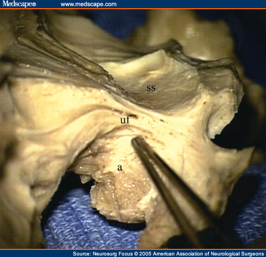
Short answer to your question is: yes, there is an anatomical homologue of temporal fenestra in humans (and to the best of my knowledge, in all the extant mammals), and no, it's not the zygomatic arch, as some redditors suggested in the comments. The temporal fenestra in mammals is reduced to a rather small opening (about 1x1 cm in humans) situated close to the fissure between the posterior part of maxilla ( tuber maxillae) and lateral parts of the sphenoid bone ( processus pterygoideus ). The superior end of this "fissure" is actually the lateral opening of the [pterygopalatine fossa] ( https://thumbor.kenhub.com/By5g-nA5GEkmM8Vx1MtTGiX4Cjs=/fit-in/800x1600/filters:watermark (/images/logo_url.png,-10,-10,0):background_color (FFFFFF):format (jpeg)/images/library/12539/l6iXeJ1PwzYeHdqTrBw_Pterygopalatine_fossa.png ), and this opening is (the remnant of) the synapsid temporal fenestra.
How do temporal fenestrae form?
Formation of temporal fenestrae proceeds by the opening of sutures between bones or the embryological failure to close sutures. Along these lines, temporal fenestrae and other cranial fenestrae associated with the jaw adductor musculature always occur between bones and never within a single bone. Essentially, the sides of fenestrae, that in time usually approach a circular geometry, afford the tendons a low angle insertion and greater area for attachment. Through dissection, it is also seen that other fenestrae in the reptilian skull and jaw, such as the palatine and external mandibular fenestrae, are associated with musculus pterygoideus and m. intramandibularis, respectively. Temporal fenestrae formation has little to do with space needed for muscle bulging during contraction, but are rather correlated with the phylogenetic expansion of jaw adductor size and thus force production. Fenestral patterns can be used systematically because they represent muscle-bone complexes and thus complex character states.
What is the temporal region of a tetrapod?
The morphology of the temporal region in the tetrapod skull traditionally has been a widely discussed feature of vertebrate anatomy. The evolution of different temporal openings in Amniota (mammals, birds, and reptiles), Lissamphibia (frogs, salamanders, and caecilians), and several extinct tetrapod groups has sparked debates on the phylogenetic, developmental, and functional background of this region in the tetrapod skull . This led most famously to the erection of different amniote taxa based on the number and position of temporal fenestrae in their skulls. However, most of these taxa are no longer recognised to represent natural groupings and the morphology of the temporal region is not necessarily an adequate trait for use in the reconstruction of amniote phylogenies. Yet, new fossil finds, most notably of parareptiles and stem-turtles, as well as modern embryological and biomechanical studies continue to provide new insights into the morphological diversity of the temporal region. Here, we introduce a novel comprehensive classification scheme for the various temporal morphotypes in all Tetrapoda that is independent of phylogeny and previous terminology and may facilitate morphological comparisons in future studies. We then review the history of research on the temporal region in the tetrapod skull. We document how, from the early 19th century with the first recognition of differences in the temporal region to the first proposals of phylogenetic relationships and their assessment over the centuries, the phylogenetic perspective on the temporal region has developed, and we highlight the controversies that still remain. We also compare the different functional and developmental drivers proposed for the observed morphological diversity and how the effects of internal and external factors on the structure of the tetrapod skull have been interpreted.
What are the amniotes?
Amniotes include mammals, reptiles and birds , representing 75% of extant vertebrate species on land. They originated around 318 million years ago in the early Late Carboniferous and their early fossil record is central to understanding the expansion of vertebrates in terrestrial ecosystems. We present a phylogenetic hypothesis that challenges the widely accepted consensus about early amniote evolution, based on parsimony analysis and Bayesian inference of a new morphological dataset. We find a reduced membership of the mammalian stem lineage, which excludes varanopids. This implies that evolutionary turnover of the mammalian stem lineage during the Early–Middle Permian transition (273 million years ago) was more abrupt than has previously been recognized. We also find that Parareptilia are nested within Diapsida. This suggests that temporal fenestration, a key structural innovation with important functional implications, evolved fewer times than generally thought, but showed highly variable morphology among early reptiles after its initial origin. Our phylogeny also addresses controversies over the affinities of mesosaurids, the earliest known aquatic amniotes, which we recover as early diverging parareptiles. A new amniote phylogeny excludes varanopids as stem-line mammals, nests Parareptilia within Diapsida and suggests that temporal fenestration evolved fewer times than previously thought.
Where are parareptiles found?
One such taxon, Macroleter poezicus is found in Middle Permian strata of the Mezen River Basin in the Arkhangel'sk Province of Russia. The cranial anatomy of Macroleter is described from four new well-preserved specimens, and 89 cranial characters are incorporated into a phylogenetic analysis of parareptiles. A single most parsimonious topology is found, consisting of 205 steps, with the novel result that Macroleter is the taxon most closely related to pareiasaurs. This result has important implications for the phylogeny of the Parareptilia as well as for the identity of a disputed element (the tabular) in the skull of pareiasaurs.
Why is there no common reference system for muscle nomenclature in vertebrates?
Up to this date, no clear common reference system for muscle nomenclature in vertebrates exists due to 1. human medical anatomy dominated traditions, 2. typological, ‘box-like’ approaches, and 3. simplifications based on the taxonomic and topographical focus of the respective authors. Hence, a large terminological and homologisation confusion in the literature is recognisable, hindering evolutionary and developmental analyses. In this paper, a comprehensive study on the cranial musculature is presented, in which more than 100 references on cranium-associated musculature of turtles were critically reviewed. Following a new traceable approach to muscular terminology, a set of 88 adult ‘muscular units’ – the smallest parts of macroscopic muscular structures – were identified across turtle species, exemplarily demonstrated in a side-necked turtle. For example, the homology of jaw muscle portions and that of epaxial and hypaxial muscular structures are defined by a comprehensive consideration of criteria such as innervation, spatial characteristics, and ontogeny. Adult muscle arrangement variability among specimens, fusions of muscular units, and drop-like apoptosis are recorded. These phenomena are the result of a fluid pattern formation – first driven by neural crest stream patterning in ontogeny. Considering this fact of ontogeny, a new discussion of the evolutionary history of turtles and of particular cranial structures is possible.
Where is the Procolophon found?
The specific composition of the genus Procolophon in Brazil, South Africa and Antarctica is discussed in the light of new data. It is found that P. pricei and P. brasiliensis, two species described from Brazil, fit within the pattern of ontogenetic variation of the type species P. trigoniceps, and they are here considered junior synonyms. The South African species P. laticeps, characterized by the presence of a temporal fenestra, is no longer considered valid. The peculiar temporal openings of this species are regarded here as an anomalous condition without taxonomic significance. The only complete skull known from Antarctica shows a unique feature consisting of an elliptical depression in the palate. The interpretation of this structure is ambiguous because it may also be attributable to individual variation, and this specimen is provisionally kept within P. trigoniceps. Therefore, only the type species, P. trigoniceps, is recognized in Gondwana. This species occupies a wide geographic range, from the Paraná Basin to the Transantarctic Mountains.
Is temporal opening a weak indication for higher taxon interrelationship?
Compared to historical classifications, there is a common consensus that temporal openings are only a weak indication for higher taxon interrelationship, although it can be informative on lower taxonomic level. Here, I present a rather morphofunctional categorization of temporal openings and provide an ontogenetic explanation on their evolutionary origins.