What does an atrial flutter feel like?
Atrial flutter is a common type of heart arrhythmia. You may have no symptoms. If present, symptoms may include a noticeable fast, steady or irregular pulse, shortness of breath, dizziness, trouble with normal activities or exercise, a feeling that your heart is pounding, or tightness in your chest.
What are some alternatives to flutter?
What is the best alternative to Flutter?
- React Native. React Native allows you to create native apps by generating native views with JavaScript instead of using a web wrapper.
- Ionic Framework. Large quantity of plugins to access native APIs without code nothing in native language. ...
- Codename One. ...
Is a flutter regular rate?
Typical atrial flutter is recognized on an electrocardiogram by presence of characteristic "flutter waves" at a regular rate of 200 to 300 beats per minute. Flutter waves may not be evident on an ECG in atypical forms of atrial flutter.
What arrives first P waves or S waves?
P Wave
- Body Waves
- P waves and S waves
- P waves. P waves, or Primary waves, are the first waves to arrive at a seismograph. ...
- S waves. S waves, or secondary waves, are the second waves to arrive during an earthquake. ...
- Difference between s waves and p waves. d=t (S-P).10 Stay tuned with BYJU’S to learn more about Physics-related concepts.
Does atrial flutter have a PR interval?
There is an absence of PR intervals in atrial flutter and fibrillation, ventricular dysrhythmias. In 3rd-degree AV heart block the PR intervals are not measurable.
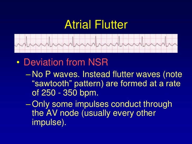
What are flutter waves?
Atrial flutter occurs when a “reentrant” circuit is present, causing a repeated loop of electrical activity to depolarize the atrium at a rate of about 250 to 350 beats per minute; the atrial rate in atrial fibrillation is 400 to 600 bpm.
What does a heart flutter look like on ECG?
Atrial flutter causes the heart to beat in a fast but regular pattern — unlike afib, which causes a fast and irregular pattern. Atrial flutter produces a distinctive "sawtooth" pattern on an electrocardiogram (EKG or ECG), a test used to monitor the heart and diagnose heart rhythm disorders.
What are flutter waves called?
Atrial Flutter and Atrial Fibrillation. Atrial Flutter and Atrial Fibrillation. With atrial flutter the 'P' waves are called flutter waves.
Do you see P waves in SVT?
Sinus tach and most SVTs have only one P wave for each QRS complex. They may or may not be buried in the preceding T waves. But there are other supra-ventricular tachycardias that have more than one P wave for each QRS or no P waves. Atrial fibrillation has no P waves.
Do you have P waves in AFib?
Diagnosis – Atrial Fibrillation. The diagnosis of atrial fibrillation is confirmed with a standard 12-lead ECG. P waves are absent, coarse “fibrillatory waves” can frequently be seen and sometimes no atrial activity can be identified. The QRS complexes are “irregularly irregular”, with varying R-R intervals.
How can you tell the difference between atrial flutter and atrial fibrillation?
Normally, the top chambers (atria) contract and push blood into the bottom chambers (ventricles). In atrial fibrillation, the atria beat irregularly. In atrial flutter, the atria beat regularly, but faster than usual and more often than the ventricles, so you may have four atrial beats to every one ventricular beat.
How can you tell the difference between atrial flutter and SVT?
But they're actually quite different. Atrial fibrillation (AFib) is a heart rhythm problem where your heart's upper chambers (the atria) beat irregularly. Supraventricular tachycardia (SVT) is a fast heart rate that begins in your atria due to abnormal electrical connections in your heart.
How do you detect atrial flutter?
ECG features of atrial flutterNarrow complex tachycardia.Regular atrial activity at ~300 bpm.Loss of the isoelectric baseline.“Saw-tooth” pattern of inverted flutter waves in leads II, III, aVF.Upright flutter waves in V1 that may resemble P waves.Ventricular rate depends on AV conduction ratio (see below)
Are P waves absent in atrial flutter?
Diagnosis – Atrial Flutter Sinus P waves are absent. The classic “sawtooth” pattern occurs, as the reentrant circuit around the tricuspid valve is large, resulting in high-amplitude P waves. Distinguishing between clockwise and counterclockwise atrial flutter was described previously.
Is aflutter regular or irregular?
In people with atrial fibrillation, the pulse is usually rapid and is always irregular. In people with atrial flutter, the pulse is usually rapid and can be regular or irregular.
What are the types of fluttering?
Type I flutter is further divided into two subtypes, known as counterclockwise atrial flutter and clockwise atrial flutter depending on the direction of current passing through the loop. Counterclockwise atrial flutter (known as cephalad-directed atrial flutter) is more commonly seen.
How serious is atrial flutter?
Atrial flutter is not life-threatening. But it can cause serious side effects, including: clots that can travel to the brain and lead to a heart attack or stroke, cardiomyopathy, which occurs when the heart muscle becomes weak and tired, and.
How do you get waves in flutter?
2:064:25How to create Wave animation in Flutter? - YouTubeYouTubeStart of suggested clipEnd of suggested clipAnd then select widget with flutter and mission controller. And then give my animation. You can seeMoreAnd then select widget with flutter and mission controller. And then give my animation. You can see most of the things are auto-generated. So here if you see my clip path.
How long can you live with atrial flutter?
Most patients with atrial flutter lead an entirely normal life with modern drugs and treatments.
Is atrial fibrillation and flutter the same?
Normally, the top chambers (atria) contract and push blood into the bottom chambers (ventricles). In atrial fibrillation, the atria beat irregularly. In atrial flutter, the atria beat regularly, but faster than usual and more often than the ventricles, so you may have four atrial beats to every one ventricular beat.
Why is the P wave narrower?
A notched P wave is usually wider (slower) because there is more tissue to pass through. The first half of the P wave before the notch represents right atrial contraction, the second half of the P wave represents left atrial contraction. A sub-type of the notched P wave is the biphasic P wave.
What is the P wave?
Definition. A P wave on an electrocardiogram represents a phase of electrical activity that causes the atria of the heart to contract. The P wave is a summation wave – electrical activity that comes from successive signaling from multiple points, causing wave-like contractions. These multiple points contain pacemaker cells ...
What Does the P Wave Represent?
The P wave represents electrical activity (in volts) that causes cardiac muscle contraction in the atria – the upper two heart chambers. When a P wave definition says it represents atrial contraction, this is not entirely incorrect.
Where do pacemaker cells send action potentials?
Pacemaker cells form and send action potentials from the sino-atrial node at the top of the right atrium. These spread throughout both atria and stimulate the muscle at the top of the heart to contract.
How long is a P wave?
Normal P wave duration is less than 0.12 seconds (120ms) – about 3 squares on an ECG printout. Depending on the number of leads and positioning of the ECG electrodes, the peak of the P wave is between 1.5 mm and 2.5 mm in height. This corresponds with 0.15 to 0.25 millivolts.
Where are pacemaker cells found?
Pacemaker cells should only be found at the sinoatrial node (SAN) and atrioventricular node (AVN).
Why is the P wave smaller than the R and T waves?
P wave: depolarization of the atria. As gravity helps blood to flow into the ventricles, less muscle contraction is required here. This is why the P wave is smaller than the R and T waves.
How fast are P waves?
There are no existing P waves, although atrial waves with “saw-tooth” pattern are spotted with rates around 300 bpm.
How to tell if a heart is fluttering?
Typical atrial flutter is easily recognised on the electrocardiogram by its well defined “saw-tooth” waves. By observing the inferior leads we can determine the direction of the stimulus and classify it further as counterclockwise atrial flutter or clockwise atrial flutter.
What is the AV ratio of flutter?
Atrial flutter is distinguishable on the electrocardiogram because it is a rhythmic tachycardia with heart rates that are divisors of 300 bpm, 150 bpm being the most frequent in untreated patients (AV conduction ratio 2:1).
What direction does the stimulus go in atrial flutter?
In 90% of the patients with typical atrial flutter the stimulus direction is as described above, but in 10% of patients the stimulus follows the opposite direction— that is, a clockwise direction.
What is the cause of a flutter in the right atrium?
Atrial flutter is an arrhythmia caused by a macro-reentry circuit in the atria (most frequently located in the right atrium) that becomes a self-perpetuating loop. During atrial flutter a very high-rated atrial impulse is originated —between 240 and 350 bpm— but, as in other types of supraventricular tachycardia, ...
What is the treatment for flutter?
The definitive typical flutter treatment is the cavotricuspid isthmus catheter ablation. This is a procedure that causes the interruption of the macro-reentry cycle, and has high success rates and few medical complications.
What is the main feature of the Saw Teeth wave?
Its main feature is a slow descending start, followed by a fast descending phase turning into a fast rise, finishing above the isoelectric line while connecting with the beginning of the following wave (creating thus the “saw-teeth” shape) 3.
What is flutter wave on ECG?
The ECG shows regular flutter waves ( F-waves; not to be confused with f-waves seen in atrial fibrillation) which gives the baseline a saw-tooth appearance. Atrial flutter is the only diagnosis causing this baseline appearance, which is why it must be recognized on the ECG. The flutter waves (on the contrary to f-waves in atrial fibrillation) have identical morphology (in each ECG lead). Flutter waves are typically best seen in leads II, III aVF, V1, V2 and V3. The exact appearance of the flutter waves will depend on the location and direction of the re-entry circuit. In the most common type of atrial flutter, the re-entry loops around the tricuspid valve in a counter-clockwise direction. This yields negative flutter waves in II, III and aVF and positive flutter waves in V1 ( Figure 1 ). If the re-entry has a clockwise direction, the flutter waves are positive in lead II, III, aVF and the P-waves typically have a notch on the apex. Please note that for most clinicians it is not necessary to be able to determine the direction of the re-entry loop.
What direction do flutter waves go?
In the most common type of atrial flutter, the re-entry loops around the tricuspid valve in a counter-clockwise direction.
What is atypical atrial flutter?
Atypical atrial flutter is a consequence of cardiac surgery or extensive ablation therapy. These interventions may cause atrial flutters with very varying ECG appearance. However, flutter waves can still be seen and a history of ablation therapy or cardiac surgery will be sufficient for diagnosing an atypical atrial flutter.
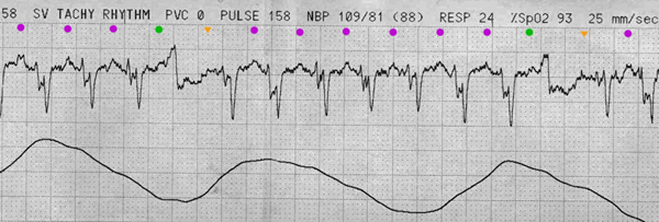
How fast is a ventricular flutter?
In typical cases of atrial flutter the atrial rate is around 300 beats per minute with a 2:1 block, which yields a ventricular rate of about 150 beats per minute. One should always consider atrial flutter when confronted with a regular tachyarrhythmia at 150 beats per minute. Note that with paper speed 25 mm/s, which is standard in the US and many other countries, a 2:1 block will be difficult to discern because the flutter wave may fuse with the preceding T-wave. Increasing the paper speed to 50 mm/s or applying carotid massage (which increases the atrioventricular block) will be helpful in such situations. Figure 2 shows another ECG with atrial flutter.
How fast is the atrial rate?
The atrial rate (i.e the rate measured between flutter waves) typically ranges between 250 and 350 beats per minute (which is slower than the atrial rate in atrial fibrillation ). The atrioventricular node is not capable of conducting all impulses, which is why some impulses will be blocked. The degree of blocking in the atrioventricular node is specified by counting the number of flutter waves preceding each QRS complex. If 3 flutter waves occur before each QRS complex then it is 3:1 block. If there are 2 flutter waves before each QRS complex then it is 2:1 block.
What are the different types of atrial flutter?
Atrial flutter tends to accompany atrial fibrillation, although some individuals may only present with atrial flutter. Similar to atrial fibrillation, atrial flutter can be classified into the following types: 1 Acute atrial flutter (includes newly diagnosed cases). 2 Paroxysmal atrial flutter. 3 Chronic atrial flutter.
Why does my atrial flutter?
This is due to the fact that atrial flutter is caused by a macro re-entry circuit (a large re-entry circuit) and re-entry circuits are vulnerable processes that usually self-terminate within minutes, hours or days.
How to diagnose atrial flutter?
Atrial flutter is diagnosed by you medical history, history of symptoms, and a physical exam. Electrocardiography (ECG or EKG) frequently makes the diagnosis by showing saw tooth flutter waves in several (II, III, aVF and/or V1) of the 12 ECG leads recorded, indicating atrial tachycardia of about 250 – 350 bpm.
What is the rate of heart flutter?
Atrial flutter is a health condition (arrhythmia) where the atria of the heart as an electrical problem (a re-entry loop) that causes the atria to beat at a rapid rate of about 242 - 360 beats per minute (bpm). Atrial flutter is a condition where the atria of the heart rapidly and regularly beat due to an anomaly in the heart's electrical system ...
What are the signs and symptoms of atrial flutter? What does it feel like?
Although a few people have no symptoms, common clinical symptoms of this arrhythmia are as follows:
What causes atrial flutter?
In individuals with these risk factors, some injury probably occurs to alter the healthy electrical pacemaker in the heart atrium that allows a reentry loop for electrical signals to follow. The sinus node sends out an electric signal, but travels along the continuous loop in atrial flutter causing the atria of the heart to contract rapidly, usually with the atria contracting faster than the ventricles although some individuals with the heart disease can have about a 1:1 conduction that results in a heartbeat of about 250 – 300 bpm.
How long does atrial flutter last?
Atrial flutter or atrial fibrillation lasts for variable times. In some people, it can convert to normal sinus rhythm often within a week or so, or it can continue constantly for weeks or months. Some patients may have flutter waves that last less than a day, spontaneously terminate, but return irregularly and are termed paroxysmal atrial flutter. Unfortunately, atrial flutter also can convert to another abnormal heart rhythm such as atrial fibrillation during the same period.
Are atrial flutter, stroke, and heart attack the same?
Atrial flutter is not the same as a stroke or heart attack. It is an abnormal heartbeat that usually is regular and faster than normal. Although it and atrial fibrillation can lead to a stroke or heart attack, it is neither. A stroke is defined as a sudden disabling attack or loss of consciousness caused by an interruption in the flow of blood to the brain. A heart attack is a sudden and sometimes fatal occurrence usually due to coronary thrombosis, resulting in death of part of the heart's muscle.
What are the risk factors for atrial flutter?
There are many risk factors for this type of flutter. The following is a list of some of the more common risk factors:
What is atrial flutter?
Atrial flutter is a re-entrant tachycardia that occurs in the atria. It can occur suddenly, and is sometimes associated with periods of atrial fibrillation. The AV node is bombarded by a regular atrial rhythm of around 300 per minute. New-onset atrial flutter is most often conducted 2:1, because that is a comfortable rate (around 150 per minute) for the AV node to conduct.
Why is ventricular flutter 2:1?
New-onset atrial flutter is most often conducted 2:1, because that is a comfortable rate (around 150 per minute) for the AV node to conduct. When we see slower ventricular rates and conduction ratios of 3:1 or more, it is usually due to medications or other causes of enhanced refractoriness of the AV node. Atrial flutter can lead to fast rates ...
What is the frequency of a P wave in front of each QRS?
This is because the P waves (flutter waves) in atrial flutter occur at about 250-350 per minute (usually around 300). At this rate, it can appear that there is a P wave in front of each QRS and a T wave after each QRS. This causes the misdiagnosis of sinus tachycardia or SVT.
How to get good at recognizing atrial flutter?
Practice rhythm interpretation. Now that you are more aware of atrial flutter with 2:1 conduction, the best way to get good at recognizing it is regular practice. Look at as many strips of confirmed 2:1 conduction as you can and your eye will become trained to see it.
What leads are used for atrial flutter?
In some leads, atrial flutter will not have a sawtooth pattern. You might consider using a Lewis lead , which enhances detection of atrial activity.
Can sinus tach cause atrial flutter?
Consider atrial flutter if the patient has no obvious reason for sinus tachycardia. Most people with sinus tach, especially over 130 bpm, will usually have a readily-apparent reason for the tachycardia, like fever, fear, pain, anxiety, exertion, drugs, hypovolemia or hypoxia.
Can you find an atrial flutter?
You won't find atrial flutter it if you aren’t looking for it. Here are 10 tips to avoid missing atrial flutter.
What is the rate of atrial flutter?
Atrial flutter occurs when a “ reentrant ” circuit is present, causing a repeated loop of electrical activity to depolarize the atrium at a rate of about 250 to 350 beats per minute; the atrial rate in atrial fibrillation is 400 to 600 bpm.
What direction does atrial flutter go?
Typical atrial flutter rotates counterclockwise in direction, from a reentrant circuit around the tricuspid valve annulus and through the cavo-tricuspid isthmus.
How many P waves are in a QRS complex?
The regularity of the QRS complexes frequently present with atrial flutter helps to distinguish it from atrial fibrillation, though atrial flutter with variable conduction of the P waves can also occur. In this situation, there may be three P waves to one QRS complex, then a quick change to two P waves to one QRS complex, and so on; any combination of P waves to QRS complexes can occur. This results in the rhythm becoming “irregularly irregular.” There are only two other rhythms that are commonly irregularly irregular, including atrial fibrillation and multifocal atrial tachycardia, or MAT.
Where does atrial flutter originate?
This appears as positively-directed flutter waves in the inferior leads. Atypical atrial flutter originates from the left atrium or areas in the right atrium, such as surgical scars, and has a variable appearance on ECG in regards to the flutter waves.
How fast is the ventricular rate?
Typically, the atrial rate will be about 300 bpm, and only every other atrial depolarization will be conducted through the AV node. In this situation, the ventricular (QRS) rate will be exactly 150 bpm and regular.
What is the difference between a fib and a flutter?
Conclusion. The major difference between a-fib and a-flutter are the saw-tooth waves in a-flutter and that in a-fib the r-waves are always irregular. The r-waves in a-flutter can be regular or irregular. Now test your knowledge and take the EKG practice test.
What is the Difference Between Atrial Fibrillation (A-fib) & Atrial Flutter (A-flutter)?
In nursing school, you will learn different types of heart dysrhythmias and will be required to identify rhythms on exams. In this article, I want to explain the difference between a-fib and a-flutter.
Why are R waves irregular?
R-waves will be irregular because of the random fibrillary waves quivering at various times. Typically there are 6-10 r-waves in a-fib in 6 seconds, BUT if the patient is having what is called a-fib with RVR (rapid ventricular response) you can have many r-waves varying from 11-200. On the heart monitor you would see a fluctuating heart rate ...
Why is PR interval not measurable?
PR interval is not measurable because you don’t have p-waves and the QRS complex is usually less than 0.12 seconds. In atrial flutter, you will always have the following: No p-waves will be present BUT a wave of f-wave called SAW-TOOTH WAVES. This will NEVER be present in a-fib.
What is the PR interval on a heart monitor?
On the heart monitor you would see a fluctuating heart rate of 110-200 when the a-flutter is not controlled. PR interval is not measurable because you don’t have p-waves and the QRS complex is usually less than 0.12 seconds.
Is the R wave irregular or regular?
R-waves tend to be regular BUT the can be irregular depending on the quivering of the atrium. You can see in the example I provided that this r-waves are irregular.
Can EKG rhythms be confused?
Many times when students are learning EKG rhythms, they get confused on atrial fibrillation and atrial flutter. In addition, they sometimes get those rhythms confused with normal sinus rhythm. However, I have identified a few steps you can take to help you avoid getting these heart rhythms confused.