
Common Causes
Treatment options
- ursodiol
- Actigall
- Urso
- Urso Forte
- Chenodal
Related Conditions
If this happens, you may develop:
- a high temperature
- more persistent pain
- a rapid heartbeat
- yellowing of the skin and whites of the eyes (jaundice)
- itchy skin
- diarrhoea
- chills or shivering attacks
- confusion
- a loss of appetite
What are treatment options for biliary colic?
Symptoms of cholecystitis may include:
- prolonged abdominal pain that doesn’t get better
- fever or chills
- nausea and vomiting
- yellowish tinge to the skin and eyes, which is known as jaundice
- tea-colored urine and pale stools
What are symptoms of biliary colic?
Biliary colic is a type of abdominal pain caused by a temporary blockage in the ducts leading out from the gallbladder. Sometimes, but not always, people who have gallstones get biliary colic. The word “colic” refers to the way the pain sometimes starts and stops abruptly, and “biliary” refers to bile or the bile ducts.
What are the characteristics of biliary colic?
Can you explain what biliary colic is?
Can bile duct stones be seen on ultrasound?
Bile duct stones can sometimes be seen on an ultrasound or CT scan, although are most reliably diagnosed by either: Magnetic resonance cholangiopancreatography (MRCP): A type of MRI, this advanced imaging technique produces very detailed images of the bile ducts, as well as the liver, gall bladder and pancreas.
Can ultrasound Miss gallbladder problems?
Gallstones in the bile duct are sometimes seen during an ultrasound scan. If they're not visible but your tests suggest the bile duct may be affected, you may need an MRI scan or a cholangiography.
What does a biliary ultrasound show?
Abdominal ultrasound: Ultrasound produces pictures of the gallbladder and bile ducts. It shows signs of inflammation or indications that there is blockage of bile flow. Ultrasound is the most common test performed to evaluate gallbladder abnormalities.
Does inflamed gallbladder show on ultrasound?
Ultrasound uses sound waves to produce pictures of the gallbladder and the bile ducts. It is used to identify signs of inflammation involving the gallbladder and is very good at showing gallstones. For information about ultrasound procedures performed on children, visit the Pediatric Abdominal Ultrasound page.
Can you have biliary colic without gallstones?
Acalculous biliary pain is biliary colic without gallstones, resulting from structural or functional disorders; it is sometimes treated with laparoscopic cholecystectomy or endoscopic sphincterotomy. (See also Overview of Biliary Function.
What can mimic gallbladder problems?
Are there other conditions that mimic gallbladder pain?Gallbladder cancer. Gallbladder cancer can cause abdominal pain, itching, bloating, and fever. ... Appendicitis. ... Heart attack. ... Pancreatitis. ... Ulcers. ... Inflammatory bowel diseases. ... Gastroenteritis. ... Kidney stones.
Is CT or ultrasound better for gallstones?
Many gallstones are not radio-opaque. As a result, CT has much lower sensitivity (39–75%) for detecting gallstones when compared to ultrasound. A recent study reported doubling in the use CT scans in the evaluation of patients presenting to the Emergency Department (ED) with abdominal pain between 2001 and 2005.
What will an ultrasound of the liver show?
A liver scan may be done to check for diseases such as liver cancer , hepatitis , or cirrhosis . Lesions such as tumors, abscesses, or cysts of the liver or spleen may be seen on a liver scan.
What causes inflamed gallbladder without stones?
In most cases, gallstones blocking the tube leading out of your gallbladder cause cholecystitis. This results in a bile buildup that can cause inflammation. Other causes of cholecystitis include bile duct problems, tumors, serious illness and certain infections.
What blood test will show gallbladder problems?
Liver function tests (LFTs): Although these tests are not done specifically for gallstone disease, a simple blood test looking at the enzyme levels in the liver can show inflammation in the gallbladder caused by gallstones.
What tests show gallbladder problems?
What tests do health care professionals use to diagnose gallstones?Ultrasound. Ultrasound is the best imaging test for finding gallstones. ... Computed tomography (CT) scan. ... Magnetic resonance imaging (MRI). ... Cholescintigraphy. ... Endoscopic retrograde cholangiopancreatography (ERCP).
What is a classic symptom of cholecystitis?
The main symptom of acute cholecystitis is a sudden, sharp pain in the upper right-hand side of your tummy (abdomen). This pain spreads towards your right shoulder. The affected part of the tummy is usually very tender, and breathing deeply can make the pain worse.
Can you have a normal ultrasound and still have gallbladder problems?
Of 41 patients with a normal HIDA scan, 57% had SOD; of 40 patients with an abnormal HIDA scan, 50% had SOD. Data from this study suggest that both SOD and gallbladder dysfunction are common in this group of patients and appear to occur independently of one another.
Can gallbladder problems go undetected?
A common gallbladder disorder, gallstones are tiny masses that form from hardened bile and cholesterol or bilirubin (a yellow substance that the body creates when it replaces old red blood cells). While gallstones may go undetected for years, they can eventually cause problems.
How often are gallstones missed on ultrasound?
Common bile duct (CBD) stones are missed frequently on transabdominal ultrasonography (sensitivity, 15%-40%).
Can you have gallbladder problems with normal blood work?
Laboratory tests and abdominal imaging — Patients with functional gallbladder disorder have normal blood tests, including aminotransferases, bilirubin, alkaline phosphatase/gamma-glutamyl transpeptidase, amylase, and lipase [11].
Where does biliary colic feel?
A person with biliary colic typically feels pain in the middle to right upper abdomen. The pain can feel sharp, crampy, or like a constant dull ache. Colic often occurs in the evening, especially after eating a heavy meal. Some people feel it after bedtime.
How long does biliary colic last?
Some people feel it after bedtime. The worst pain of biliary colic commonly lasts for 30 minutes to an hour, but may continue at a lower intensity for several more hours. The pain stops when the gallstone breaks free of the bile duct and passes into the intestine.
What is bile?
Bile and digestive enzymes are carried by the bile ducts from the liver, gallbladder, and pancreas to the small intestine.
Why do gallstones form in the gallbladder?
They can be small and numerous, or large and few. Gallstones form due to chemical imbalances in bile or infrequent or incomplete emptying of the gallbladder.
What is it called when you have a gallstone in your pancreas?
That is called cholecystitis. Also, a gallstone that blocks the duct from the pancreas to the intestine can cause inflammation of the pancreas, called gallstone pancreatitis. Gallstone pancreatitis is potentially life-threatening.
How to treat gallstones with pain?
The usual treatment for chronic gallstones with pain is removal of the gallbladder. This organ is not essential to digestive health.
What are the health risks of a blocked bile duct?
Health risks. Prolonged blockages of the bile ducts can lead to serious complications, such as damage and infection in the gallbladder, bile ducts, or liver. One serious complication is swelling or inflammation in the gallbladder. That is called cholecystitis.
What is biliary colic?
Biliary colic is a common presentation of a stone in the cystic duct or common bile duct of the biliary tree. Colic refers to the type of pain that "comes and goes," typically after eating a large, fatty meal which causes contraction of the gallbladder. However, the pain is usually constant and not colicky. Treatment of this disease is primarily surgical, involving removal of the gallbladder, typically using a laparoscopic technique. This medical condition does not typically require hospital admission. [1][2] Biliary colic generally refers to the pain that occurs from a temporary obstruction of the biliary tree which resolves on its own. Prolonged obstruction of the biliary tree or complete impaction of a stone within the biliary tree will eventually lead to cholecystitis or cholangitis, at which pain the pain will constant and increasing.
How to manage biliary colic?
Medical management of biliary colic involves strict maintenance of a low-fat diet and supportive management with antiemetics and pain control , however since patients typically have multiple stones the risk for recurrence of their biliary colic is high. There is no role for antibiotics in biliary colic as there is no infectious etiology, such as in acute cholecystitis or cholangitis. Oral ursodeoxycholic acid has also been used to help dissolve gallstones. Surgical intervention with laparoscopic cholecystectomy remains the gold standard. In patients who are poor surgical candidates, extracorporeal shockwave lithotripsy may be considered, but there is a considerable chance of stone recurrence. Open cholecystectomy is a less common approach, used in patients who are not candidates for laparoscopic surgery. [10][11]
What is the first radiologic test for biliary pathology?
RUQ abdominal ultrasound is the first radiologic test to evaluate suspected biliary pathology. HIDA scans are useful in evaluating acute or chronic cholecystitis and biliary dyskinesia. Abdominal CT is less sensitive than ultrasound at evaluating stones within the gallbladder. However, CT scans are a common modality used by emergency room physicians for nonspecific severe abdominal pain, which may find gallstones present. MRCP may be used for better visualization of the biliary tree, especially when evaluating for choledocholithiasis. Endoscopic retrograde cholangiopancreatography (ERCP) can be used to evaluate for common bile duct stones if all other imaging is equivocal. ERCP is also a therapeutic intervention for choledocholithiasis.
What blood test is needed for gallbladder disease?
Laboratory tests to be ordered include a complete blood count (CBC) and a metabolic panel with liver function tests. It is important to have these tests to rule out more serious gallbladder pathology such as acute cholecystitis or cholangitis. With an elevated white blood cell (WBC) count, the suspicion of acute cholecystitis or cholangitis rises. Elevated liver enzymes such as direct bilirubin, AST, ALT, ALP, and GGT suggest a stone or blockage in the common bile duct. Stones within the gallbladder or cystic duct typically do not produce any laboratory abnormalities unless it has progressed from biliary colic to cholecystitis in which case leukocytosis may be seen. [2][9]
What is gallstone pain?
Gallstones are formed within the gallbladder and may be composed of either cholesterol or bilirubin. These stones may stay in the gallbladder and remain asymptomatic or may enter the cystic duct or common bile duct where they may become lodged and cause pain when the gallbladder contracts. The pain typically arises after fatty meals, when the gallbladder contracts to release bile into the duodenum to aid in digestion by emulsifying fats. Stones commonly exist within the gallbladder without symptoms, referred to as asymptomatic cholelithiasis. Asymptomatic cholelithiasis typically requires no medical or surgical treatment and may be managed expectantly and does not require further follow up. However, if pain, nausea, or vomiting do present, most commonly as right upper quadrant (RUQ) abdominal pain, the patient may be diagnosed with symptomatic cholelithiasis and will require surgical evaluation. [3][4] If the pain resolves on its own, typically by the stone either passing through the common bile duct and into the duodenum or by falling back into the gallbladder after obstructing the cystic duct, then it is termed biliary colic.
What causes gallstones in the gallbladder?
Gallstones are formed in the gallbladder and can be composed of cholesterol or bilirubin. Fatty meals cause the release of cholecystokinin (CCK) from the duodenum, which subsequently causes contraction of the gallbladder. This contraction can expel stones from the gallbladder into the cystic duct or common bile duct. Less commonly, stones may also be formed within the common bile duct (CBD) and are referred to as primary CBD stones. These stones irritate the lining of the ducts, causing pain, which notably is present during times of gallbladder and duct contraction. [7][8] The stones may also become impacted in the cystic duct or common bile duct, with pain resulting when the gallbladder contracts against the obstruction.
Is cholecystectomy for gallbladder colic a postoperative procedure?
Cholecystectomy for biliary colic is an elective procedure and thus has a very favorable postoperative course. Since biliary colic by definition precedes the more serious inflammation that accompanies cholecystitis the dissection and removal of the gallbladder is typically much easier. Patients can often be discharged the same day or after a single day in the hospital provided they can tolerate appropriate oral intake of hydration and nutrition and their pain is well controlled.
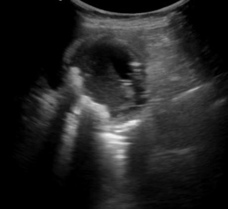
What is the ultrasonographic sign of the gallbladder?
This ultrasonographic sign occurs when the gallbladder is contracted around a stone or collection of stones. As a result, the shadowing from the stone (s) obscures the gallbladder posterior to the stone (s). Thus, the anterior gallbladder wall is visualized, followed by the hyperechoic stone (s), and then the stone (s) shadow
What is bedside ultrasound?
Bedside ultrasound can be quickly used to either narrow a differential diagnosis – for example in a patient presenting with fever and abdominal tenderness – or to evaluate biliary structures in a patient with clinical suspicion for biliary disease.
Why is it important to reposition a patient while visualizing gallstones?
Repositioning the patient while visualizing gallstones allows a clinician to evaluate if stones are impacted or freely mobile
Can you visualize the neck of the gallbladder?
Failure to visualize the neck of the gallbladder may result in missing pathology such as obstructing stones. Inability to locate the CBD. The CBD may be difficult to visualize on ultrasound and is often the most difficult part of the study for novice ultrasonographers.
Is bedside ultrasonography effective?
Bedside ultrasonography in the Emergency Department has been shown to be effective in detecting choledocholithiasis and cholecystitis, as well as reducing patient length of stay (Blaivas 1999, PMID: 10530660; Summers 2010, PMID: 20138397; Ross 2011, PMID: 21401784)
Can a gallbladder wall thicken?
A number of normal states can also cause gallbladder wall thickening – including congestive states, ascites, and in the setting of a contracted gallbladder. Ultrasound findings must be interpreted in conjunction with clinical reasoning
What is biliary microlithiasis?
Biliary microlithiasis refers to gallstones <3 mm in diameter. The term biliary microlithiasis is occasionally used as a synonym for sludge, however, this is not strictly correct. Sludge may include these microliths in its composition, but these are only one element of a variable mixture of crystals, proteinaceous debris, lysed cells and mucin 12 .
What is the Collins sign for gallstone pain?
Abdominal pain is often referred to the right shoulder. Patients may demonstrate this radiation to the tip of the scapula by placing their hand behind the back and thumb pointing upwards: " Collins sign ". This may be useful in distinguishing gallstone pain from esophagitis, gastritis, or duodenal ulcer in ~50% of patients 5 .
Where do gallstones occur?
Gallstones, also called cholelithiasis, are concretions that may occur anywhere within the biliary system, most commonly within the gallbladder .
What is the gold standard for detecting gallstones?
Ultrasound is considered the gold standard for detecting gallstones 6:
Can you see radiopaque gallstones on plain film?
Some radiopaque gallstones may be seen on plain film:
Does genetics affect gallstones?
Genetics may have an important role in gallstone formation . Several studies have shown an association between age-adjusted prevalence of gallstone, ethnicity 4 and family history of gallstones:
Is gallstone radiopaque?
gallstones are radiopaque only in 15-20% of cases 3. may be laminated (a.k.a. lamellated): radiopaque outline with lucent center. may have a faceted outline. may show a Mercedes-Benz sign: triradiate pattern of gas lucency.
