What diseases are caused by the lymphatic system?
- Multiple sclerosis (MS) – destroys the myelin sheaths of the brain and spinal cord
- Myasthenia gravis – impairs communication between nerves and skeletal muscles; as a result, muscles are weakened (drooping eyelids, difficulty in swallowing, talking, overall muscle fatigue)
- Type 1 diabetes – destroys pancreatic Beta cells that produce insulin
How does lymph get into the lymphatic capillaries?
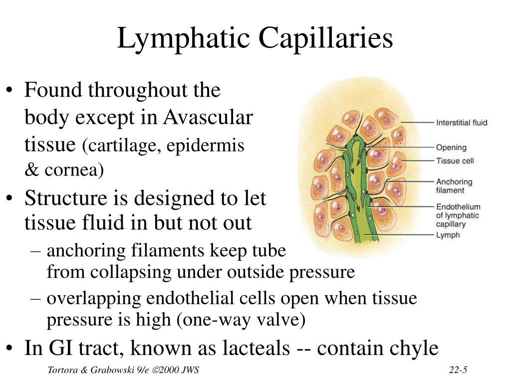
When pressure is greater in the interstitial fluid than in lymph, the cells separate slightly, like the opening of a one-way swinging door, and interstitial fluid enters the lymphatic capillary. When pressure is greater inside the lymphatic capillary, the cells adhere more closely, and lymph cannot escape back into the interstitial fluid. Attached to the lymphatic capillaries are anchoring filaments, which contain elastic fibers. They extend out from the lymphatic capillary, attaching ...
What are the 6 lymphatic organs?
What are the organs of lymphatic system?
- Lymphoid organs. The immune system is made up of organs that control the production and maturation of certain defense cells, the lymphocytes.
- Bone marrow.
- Thymus.
- Lymph nodes.
- Spleen.
- Tonsils.
- Lymphatic tissue in the bowel and in other mucous membranes in the body.
- Sources.
Where do lymphatic capillaries allow fluid to flow?
The role of the lymphatic capillaries is to pick up this excess fluid and return it to the cardiovascular system to maintain blood flow. The lymphatic vessels are therefore found intertwined between the blood capillaries, where they pick up the excess fluid and empty it into lymphatic vessels.
What is the structure of lymphatic capillaries?
What is the lymphatic system?
What are the features of lymphatic endothelial cells?
About this website
Where do lymphatic capillaries originate?
Lymph capillaries of the superficial system originate near blood capillaries and cover the entire surface of the body. Lymph capillaries are closed tubelike structures in the interstitial space that are formed by a single layer of continuous or overlapping endothelial cells.
How lymphatic vessels are formed?
100 yr ago, Florence Sabin proposed that the lymphatic system develops by the sprouting of endothelial cells from embryonic veins, leading to the formation of primitive lymph sacs from which lymphatic endothelial cells then sprout into surrounding organs to form mature lymphatic networks (5, 6).
How is lymph formed from blood capillaries?
Formation of the lymph fluid is dependent on pressure gradients in the capillary beds and the composition of the endothelial cell glycocalyx, which acts as a molecular sieve. Fluid propulsion toward the draining node is dependent on the intrinsic pumping mechanism of the lymphangions and their unidirectional valves.
What tissue makes up the lymphatic capillaries?
Endothelial cellsLymphatic capillaries consist of: Endothelial cells, which line the capillary walls. Basement membrane, which supports the endothelial cells. Mini-valves, which let lymph flow into the capillaries but not out of them.
What are lymphatic capillaries?
Lymph or lymphatic capillaries are tiny thin-walled vessels, closed at one end and located in the spaces between cells throughout the body, except in the central nervous system and non-vascular tissues. Lymphatic capillaries are slightly larger in diameter and have greater oncotic pressure than blood capillaries.
How is lymph formed quizlet?
Lymph is formed from the absorbed fluids from cells in the capillary beds. The lymph vessels return this fluid, after undergoing filtration in lymph glands, into the subclavian veins. Lymphatic organs are connective tissue capsules at well defined sites.
What is the difference between blood capillaries and lymphatic capillaries?
Lymphatic capillaries are the tiny vessels of the lymphatic system that carry lymph. Blood capillaries are the smallest blood vessels of the circulatory system and they carry blood.
What is the formation of lymph called?
Lymph is the clear watery-appearing fluid found in lymphatic vessels and is formed by the passage of substances from blood capillaries into tissue spaces. This process is known as transudation which involves the processes of diffusion and filtration.
Where is the lymphatic vessels?
Location. Lymphatics span throughout most of the body, except for the bone marrow, brain, and spinal cord. 2 Lymph nodes are distributed along the lymphatic pathway, connected by vessels. Clusters of lymph nodes are found in the armpit, groin, and neck.
What is the structure of lymphatic vessels?
General Collecting Lymphatic Structure. The collecting lymphatic vessel wall has an inner endothelium surrounded by a medial layer of circular smooth muscle cells.
What are the vessels of the lymphatic system?
Lymphatic vessels: Lymphatic vessels are the network of capillaries (microvessels) and a large network of tubes located throughout your body that transport lymph away from tissues. Lymphatic vessels collect and filter lymph (at the nodes) as it continues to move toward larger vessels called collecting ducts.
What is the function of lymphatic vessel?
The general functions of lymphatic vessels in fluid transport and immunosurveillance are well recognized, as is their specialization into capillaries, serving as an entrance point of interstitial components and immune cells and collecting vessels that deliver lymph to lymph nodes (LNs) and blood circulation.
How does the structure of lymphatic capillaries correlate - Classy Writers
How does the structure of lymphatic capillaries correlate with their function? What are some differences between lymphatic and blood capillaries? What
Lymphatic Capillaries: Function, Anatomy and Structure - Cleveland Clinic
Conditions and Disorders What conditions affect the lymphatic capillaries? Conditions that affect the lymphatic capillaries include: Adult Hodgkin’s lymphoma and adult non-Hodgkin’s lymphoma: These two types of cancer develop in the lymph system. They begin in white blood cells called lymphocytes.
1. How does the structure of lymphatic capillaries correlate with...
Get more out of your subscription* Access to over 100 million course-specific study resources; 24/7 help from Expert Tutors on 140+ subjects; Full access to over 1 million Textbook Solutions
[Structure and function of lymphatic capillaries in synovial joint]
The review article focuses on the structure and function of lymphatic capillaries in connective tissues of skin, muscles and synovial membrane. Lymphatic capillaries (initial lymphatics) are formed from endothelial cells mutually arranged so that their intercellular junctions have different structur …
What is the structure of lymphatic capillaries?
The structure of lymphatic capillaries in lymph formation. The lymphatic vascular system consists of endothelial lined vessels which begin as blind-end tubes or saccules that are located within the connective tissue areas.
What is the lymphatic system?
The lymphatic vascular system consists of endothelial lined vessels which begin as blind-end tubes or saccules that are located within the connective tissue areas. This system serves as a one-way drainage apparatus for the removal of diffusible substances as well as plasma proteins that escape the blood capillaries.
What are the features of lymphatic endothelial cells?
Another salient feature of the lymphatic endothelial cell includes the presence of numerous cytoplasmic filaments, which are similar in morphology to the actin filaments observed in a variety of cell types.
How do lymphatic vessels work?
Like veins, skeletal muscle contraction exerts pressure on the lymph vessels and forces the lymph forward through them. Lymph vessels are like one-way roads, with the lymph being collected at the capillary beds and travels through the body into the thoracic cavity. Lymph is deposited in one of two large ducts in the chest region: the right lymphatic duct and the thoracic duct. The lymph then travels from these ducts into venous circulation via the subclavian and jugular veins.
What are the structures of the lymphatic system?
The lymphatic system contains both capillaries and vessels . Lymphatic vessels begin as capillaries. Both of these structures are thin walled, which allows lymph to be transported across the membrane and collected in the vessels. Lymphatic capillaries have greater permeability than blood capillaries and can absorb large molecules such as proteins and lipids. The endothelial cells that make up the wall of a lymphatic capillary lack a basement membrane, loosely attach to each other and slightly overlap. Interstitial fluid enters the lymphatic vessel when the pressure is greater in the interstitial fluid than in lymph and nothing in the interstitial fluid is excluded from entering the lymphatic capillaries. When pressure is greater inside the lymphatic capillary, the endothelial cells prevent lymph from passing back into the interstitial spaces by acting like a one-way swinging door. Lymphatic capillaries are found wherever blood capillaries are located except in the central nervous system and bone marrow.
What causes lymph node swelling?
Lymphedema is a condition of localized fluid retention and a tissue swelling caused by a compromised lymphatic system. Lymphedema can be primarily caused genetically or secondarily due to injury or obstruction of lymphatic vessels. It is most frequently seen after lymph node dissection, surgery and/or radiation, in which lymphatic system damage is caused during the treatment of cancer, usually breast cancer. Lymphedema may also be associated with parasitic infections in which parasites obstruct lymph vessels. Symptoms may include fatigue, a swollen limb or localized fluid accumulation in other body areas, including the head and neck, discoloration of the skin overlying the swollen tissue and eventually deformity (elephantiasis).
What is the process of filtration of blood?
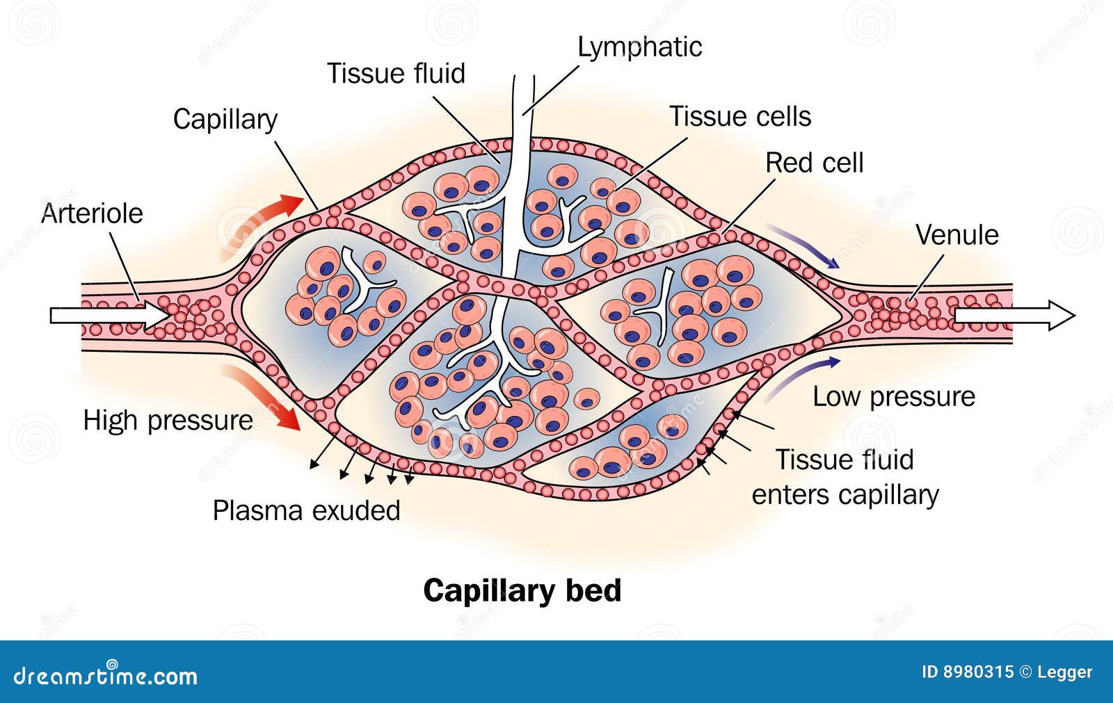
On its way through the capillaries, some of the fluid passes out across the capillary wall and into the interstitial fluid in a process called capillary filtration. This filtration tends to occur across the arterial end of the capillary, with most of the filtered fluid being reabsorbed at the venous end of the capillary. This leaves a small amount of fluid that remains in the interstitial spaces between cells. This filtered fluid is mostly plasma plus any plasma proteins that might have leaked from the blood vessel as well. This excess interstitial fluid is collected by the lymphatic system. The fluid flows through the lymphatic vessels until it is returned to the circulatory system to again become a component of blood. Once interstitial fluid passes into lymphatic vessels, it is called lymph. Lymph is a clear, pale-yellow fluid connective tissue.
Where does fluid flow?
Fluid moves from blood capillaries into the interstitial spaces. Most of the fluid returns to the blood, but some of the fluid moves from the interstitial spaces into lymphatic capillaries to become lymph. To collect the lymph from the interstitial space, lymph capillaries originate in the blood capillary beds, and lymph vessels run parallel to the veins. At intervals along the lymphatic vessels, lymph flows through lymph nodes. Fluid collected in the lymph system is returned to the heart via veins in the chest. Unlike the circulatory system, the lymphatic system does not flow through a closed, circular system. There are no lymph arteries. Lymph fluid is not pumped around the body. Instead, the lymph system collects the lymph into vein-like structures called lymph vessels and returns it to the bloodstream.
Does lymphatic circulation have a pump?
Unlike the cardiovascular circulation, the lymphatic circulation lacks a pump like the heart. Lymphatic vessels are low pressure vessels similar to veins and the same muscle pump and respiratory pump that promote venous return also facilitate lymph flow. Therefore, even though there is some smooth muscle in lymphatic vessels, movement of the body is important to lymph circulation.
How do lymphatic capillaries grow?
During wound healing lymphatic capillaries grow by sprouting from existing lymphatics, much in the same way as new blood capillaries sprout from existing capillaries or postcapillary veins during angiogenesis. The appearance of new lymphatic capillaries is always secondary to that of blood capillaries, although linear growth occurs at comparable speed ( Clark, 1922; Clark and Clark, 1932 ). Paavonen et al. (2000) studied VEGFR-3 expression in experimental wounds made in dorsal skin of pigs. VEGFR-3-positive vessels were observed in the granulation tissue from day 5 onward, and very few VEGFR-3-positive lymphatic vessels persisted on day 9 and none on day 14. These results suggest that transient lymphangiogenesis occurs parallel with angiogenesis in healing wounds. Witmer et al. (1991) demonstrated that in granulation tissue, VEGFR-3 staining was observed in the proliferative superficial zone in plump blood vessel sprouts, in the intermediate zone in blood vessels and long lymphatic sprouts, and in deeper fibrous zone in large lymphatics, in a pattern demonstrating that lymphangiogenesis follows behing blood vessel angiogenesis.
What are the capillaries of lymphatic cells?
Lymphatic capillaries start as blind-ended sacs in the periphery and merge to collecting lymphatic vessels, which are surrounded by pericytes and have valves to direct the flow into the draining lymph nodes ( Figure 1 ). Initial lymphatic vessels in the periphery either lack the basement membrane totally or it is incomplete. The abluminal walls of lymphatic endothelial cells are anchored to the extracellular matrix via anchoring filaments ( Figure 1 ). Anchoring filaments consist of microfibrils, which are composed mainly of fibrillin. Fibrillin microfibrils are characteristic of lymphatic capillaries, as they are not found in blood capillaries. However, they are present in larger blood vessels. The microfibrils of lymphatic and blood endothelial cells, however, differ from each other as lymphatic endothelial cells form irregular patterns, while in blood endothelial cells they are in a honeycomb organization most likely reflecting their different functional requirements ( Weber et al., 2002 ). Anchoring filaments are connected to lymphatic endothelial cells via focal adhesions, which contain molecules such as focal adhesion kinase, talin, vinculin, cytoskeletal beta actin, and clusters of integrins. Anchoring filaments–focal adhesion complexes are thought to be important in cell-matrix communication ( Weber et al., 2002 ). Moreover, they regulate endothelial permeability and interstitial fluid drainage by preventing the vessels from collapsing in conditions of high interstitial pressure ( Langheinrich et al., 2012 ).
What are the structures of large lymphatic vessels?
Larger lymphatic vessels have a structure similar to that of veins; the tunicae intima, media and adventitia can all be defined. Sacchi et al. (1997) described precollector vessels between lymphatic capillaries and larger lymphatic vessels. The smooth muscle layer of the walls of these vessels is incomplete, exposing sections where the endothelium is essentially naked and takes part in fluid exchange. Large lymphatic vessels often run with blood vessels; a plexus of small lymphatic vessels often forming around adjacent blood vessels.
How does lymphatic flow to lymph node?
The steering of the lymph flow toward the node is also facilitated by a series of bicuspid valves organized by layers of connective tissue overlaid by LEC ( Schmid-Schonbein, 1990; Vittet, 2014 ). The valves are unidirectional with the lymph flow and are placed at anatomical intervals along the collectors. The vessel area between two sets of valves is known as a lymphangion. In close proximity to the valves, specialized muscle cells initiate muscle contraction through a Ca ++ ion-mediated depolarization that drives the muscle action potential. These cells act as the lymphatic collector pacemaker, and initiate and propagate the contraction to all other muscle cells. The contraction originates from the more peripheral lymphangion and propagates toward those nearer to the lymph node. The valves open and close in synchrony with vessel contraction. By coordinating the lymphangion pumping activity with the opening and closing of the valves, the lymph is precluded from backflow and will move unidirectionally to the lymph node ( Davis et al., 2008; Gashev, 2002, 2008, 2010; Muthuchamy and Zawieja, 2008; Schmid-Schonbein, 1990 ).
What is the lymphatic system?
The General Anatomy of the Lymphatic System. Lymphatic capillaries form a network beneath the epithelial surfaces of the epidermis and the mucosa of the gut, respiratory and genitourinary systems.
Why is lymphatic fluid increased?
In contrast, under inflammatory conditions, associated with pathogen invasion, or sterile inflammation as observed in autoimmune diseases, an increased lymph formation is observed, due to increased tissue edema . The increased amount of interstitial fluid and, by default lymph fluid, is also associated with neolymphangiogenesis (generation of newly developed lymphatic vessels from preexisting ones), increased immune cell trafficking, from the periphery to the draining lymph node, and upregulation of proinflammatory mediators, which alter lymphatic permeability and contractility ( Cromer et al., 2014; Rahbar et al., 2014; Swartz and Randolph, 2014 ).
How much lymph flow does a sheep have?
Under physiological conditions, in sheep, it was originally reported that the prenodal and postnodal lymph flow run at about 1–5 mL/h. However, several differences have been reported according to the location of measurement, animal body mass, and physiological state. For example in the mesentery of fasting rats, prenodal lymph flow has been calculated to be around 15 μL/h, and the postnodal lymph flow of the mesenteric duct around 1.3 mL/h ( Dixon et al., 2006 ).
Why is the lymph capillary white?
The lymph within these capillaries, called chyle, has a creamy white color (rather than clear) due to the presence of fats. Lymphatic‐collecting vessels form as lymph capillaries merge. Collecting vessels have the following characteristics: Valves are present to prevent the backward flow of lymph (as in veins).
What are the characteristics of lymphatic vessels?
Lymphatic‐collecting vessels form as lymph capillaries merge. Collecting vessels have the following characteristics:#N#Valves are present to prevent the backward flow of lymph (as in veins).#N#The walls of collecting vessels consist of the same three tunics (layers) that characterize veins, but the layers are thinner and poorly defined. 1 Valves are present to prevent the backward flow of lymph (as in veins). 2 The walls of collecting vessels consist of the same three tunics (layers) that characterize veins, but the layers are thinner and poorly defined.
What is the smallest lymphatic vessel?
Lymph capillaries, the smallest lymphatic vessels, begin as dead‐end vessels. They resemble blood capillaries, but are much more porous to surrounding fluids due to the following two features: Valvelike openings form at the juncture of adjacent endothelial cells.
What happens when the pressure inside the capillary exceeds the pressure outside?
When pressure inside the capillary exceeds the pressure outside, the spaces between the endothelial cells close, holding fluids inside the capillary. Anchoring filaments attach the endothelial cells of the lymphatic vessels to surrounding collagen. When interstitial fluid pressure increases, the anchoring filaments prevent ...
Where does lymph go in the thoracic duct?
The thoracic duct collects lymph from the left side of the body and regions of the right side of the body below the thorax. It ultimately drains lymph into the left subclavian vein. It begins at the cisterna chili, an enlarged region of the lymphatic vessel that forms following the union of the intestinal trunk and right and left lumbar trunks.
Which lymphatic ducts drain blood into the neck?
Lymphatic ducts are the largest lymphatic vessels. These two ducts drain lymph into veins in the neck (the right and left subclavian veins at their junctures with the internal jugular veins). Valves in the lymphatic ducts at their junctures with the veins prevent the entrance of blood into the lymphatic vessels.
Where does lymph go in the body?
The right thoracic duct collects lymph from the upper right side of the body (right arm and right regions of thorax, neck, and head), a much smaller area than that serviced by the thoracic duct. It ultimately drains lymph into the right subclavian vein.
What is the structure of lymphatic capillaries?
The structure of lymphatic capillaries in lymph formation. The lymphatic vascular system consists of endothelial lined vessels which begin as blind-end tubes or saccules that are located within the connective tissue areas.
What is the lymphatic system?
The lymphatic vascular system consists of endothelial lined vessels which begin as blind-end tubes or saccules that are located within the connective tissue areas. This system serves as a one-way drainage apparatus for the removal of diffusible substances as well as plasma proteins that escape the blood capillaries.
What are the features of lymphatic endothelial cells?
Another salient feature of the lymphatic endothelial cell includes the presence of numerous cytoplasmic filaments, which are similar in morphology to the actin filaments observed in a variety of cell types.