How are the bones of the skull held together?
The bones of the skull are connected to each other with the help of fibrous junctions called the sutures. The bones of the skull grow and fuse together during fetal and childhood development, forming a single skull. However, the mandible remains separate from the rest of the skull.
What are the sutures of the skull?
Mar 22, 2022 · Most of the bones of the skull are held together by firm, immovable fibrous joints called sutures or synarthroses. These joints allow the developing skull to grow both pre- and postnatally. The sutures of the skull are morphologically distinct, being divided into three main groups based on the margins of the articulating bones.
What is the skull made up of?
Except for the mandible, all skull bones are joined together by sutures —synarthrodial (immovable) joints. The skull contains air-filled cavities called sinuses. Their functions are debatable, but may be related to lessening skull weight, contributing to voice resonance, and warming and moistening inspired air. Key Terms
What are the different views of the skull joints?
Apr 05, 2022 · All the bones of the skull are joined together except for the mandible or jaw bone. These bones get separated early which provides the fetal skull with the flexibility necessary to pass through the type confines of the birth canal. The skull bones stay relatively separated during childhood development, allowing the brain and skull to expand, and when the kid reaches …
How are the bones of the skull joined?
Your cranial bones are held together by unique joints called sutures, which are made of thick connective tissue. They're irregularly shaped, allowing them to tightly join all the uniquely shaped cranial bones.May 24, 2018
How are most bones in the skull held together?
Most of the bones of the skull are held together by firm, immovable fibrous joints called sutures or synarthroses. These joints allow the developing skull to grow both pre- and postnatally.
What type of joint is found in between skull bones?
Sutures(1) Sutures are nonmoving joints that connect bones of the skull. These joints have serrated edges that lock together with fibers of connective tissue.
Why are the bones of the skull joined tightly together?
All the bones of the skull, except for the mandible, are joined to each other by a fibrous joint called a suture. The fibrous connective tissue found at a suture (“to bind or sew”) strongly unites the adjacent skull bones and thus helps to protect the brain and form the face.
Do the bones on top of the skull move?
' Many state that there is 'no research' supporting this idea. This statement is incorrect. There may not be sufficient evidence at this time supporting this idea. However, there is much more research showing that there the bones of the head can than there is research showing that the bones of the head do not move.
What joins the bones of the skull together how is this different in newborn babies?
These bones are held together by strong, fibrous, elastic tissues called sutures. The spaces between the bones that remain open in babies and young children are called fontanelles. Sometimes, they are called soft spots. These spaces are a part of normal development.
What connects the bones in cartilaginous joints?
Cartilaginous joints are connected entirely by cartilage (fibrocartilage or hyaline). Cartilaginous joints allow more movement between bones than a fibrous joint but less than the highly mobile synovial joint.
When bones are brought closer together?
Your joints are powered by muscle to move your body. And there are two main ways your joints move. When the bones in a joint move away from each other (like when you spread out your fingers and open your hand) it's called extension. Flexion happens when your bones are brought closer together (making a fist).Feb 18, 2019
What are the 3 joints in the skull?
There are three types of fibrous joints: sutures, syndesmoses, and gomphoses. Sutures are found only in the skull and possess short fibers of connective tissue that hold the skull bones tightly in place. Sutures: Sutures are fibrous joints found only in the skull.
What holds skull sutures together?
Sutures are bound together by a matrix of connective tissues called Sharpey's fibers, which grow from each bone into the adjoining one. A tiny amount of movement is permitted at sutures, which contributes to the compliance and elasticity of the skull. These joints are synarthroses (immovable joints).
What is the Squamosal suture?
The squamosal suture is one of the lateral minor skull sutures, separating the parietal and squamous temporal bones. While the phenotypic appearances and sequelae of synostosis of the major cranial vault sutures are well documented, little is reported concerning synostosis of the squamosal suture (SQS).
What is syndesmosis held together by?
In a syndesmosis, the bones are kept joined together by ligaments or fibroelastic membranes, as occurs, for example, between the metacarpal (Fig.
How are skulls formed?
The skull is a complex structure; its bones are formed both by intramembranous and endochondral ossification. The skull roof bones, comprising the bones of the facial skeleton and the sides and roof of the neurocranium, are dermal bones formed by intramembranous ossification, though the temporal bones are formed by endochondral ossification. The endocranium, the bones supporting the brain (the occipital, sphenoid, and ethmoid) are largely formed by endochondral ossification. Thus frontal and parietal bones are purely membranous. The geometry of the skull base and its fossae, the anterior, middle and posterior cranial fossae changes rapidly. The anterior cranial fossa changes especially during the first trimester of pregnancy and skull defects can often develop during this time.
What are the parts of the skull?
The skull consists of three parts, of different embryological origin—the neurocranium, the sutures, and the facial skeleton (also called the membraneous viscerocranium ). The neurocranium (or braincase) forms the protective cranial cavity that surrounds and houses the brain and brainstem.
What is the skull?
Anatomical terminology. The skull is a bone structure that forms the head in vertebrates. It supports the structures of the face and provides a protective cavity for the brain. The skull is composed of two parts: the cranium and the mandible.
How many bones are there in the skull?
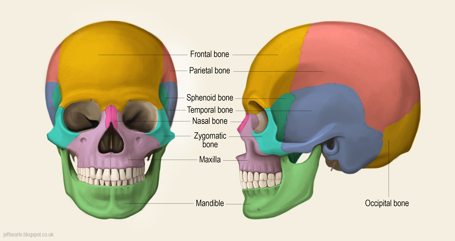
The human skull is generally considered to consist of twenty-two bones —eight cranial bones and fourteen facial skeleton bones. In the neurocranium these are the occipital bone, two temporal bones, two parietal bones, the sphenoid, ethmoid and frontal bones .
Where does the word "skull" come from?
The English word "skull" is probably derived from Old Norse "skulle", while the Latin word cranium comes from the Greek root κρανίον ( kranion ). The skull is made up of a number of fused flat bones, and contains many foramina, fossae, processes, and several cavities or sinuses.
What are the two parts of the human body?
In humans, these two parts are the neurocranium and the viscerocranium ( facial skeleton) that includes the mandible as its largest bone. The skull forms the anterior-most portion of the skeleton and is a product of cephalisation —housing the brain, and several sensory structures such as the eyes, ears, nose, and mouth.
What are the functions of the skull?
Functions of the skull include protection of the brain, fixing the distance between the eyes to allow stereoscopic vision, and fixing the position of the ears to enable sound localisation of the direction and distance of sounds.
What bones are in the skull?
The facial skeleton contains the vomer, two nasal conchae, two nasal bones, two maxilla, the mandible, two palatine bones, two zygomatic bones, and two lacrimal bones.
What are the two parts of the skull?
The adult human skull consists of two regions of different embryological origins: the neurocranium and the viscerocranium. The neurocranium is a protective shell surrounding the brain and brain stem. The viscerocranium (or facial skeleton) is formed by the bones supporting the face. Except for the mandible, all skull bones are joined together by ...
How many bones are in the neurocranium?
Key Points. The eight bones of the neurocranium form major portions of the skull and protect the brain. The neurocranium consists of two temporal bones situated to the base and side of the skull, and two parietal bones that make up the roof of the skull.
Which bone forms the base of the skull?
A single occipital bone forms the base of the skull, and the frontal bone forms the forehead. The sphenoid and ethmoid bones located to the front of the skull form parts of the orbital sockets and nasal cavity; they also support and protect key organs found in the skull.
Where are the temporal bones located?
The temporal bones are situated at the base and sides of the skull, lateral to the temporal lobes of the brain. The temporal bones consist of four regions the squamous, mastoid, petrous and tympanic regions. The squamous region is the largest and most superior region.
What is the skull?
The skull supports the musculature and structures of the face and forms a protective cavity for the brain. The skull is formed of several bones which, with the exception of the mandible, are joined together by sutures—synarthrodial (immovable) joints.
Which bone is the most complex?
The sphenoid bone is one of the most complex in the body due to its interactions with numerous facial bones, ligaments, and muscles. The body that forms the middle of the sphenoid bone articulates with the ethmoid and occipital bone and forms a key part of the nasal cavity; it also contains the sphenoidal sinuses.
What is cranial synostosis?
What is Craniosynostosis? Craniosynostosis is a birth defect in which the bones in a baby’s skull join together too early. This happens before the baby’s brain is fully formed. As the baby’s brain grows, the skull can become more misshapen.
When is craniosynostosis surgery performed?
When needed, a surgical procedure is usually performed during the first year of life. But, the timing of surgery depends on which sutures are closed and whether the baby has one of the genetic syndromes that can cause craniosynostosis. Babies with very mild craniosynostosis might not need surgery.
Why do babies have craniosynostosis?
Some babies have a craniosynostosis because of changes in their genes. In some cases, craniosynostosis occurs because of an abnormality in a single gene, which can cause a genetic syndrome. However, in most cases, craniosynostosis is thought to be caused by a combination of genes and other factors, such as things the mother comes in contact with in her environment, or what the mother eats or drinks, or certain medications she uses during pregnancy.
How to tell if a baby has craniosynostosis?
Sometimes, it is diagnosed later in life. Usually, the first sign of craniosynostosis is an abnormally shaped skull. Other signs may include: No “soft spot” on the baby’s skull. A raised firm edge where the sutures closed early.
What are the different types of craniosynostosis?
Types of Craniosynostosis. The types of craniosynostosis depend on what sutures join together early. Sagittal synostosis– The sagittal suture runs along the top of the head, from the baby’s soft spot near the front of the head to the back of the head.
What happens when a suture closes?
When a suture closes and the skull bones join together too soon, the baby’s head will stop growing in only that part of the skull. In the other parts of the skull where the sutures have not joined together, the baby’s head will continue to grow.
Why do babies have developmental delays?
Some children, however, have developmental delays or intellectual disabilities, because either the craniosynostosis has kept the baby’s brain from growing and working normally, or because the baby has a genetic syndrome that caused both craniosynostosis and problems with how the brain works.
Where are the fontanelles located?
There are normally several fontanelles on a newborn's skull. They are located mainly at the top, back, and sides of the head. Like the sutures, fontanelles harden over time and become closed, solid, bony areas.
Why do fontanelles turn sunken?
Causes. Expand Section. Reasons a child may have sunken fontanelles include: Dehydration (not enough fluid in the body) Malnutrition. When to Contact a Medical Professional. Expand Section. A sunken fontanelle can be a medical emergency. A health care provider should check the infant right away.
When does the fontanelle close?
The fontanelle at the top of the head ( anterior fontanelle) most often closes within 7 to 19 months. The fontanelles should feel firm and should curve inward slightly to the touch. A noticeably sunken fontanelle is a sign that the infant does not have enough fluid in their body.
What is a sunken fontanelle?
Sunken fontanelles are an obvious curving in of the "soft spot" in an infant's head.

Overview
The skull is a bone structure that forms the head in vertebrates. It supports the structures of the face and provides a protective cavity for the brain. The skull is composed of two parts: the cranium and the mandible. In humans, these two parts are the neurocranium and the viscerocranium (facial skeleton) that includes the mandible as its largest bone. The skull forms the anterior-most portion of the skeleton and is a product of cephalisation—housing the brain, and several sensorystructure…
Structure
The human skull is the bone structure that forms the head in the human skeleton. It supports the structures of the face and forms a cavity for the brain. Like the skulls of other vertebrates, it protects the brain from injury.
The skull consists of three parts, of different embryological origin—the neurocranium, the sutures, and the facial skeleton(also called the membraneo…
Development
The skull is a complex structure; its bones are formed both by intramembranous and endochondral ossification. The skull roof bones, comprising the bones of the facial skeleton and the sides and roof of the neurocranium, are dermal bones formed by intramembranous ossification, though the temporal bones are formed by endochondral ossification. The endocranium, the bones supporting the brai…
Clinical significance
Craniosynostosis is a condition in which one or more of the fibrous suturesin an infant skull prematurely fuses, and changes the growth pattern of the skull. Because the skull cannot expand perpendicular to the fused suture, it grows more in the parallel direction. Sometimes the resulting growth pattern provides the necessary space for the growing brain, but results in an abnormal head shape and abnormal facial features. In cases in which the compensation does not effectiv…
Society and culture
Artificial cranial deformation is a largely historical practice of some cultures. Cords and wooden boards would be used to apply pressure to an infant's skull and alter its shape, sometimes quite significantly. This procedure would begin just after birth and would be carried on for several years.
Like the face, the skull and teeth can also indicate a person's life history and or…
Terminology
• Chondrocranium, a primitive cartilaginous skeletal structure
• Endocranium
• Epicranium
• Pericranium, a membrane that lines the outer surface of the cranium
History
Trepanning, a practice in which a hole is created in the skull, has been described as the oldest surgical procedure for which there is archaeological evidence, found in the forms of cave paintings and human remains. At one burial site in France dated to 6500 BCE, 40 out of 120 prehistoric skulls found had trepanation holes.
See also
• Craniometry
• Crystal skull
• Head and neck anatomy
• Human skull symbolism
• Memento mori