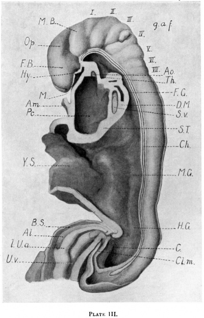
Somite formation begins as paraxial mesoderm cells become organized into whorls of cells called somitomeres. The somitomeres become compacted and bound together by an epithelium, and eventually separate from the presomitic paraxial mesoderm to form individual somites.
How do somites become specific tissue and cell types?
These somites then separate into cranial and caudal portions, and the cranial portion of each fuses with the caudal portion of the somite directly anterior to it in a process known as metametric shifting. Distinct regions of each somite become specific tissue and cell types as the body matures.
What is the wave of somites?
The wave is a gradient of the FGF protein that is rostral to caudal (nose to tail gradient). Somites form one after the other down the length of the embryo from the head to the tail, with each new somite forming on the caudal (tail) side of the previous one.
What is the angle of the first somite formation?
Finally, the angle of the first somite formation was defined as the angle between the vector orthogonal to the LR segment and the tangent vector in the intersecting position P along the midline (Supplementary Fig. 13d ).
Where do we get somite images from?
For the somites of human embryos, the images were obtained from the Virtual Human Embryo project 20 and the Kyoto collection, and the first 4–5 rows of somites were measured. For the somites of mouse trunk-like structures (TLSs) and CHIR99021- and LDN193189-treated TLSs (TLS-CLs) 15, the DAPI images were provided by the authors.

What are the somites?
Somites are precursor populations of cells that give rise to important structures associated with the vertebrate body plan and will eventually differentiate into dermis, skeletal muscle, cartilage, tendons, and vertebrae.
What phase are somites formed?
Somites begin formation as discrete clusters of mesenchymal cells that undergo segmentation in a cranial to caudal progression beginning around the third week of intrauterine life; this stage is termed “compaction.” A single somite can be described as having six faces, like a cube, with each facet having a slightly ...
What germ layer are somites derived from?
somite, in embryology, one of a longitudinal series of blocklike segments into which the mesoderm, the middle layer of tissue, on either side of the embryonic spine becomes divided. Collectively, the somites constitute the vertebral plate.
What are somites quizlet?
Somites. Somites are transient structures that do not exist in the adult. They disappear as organogenesis proceeds. Somite Functions. -They are extremely important in organizing the segmental patterns of the mammalian (human) body plan.
Are somites stem cells?
Somites (SMs) comprise a transient stem cell population that gives rise to multiple cell types, including dermatome (D), myotome (MYO), sclerotome (SCL) and syndetome (SYN) cells.
What do somites formed from mesoderm during somitogenesis go on to form?
What do somites, formed from mesoderm during somitogenesis, go on to form? Explanation: Somites go on to form skeletal muscle, vertebrae, and the dermis.
How many somites does a human embryo have?
In embryos with 30 somites, the cells of the neural crest have developed and they push the myotomes in a ventral direction. The spinal ganglia that increase in volume form a relief on the surface of the embryo. The segmentation that is now visible is ganglionic and no more somitic.
How many somites do humans have?
The somite formation, or somitogenesis, starts around day 20 after fertilization (Carnegie stage 9) in human embryos, and a total of ~40 pairs of somites are formed4.
What are the three layers of blastocyst?
Gastrulation is a complex process involving the embryo reorganizing itself to generate the three embryonic germ layers, endoderm, mesoderm, and ectoderm.
What is the process of neurulation?
Neurulation is a process in which the neural plate bends up and later fuses to form the hollow tube that will eventually differentiate into the brain and the spinal cord of the central nervous system. From: Current Topics in Developmental Biology, 2012.
What process is responsible for the formation of the three germ layers?
Gastrulation leads to the formation of the three germ layers that give rise during further development to the different organs in the animal body. This process is called organogenesis. Organs develop from the germ layers through the process of differentiation.
What is the process of somitogenesis?
Somitogenesis is the process by which somites form. Somites are bilaterally paired blocks of paraxial mesoderm that form along the anterior-posterior axis of the developing embryo in segmented animals. In vertebrates, somites give rise to skeletal muscle, cartilage, tendons, endothelium, and dermis.
How do somites develop?
The wave is a gradient of the FGF protein that is rostral to caudal (nose to tail gradient). Somites form one after the other down the length of the embryo from the head to the tail, with each new somite forming on the caudal (tail) side of the previous one.
What are somites?
In vertebrates, somites subdivide into the sclerotomes, myotomes, syndetomes and dermatomes that give rise to the vertebrae of the vertebral column, rib cage and part of the occipital bone; skeletal muscle, cartilage, tendons, and skin ( of the back). The word somite is sometimes also used in place of the word metamere.
What is the term for a set of bilaterally paired blocks of paraxial mesoderm?
The somites (outdated term: primitive segments) are a set of bilaterally paired blocks of paraxial mesoderm that form in the embryonic stage of somitogenesis, along the head-to-tail axis in segmented animals. In vertebrates, somites subdivide into the sclerotomes, myotomes, syndetomes and dermatomes that give rise to the vertebrae of the vertebral column, rib cage and part of the occipital bone; skeletal muscle, cartilage, tendons, and skin (of the back).
What is the dermatome?
The dermatome is the dorsal portion of the paraxial mesoderm somite which gives rise to the skin ( dermis ). In the human embryo it arises in the third week of embryogenesis. It is formed when a dermamyotome (the remaining part of the somite left when the sclerotome migrates), splits to form the dermatome and the myotome. The dermatomes contribute to the skin, fat and connective tissue of the neck and of the trunk, though most of the skin is derived from lateral plate mesoderm.
How do Hox genes specify somites?
The Hox genes specify somites as a whole based on their position along the anterior-posterior axis through specifying the pre-somitic mesoderm before somitogenesis occurs. After somites are made, their identity as a whole has already been determined, as is shown by the fact that transplantation of somites from one region to a completely different region results in the formation of structures usually observed in the original region. In contrast, the cells within each somite retain plasticity (the ability to form any kind of structure) until relatively late in somitic development.
What happens to presomitic mesoderm?
The pre-somitic mesoderm commits to the somitic fate before mesoderm becomes capable of forming somites. The cells within each somite are specified based on their location within the somite. Additionally, they retain the ability to become any kind of somite-derived structure until relatively late in the process of somitogenesis.
What is the myotome?
The myotome is that part of a somite that forms the muscles of the animal. Each myotome divides into an epaxial part ( epimere ), at the back, and a hypaxial part ( hypomere) at the front. The myoblasts from the hypaxial division form the muscles of the thoracic and anterior abdominal walls.
Where does somite occur?
A somite is added either side of the notochord ( axial mesoderm) to form a somite pair. The segmentation does not occur in the head region, and begins cranially (head end) and extends caudally (tailward) adding a somite pair at regular time intervals.
What are the developmental dynamics of somites?
Developmental dynamics of occipital and cervical somites "Development of somites leading to somite compartments, sclerotome, dermomyotome and myotome, has been intensely investigated. Most knowledge on somite development, including the commonly used somite maturation stages, is based on data from somites at thoracic and lumbar levels. Potential regional differences in somite maturation dynamics have been indicated by a number of studies, but have not yet been comprehensively examined. Here, we present an overview on the developmental dynamics of somites at occipital and cervical levels in the chicken embryo. We show that in these regions, the onset of sclerotomal and myotomal compartment formation is later than at thoracolumbar levels, and is initiated simultaneously in multiple somites, which is in contrast to the serial cranial- to- caudal progression of somite maturation in the trunk."
What is the segmentation clock?
In vitro characterization of the human segmentation clock "The segmental organization of the vertebral column is established early in embryogenesis, when pairs of somites are rhythmically produced by the presomitic mesoderm (PSM). The tempo of somite formation is controlled by a molecular oscillator known as the segmentation clock1,2. Although this oscillator has been well-characterized in model organisms1,2, whether a similar oscillator exists in humans remains unknown. Genetic analyses of patients with severe spine segmentation defects have implicated several human orthologues of cyclic genes that are associated with the mouse segmentation clock, suggesting that this oscillator might be conserved in humans3. Here we show that human PSM cells derived in vitro-as well as those of the mouse4-recapitulate the oscillations of the segmentation clock. Human PSM cells oscillate with a period two times longer than that of mouse cells (5 h versus 2.5 h), but are similarly regulated by FGF, WNT, Notch and YAP signalling5. Single-cell RNA sequencing reveals that mouse and human PSM cells in vitro follow a developmental trajectory similar to that of mouse PSM in vivo. Furthermore, we demonstrate that FGF signalling controls the phase and period of oscillations, expanding the role of this pathway beyond its classical interpretation in 'clock and wavefront' models1. Our work identifying the human segmentation clock represents an important milestone in understanding human developmental biology."
What is the term for the process of segmentation of the paraxial mesoderm within the trilaminar?
Introduction. Human embryo ( week 4, Carnegie stage 11) Somites. The term somitogenesis is used to describe the process of segmentation of the paraxial mesoderm within the trilaminar embryo body to form pairs of somites, or balls of mesoderm. In humans, the first somite pair appears at day 20 and adds caudally at 1 somite pair/90 minutes ...
What is the purpose of the mesenchymal to epithelial transition?
A mesenchymal to epithelial transition defines the outer cellular "shell" of the developing somite, with the core cells remain as a mesenchymal organisation.
Which sclerotomes engulf the notochord?
The left and right sclerotomes from the same segmental level engulf the notochord.
Which layer of the cell is formed during segmentation?
During segmentation the outer cell layer forms an epithelial layer over a still mesenchymal organization of cells at the core.
What is a somite?
Somites are transient structures that do not exist in the adult. They disappear as organogenesis proceeds.
Which part of somite is the direct expression of Myf5 and MyoD?
from lateral mesoderm direct expression of Myf5 and MyoD in dorsolateral part of somite initiating limb and body wall muscles.
When does paraxial mesoderm begin to organize in segments?
paraxial mesoderm begins to organize in segments by Wk 3
When does mesoderm thicken?
The initial sheet of mesoderm thickens near midline by Day 17
How many primary sources does the skeleton develop from?
The skeleton develops from three primary sources?
Which epiblast cells lie between the ectoderm and endoderm?
Invaginating epiblast cells that lie between developing ectoderm and endoderm will become mesoderm.
Where do ribs develop?
Ribs develop from sclerotome of thoracic vertebrae (costal processes).
Which muscle arises directly from somitomeres?
Skeletal muscles of the head and neck; arise directly from somitomeres.
Where is the myotome layer?
3. The myotome layer forms by migration of some dermamyotomal cells to a position in between the sclerotome and dermamyotome.
Which sclerotome migrates medially and dorsally to surround the neural tube in cranial?
Somites 6+: Dorsal sclerotome migrates medially and dorsally to surround the neural tube in cranial to caudal progression. Depends on signals from neural tube, surface ectoderm.
What is the term for cartilage gradually replaced by bone?
Endochondrous ossification: cartilage gradually replaced by bone; most bones of the body.
What is the name of the notochord in the intervertebral disc?
Annulus fibrosus = intervertebral disc. Nucleus pulposus = the notochord, which remains present as the core of the intervertebral disc.
