How many cases of lentigo maligna melanoma are there?
[Lentigo maligna and lentigo maligna melanoma: a clinicopathologic analysis of twenty-four cases]. [Zhonghua Bing Li Xue Za Zhi. 2...]
What is the average age of diagnosis for lentigo maligna?
The majority of patients with lentigo maligna are older than 40 years, and the peak age of diagnosis is be between 60 and 80 years. Unlike superficial spreading melanoma, lentigo maligna is not related to the number of melanocytic naevi ( moles) or atypical naevi.
What is lentigo maligna (lentigo)?
Lentigo maligna is also known as Hutchinson melanotic freckle. Lentigo maligna is an early form of melanoma in which the malignant cells are confined to the tissue of origin, the epidermis, hence it is often reported as ‘ in situ ’ melanoma.
What are the risk factors for lentigo maligna?
Risk factors for development of lentigo maligna include the following: a history of sunburns, a history of nonmelanoma skin cancers, advanced age, lighter skin types, and tendency to form solar lentigines.
How serious is lentigo maligna?
Lentigo maligna is not dangerous; it only becomes potentially life threatening if an invasive melanoma develops within it. Long term follow-up involves reviewing the treated area and full skin examination to identify new lesions of concern.
How often does lentigo maligna turn into melanoma?
The percentage of lentigo maligna that progress to lentigo maligna melanoma remains unknown, but estimates of the lifetime risk of developing lentigo maligna melanoma in patients diagnosed with lentigo maligna at age 45 years appears to be 5%.
Is lentigo maligna slow growing?
Lentigo maligna melanoma These melanomas develop from very slow growing coloured patches of skin called lentigo maligna or Hutchinson's melanotic freckle. The lentigo maligna is flat and grows outwards in the surface layers of the skin. It might slowly get bigger over several years and might change shape or colour.
Is lentigo maligna curable?
Diagnosis and Treatment of Lentigo Maligna Melanoma LM and LMM are highly curable when diagnosed early.
Is lentigo maligna fast growing?
Lentigo maligna grows slowly and is usually harmless, but lentigo maligna melanoma can spread aggressively. It's important to recognize the symptoms of lentigo maligna melanoma so you can seek treatment early on.
Is lentigo maligna precancerous?
Lentigo malignas may remain precancerous for up to 50 years. Lentigo maligna may look similar to benign (noncancerous) skin lesions, such as solar lentigo, or “liver spots,” which are harmless. If lentigo maligna is misdiagnosed as a benign or noncancerous condition, it may delay treatment.
Can lentigo maligna spread?
Lentigo maligna melanoma is an invasive form of skin cancer, but it is treatable if a doctor or dermatologist diagnoses it early. A person should, therefore, take any early signs or symptoms seriously and seek medical advice. In its early form, lentigo maligna, the cancer has not yet spread.
What is the difference between lentigo and lentigo maligna?
Lentigo maligna (LM), first described by Hutchinson in 1890, is the noninvasive counterpart to lentigo maligna melanoma (LMM). The latter (LMM) refers to invasive melanoma associated with a LM. LM and LMM occur on chronically sun-damaged skin, most commonly on the head and neck.
Can lentigo maligna regress?
These skin lesions grow radially and may grow/regress in a pattern that makes the LM/LMM appear to “move across” the skin [1, 3]. The skin surrounding the LM/LMM may also show signs of chronic solar damage [solar elastosis, solar lentigines, actinic keratosis (AK)].
Is lentigo maligna the same as melanoma in situ?
Lentigo maligna is a type of melanoma in situ. It is a slow growing lesion that appears in areas of skin that has a lot of sun exposure such as the face or upper body. Lentigo maligna grows slowly and can take years to develop.
How do you biopsy lentigo maligna?
Excisional biopsy is ideal for diagnosis of lentigo maligna [40]. In theory, excisional biopsy removes the whole clinical lesion down to subcutaneous fat with a 1–3 mm margin. This potentially allows for complete evaluation of depth and peripheral involvement.
How do I get rid of lentigo?
Your dermatologist might recommend one of these treatments:medicines like bleaching creams containing hydroquinone or retinoids (tretinoin)chemical peels.skin resurfacing.laser or intense pulse light therapy to destroy melanocytes.freezing (cryotherapy) to destroy melanocytes.
What is a lentigo maligna?
Lentigo maligna is an early form of melanoma in which the malignant cells are confined to the tissue of origin, the epidermis, hence it is often reported as ‘ in situ ’ melanoma. It occurs in sun damaged skin so is generally found on the face or neck, particularly the nose and cheek.
What are the characteristics of lentigo maligna?
The characteristics of lentigo maligna include: Large size: >6 mm and often several centimetres in diameter at diagnosis. Irregular shape.
How much margin is needed for melanoma in situ?
The ideal margin for all forms of melanoma in situ is 5-10mm, depending on how well defined are the edges of the lesion.
How much margin is needed for a biopsy of lentigo maligna?
If a skin lesion is clinically suspicious of lentigo maligna, it is best cut out ( excision biopsy) with a 2–3 mm margin. Partial biopsy is less accurate than complete excision biopsy, as a single small biopsy could miss a malignant focus.
How many invasive melanomas were diagnosed in 2008?
According to New Zealand Cancer Registry data, 2256 invasive melanomas were diagnosed in 2008 and about 10% were pathologically lentigo maligna melanoma. Rates of the precursor, lentigo maligna, are not reported by national cancer registries but it is thought to be the most common variant of melanoma in situ in New Zealand and Australia.
How long does it take for a lentigo maligna to grow?
It grows slowly in diameter over 5 to 20 years or longer. Lentigo maligna melanoma is diagnosed when the melanoma cells have invaded into the dermis and deeper layers of skin. Lentigo maligna has a lower rate of transformation to invasive melanoma than the other forms of melanoma in situ (under 5% overall).
What is a melanoma staging?
Melanoma staging means finding out if the melanoma has spread from its original site in the skin. Most melanoma specialists refer to the American Joint Committee on Cancer (AJCC) cutaneous melanoma staging guidelines (2009). In essence, the stages are:
Where is LM/LMM most likely to occur?
LM/LMM is also most likely to occur on the face, a site of chronic sun damage, whereas SSM and NM are more likely to occur on the trunk in men and legs in women, sites which are more often protected.[6]
What is LM skin?
LM often presents as an irregular brown macule or patch on chronically sun-damaged skin in the elderly. Lesions are light-brown to black, may display color variegation, may appear asymmetric, and tend to have an ill-defined border. As lesions enlarge, they may develop skip areas with a patchy, non-contiguous pattern.
What is LM in medical terms?
Lentigo maligna ( LM) is a slow-growing subtype of melanoma that often occurs on chronically sun-damaged skin, particularly the head and neck, in the elderly. Due to its clinical and histopathologic mimicry of benign lesions, it is often misdiagnosed for years or even decades, which can lead to increased morbidity and mortality.
What is a LM?
Lentigo maligna (LM) is a subtype of melanoma and commonly presents as an irregular brown macule on chronically sun-damaged skin, particularly the head and neck, in the elderly.[1] .
What happens to junctional nests as LM becomes more invasive?
As LM becomes more invasive (i.e., progresses to LMM), junctional nests become larger and more spindled. Invasion is typically superficial, consisting of isolated or small aggregates of spindled cells in the papillary dermis.
Is LM a genetic condition?
LM is also more likely to occur in genetic conditions that predispose to sun sensitivity, including oculocutaneous albinism, xeroderma pigmentosum, Werner syndrome, and porphyria cutanea tarda.[2] There have been no associations demonstrated with smoking or alcohol. [17]
Is LM a smooth or a palpable lesion?
Due to its in situ nature, LM is typically smooth and non-palpable. If the lesion becomes invasive (LMM), then a papular or dermal component may be felt. LM exhibits slow radial growth and might be misdiagnosed for years or even decades as solar lentigo or other benign lesions (see Differential Diagnosis section).[5] .
Where does lentigo maligna most commonly present?
Clinical presentation, risk factors, and genetics. Lentigo maligna most commonly presents on the head and neck region of elderly patients, with the highest incidence in the seventh and eighth decades of life.
What is lentigo maligna?
Lentigo maligna is a melanocytic neoplasm occurring on sun-exposed skin, usually on the head and neck, of middle-aged and elderly patients. It is thought to represent the in situ phase of lentigo maligna melanoma. The ill-defined nature and potentially large size of lesions can pose significant diagnostic and treatment challenges. The goal of therapy is to cure the lesions in order to prevent development of invasive disease, and surgical excision is the treatment of choice to achieve clear histological margins. Nonsurgical treatment modalities have been reported; however, evidence is lacking to support their use. Age, general health, and comorbidities need to be taken into account when deciding the right treatment modality for each individual patient.
What is the treatment for lentigo maligna?
Radiation therapy . Radiotherapy, like imiquimod, is a noninvasive treatment option that has been used as a primary treatment for lentigo maligna in patients who are poor surgical candidates. Studies have used Grenz ray therapy for treatment of lentigo maligna [102–104].
What is the best biopsy for lentigo maligna?
Excisional biopsy is ideal for diagnosis of lentigo maligna [ 40 ]. In theory, excisional biopsy removes the whole clinical lesion down to subcutaneous fat with a 1–3 mm margin. This potentially allows for complete evaluation of depth and peripheral involvement. Excisional biopsy , however, is often not feasible for lentigo maligna because the lesions are typically ill-defined, widespread, and located in cosmetically sensitive areas. If the size of the lesion limits the ability to perform an excisional biopsy, scouting shave or punch biopsies can be performed ( Figure 1B ). Scouting biopsies should include samples from the darkest part, or most concerning part of the lesion, which will minimize the sampling error. They can also be taken from the periphery of the lesion to help delineate the peripheral margin involvement ( Figure 1B ).
Why is woods light used in lentigo maligna?
Since the true margins of lentigo maligna can exist far beyond the margins seen with visible light, the Wood’s light is used to improve margin delineation.
Can you do an excisional biopsy on lentigo maligna?
Excisional biopsy, however, is often not feasible for lentigo maligna because the lesions are typically ill-defined, widespread, and located in cosmetically sensitive areas. If the size of the lesion limits the ability to perform an excisional biopsy, scouting shave or punch biopsies can be performed (Figure 1B).
Can lentigo maligna develop de novoor?
Lentigo maligna can develop de novoor within a pre-existing solar lentigo. Patients typically present with a chief complaint of a new, asymptomatic pigmented macule or patch on the head or neck region, or a freckle that has changed in size, shape, or color. Open in a separate window. Figure 1A.
Where does lentigo maligna develop?
It develops from lentigo maligna, also known as Hutchinson’s melanotic freckle, which is a slow growing type of melanoma. This cancer typically starts on the surface of the skin on parts of the body that get exposed to too much sun, which tend to include the face, forearms, and neck. When the cancer goes beneath the surface of the skin, ...
What causes lentigo maligna?
The most common risk factors for lentigo maligna melanoma include: 1 chronic exposure to sunlight, which causes skin damage 2 a history of sunburns 3 lighter skin 4 the tendency to form harmless patches of darkened skin, known as solar lentigines 5 a history of nonmelanoma skin cancers 6 certain genetic mutations, most commonly P53 mutations
What does it mean when a doctor diagnoses lentigo maligna melanoma?
When a doctor diagnoses lentigo maligna melanoma, this means that the cancer has spread from the skin to other tissue in the body. Last medically reviewed on February 17, 2020. Dermatology. Cancer / Oncology. Melanoma / Skin Cancer.
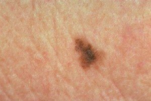
What is the name of the dark mole on the head?
Signs and symptoms. Lentigo maligna melanoma typically presents as a dark mole that changes in shape, color, or size. Initially, it tends to appear as an irregular brown macule or lesion on the head or neck. As it progresses, it can become one of several different colors, including black.
How to prevent lentigo maligna?
Some practical tips for preventing sun damage include: avoiding spending prolonged periods in the sun. wearing a hat and protective gear to avoid sun exposure. wearing and regularly reapplying sunblock that is SPF 15 or higher.
What is the name of the cancer that grows faster than lentigo maligna?
When the cancer goes beneath the surface of the skin, it becomes lentigo maligna melanoma. is an aggressive form of cancer that is faster growing than lentigo maligna and may spread to other areas of the body. In this article, we look at the signs and symptoms of lenti go maligna melanoma , as well as the causes and risk factors.
What kind of doctor would you see for moles?
A doctor will often refer a person to a dermatologist for a diagnosis. A dermatologist can better distinguish between a benign skin growth, such as a mole, and more serious conditions. This specialist will typically ask the person about their symptoms and what they have observed over time.
What is lentigo maligna melanoma?
Lentigo maligna melanoma is a rare type of melanoma skin cancer, accounting for about 5 percent of all melanomas, according to the NCI. It’s also sometimes called Hutchinson’s melanotic freckle.
Causes of lentigo maligna melanoma
Changes to the genetic material in your cells over your lifetime are the main cause of lentigo maligna melanoma. These are called gene mutations.
Symptoms of lentigo maligna melanoma
If a lesion hasn’t yet grown into the surrounding layers of skin, it’s simply referred to as lentigo maligna. Once it becomes invasive, it’s called lentigo maligna melanoma.
Risk factors of lentigo maligna melanoma
Risk factors increase the chance that you’ll develop lentigo maligna melanoma. Knowing these risk factors may help you and your doctor discuss steps for cancer prevention.
How lentigo maligna melanoma is diagnosed
Due to the similarities between lentigo maligna and benign skin lesions like liver spots, accurate diagnosis is key.
How lentigo maligna melanoma is treated
The main treatment for lentigo maligna or lentigo maligna melanoma is to remove the lesion. However, as most lentigo malignas occur on the head or neck, there may be cosmetic implications, especially on the face. Ask your care team about surgical techniques or tools that may help minimize scarring and long-term damage to the skin in that area.
Survival rates for lentigo maligna melanoma
The survival rates for lentigo maligna melanoma are good, especially when diagnosed while it’s still localized to the tissue where it began. The five-year survival rate represents what percentage of people are still alive five years after a cancer diagnosis.
What is lentigo maligna?
Lentigo maligna is a type of melanoma in situ and is a precursor to lentigo maligna melanoma, a potentially serious form of skin cancer. Lentigo maligna is also known as Hutchinson melanotic freckle.
What are the characteristics of lentigo maligna?
The clinical characteristics of lentigo maligna include: Large size: > 6 mm and often several centimetres in diameter at diagnosis. An irregular shape.
What are the symptoms of melanoma?
Invasive melanoma is reported to arise within lentigo maligna in 3–10% of cases. The following features are very suspicious of invasion: 1 Thickening of part of the lesion 2 An increasing number of colours, especially blue or black 3 Ulceration or bleeding 4 Itching or stinging.
Is Lentigo maligna invasive?
Lentigo maligna melanoma is diagnosed when the malignant melanoma cells have invaded into the dermis and deeper layers of skin. Lentigo maligna has a comparatively low rate of transformation to invasive melanoma (under 5%). The risk of invasive melanoma is greater in larger lesions, with up to 50% of those with a diameter ...

Prognosis
- Lentigo maligna grows slowly and is usually harmless, but lentigo maligna melanoma can spread aggressively. Its important to recognize the symptoms of lentigo maligna melanoma so you can seek treatment early on. Untreated lentigo maligna melanoma can eventually spread throughout …
Symptoms
- The visual symptoms of lentigo maligna melanoma are very similar to those of lentigo maligna. Both look like a flat or slightly raised brown patch, similar to a freckle or age spot. They have a smooth surface and an irregular shape. While theyre usually a shade of brown, they can also be pink, red, or white. It can be hard to tell lentigo maligna melanoma from a freckle or age spot by l…
Signs and symptoms
- Compared to other types of skin cancer, lentigo maligna and lentigo maligna melanoma are on the larger side. They tend to be at least 6 millimeters (mm) wide and can grow to several centimeters. Most people with either condition have it on their neck or face, especially their nose and cheeks.
Diagnosis
- Its also hard to visually tell the difference between lentigo maligna and lentigo maligna melanoma. Keep an eye out for these signs that may indicate lentigo maligna melanoma: After reviewing your medical history and doing a physical exam, your doctor may refer you to a dermatologist or other specialist. They may use a dermatoscope, which combines a magnifying …
Causes
- The exact cause of lentigo maligna melanoma is unknown, but sun exposure is the biggest risk factor for developing it. That puts people with sun-damaged skin and those who spend a lot of time outside at a higher risk. Other risk factors for developing lentigo maligna melanoma include:
Treatment
- The most common treatment for lentigo maligna melanoma is to remove the spot with surgery. Lentigo maligna melanoma is more likely to return than some other types of skin cancer, so your doctor may also remove some of the skin surrounding the spot to prevent this. If its spread to your lymph nodes, your doctor may choose to remove those as well. If you have other condition…
Prevention
- The best way to prevent lentigo malignant melanoma is to limit your exposure to UV rays from the sun and tanning beds. When you do spend time in the sun, use a high-SPF sunscreen and wear a large hat that protects your face and neck.