Cones are packed into a part of the retina directly behind the retina called the fovea, which is responsible for sharp central vision. When light strikes either the rods or the cones of the retina, it's converted into an electric signal that is relayed to the brain via the optic nerve.
What is the function of a cone in the eye?
Cone cells, or cones, are photoreceptor cells in the retinas of vertebrate eyes (e.g. the human eye).They respond differently to light of different wavelengths, and are thus responsible for color vision and function best in relatively bright light, as opposed to rod cells, which work better in dim light.
What do cones allow your eyes to see in?
- A Warmer Future for National Parks?
- A Win-Win with Wind
- Aerial Ant Acrobatics
- Always Judge a Grouse by its Cover
- Amoeba Families Stick Together
- Are Plankton Ocean Super Stars?
- Batty for Food
- Benefits of Being Choosy
- Birds of a Feather Change Together
- Blurring the Line Between Plants and Animals
What are the cones in the eye responsible for?
What Are Eye Cones?
- Structure of Eye Cones. These light-sensitive cones are mostly concentrated into a portion of the eye's retina known as the fovea, which enables small details to come into sharp focus ...
- Types of Cones
- Function of Cones. ...
- Color Vision. ...
- Problems With Eye Cones. ...
- Color Blindness. ...
What part of the eye contains only cones?
- Retina The thin lining at the back of the eye that is responsible for vision. ...
- Macula The central portion of the retina responsible for the sharp, detailed vision.
- Vitreous The clear jelly-like substance that fills the rear two-thirds of the eye. ...

How do cones and rods work in the eye?
Rods are responsible for vision at low light levels (scotopic vision). They do not mediate color vision, and have a low spatial acuity. Cones are active at higher light levels (photopic vision), are capable of color vision and are responsible for high spatial acuity. The central fovea is populated exclusively by cones.
How are cones activated?
As is the case for rods, when a cone is activated by light it is in a hyperpolarized state (as opposed to depolarized state). While at rest, cone cells transmit a steady inhibitory input to the bipolar cells. The transduction process, as it occurs in the rods of the retina, occurs in a similar manner in the cone cells.
How do colored cones work?
2:195:23Cone Cells and the Color Vision - YouTubeYouTubeStart of suggested clipEnd of suggested clipAnd these filter curls are preceded by cone cells. And give us a colored region as you already knowMoreAnd these filter curls are preceded by cone cells. And give us a colored region as you already know visible spectrum gives no more spectral colors. And rest of the colors are being made by mixing the
How do cones detect light?
Cones respond to light that has passed through the lens and onto the fovea. As each cone absorbs its color of light, it produces an electrical signal. These signals travel to the brain, filling our worlds with color.
What do cones do?
Cones Allow You To See Color The cone is made up of three different types of receptors that allow you to see color. These three different receptors are aptly named the short, medium, and long-wavelength cones. This size difference represents each receptor's sensitivity to light.
Do cones respond dim light?
Signals from the cones are sent to the brain which then translates these messages into the perception of color. Cones, however, work only in bright light. That's why you cannot see color very well in dark places. So, the cones are used for color vision and are better suited for detecting fine details.
What happens if cone cells are absent in eye?
Rod monochromacy: Also known as achromatopsia, it's the most severe form of color blindness. None of your cone cells have photopigments that work. As a result, the world appears to you in black, white, and gray. Bright light may hurt your eyes, and you may have uncontrollable eye movement (nystagmus).
What colours do cones detect?
Scientists have known for decades that some cells — known as cones — detect color. They are part of the retina inside the back of the eye. Cone cells can sense red, green or blue light. But Ramkumar Sabesan discovered that some of them sense white light — and only white light.
Why are cones important?
Since the cone requires a high level of light in order to send signals, the cones are primarily responsible for your visual acuity (your ability to see objects in fine detail). Defective cones won’t enable you to focus on a certain object or perceive its color correctly, if at all.
Why is the cone more sensitive than the rod?
The most important difference between the cone and the rod is that the cone is more light-sensitive than the rod, and the cone requires much more light to enter it in order to send signals to the brain. This is the reason that you are unable to differentiate colors in dim light conditions.
What are the three types of receptors that allow you to see color?
Cones Allow You To See Color. The cone is made up of three different types of receptors that allow you to see color. These three different receptors are aptly named the short, medium and long wavelength cones. This size difference represents each receptor’s sensitivity to light.
What parts of the eye let you see the object you are looking at?
The eye is made up of a network of vessels, muscles, and nerves, but what parts of the eye actually let you see the object you are looking at? Enter the cone and the rod. To learn more about cones and rods, we have to zoom in on one of the most important parts of the eye, the retina.
How long does it take for a rod to adjust to light?
That’s because it usually takes the rod about 30 minutes to fully adjust to the absence of light. In addition to being the receptor that allows you to see in the dark, the rod is also the better motion sensor since it is more sensitive in nature, and has more individual receptors than the cone.
What type of cell is the cone?
Cones are a type of photoreceptor cell in the retina. They give us our color vision. Cones are a type of photoreceptor cell in the retina. They give us our color vision.
How many cones are there in the retina?
The retina has approximately 120 million rodsand 6 million con es. There are three types of cone cells:
Why do cones have a higher visual acuity?
Cones also tend to possess a significantly elevated visual acuity because each cone cell has a lone connection to the optic nerve, therefore, the cones have an easier time telling that two stimuli are isolated. Separate connectivity is established in the inner plexiform layer so that each connection is parallel.
What is the role of S cones in the circadian system?
It is possible that S cones may play a role in the regulation of the circadian system and the secretion of melatonin but this role is not clear yet. The exact contribution of S cone activation to circadian regulation is unclear but any potential role would be secondary to the better established role of melanopsin.
How long does it take for a retina to become white?
This is done by exposing dark-adapted retina to a certain wavelength of light that paralyzes the particular type of cone sensitive to that wavelength for up to thirty minutes from being able to dark-adapt making it appear white in contrast to the grey dark-adapted cones when a picture of the retina is taken.
What is the wavelength of light in the S cone?
S Cones are most sensitive to light at wavelengths around 420 nm. However, the lens and cornea of the human eye are increasingly absorptive to shorter wavelengths, and this sets the short wavelength limit of human-visible light to approximately 380 nm, which is therefore called ' ultraviolet ' light.
What are the three types of cones?
Cones are normally one of the three types, each with different pigment, namely: S-cones, M-cones and L-cones. Each cone is therefore sensitive to visible wavelengths of light that correspond to short-wavelength, medium-wavelength and longer-wavelength light. Because humans usually have three kinds of cones with different photopsins, ...
What is the synaptic terminal of a cone cell?
The synaptic terminal forms a synapse with a neuron such as a bipolar cell. The inner and outer segments are connected by a cilium.
How are cone cells different from rods?
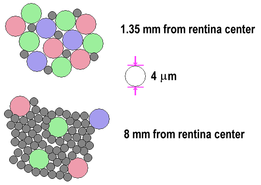
Cone cells are somewhat shorter than rods, but wider and tapered, and are much less numerous than rods in most parts of the retina, but greatly outnumber rods in the fovea. Structurally, cone cells have a cone -like shape at one end where a pigment filters incoming light, giving them their different response curves. They are typically 40–50 µm long, and their diameter varies from 0.5 to 4.0 µm, being smallest and most tightly packed at the center of the eye at the fovea. The S cone spacing is slightly larger than the others.
How Our Eyes Work
To process vision, the light reflected from an object in our field of view is gathered by the cornea. The cornea then refracts the light rays through the pupil (the center of the iris where light enters the eye). The iris then passes the image onto the crystalline lens.
Rods and Cones: Color Vision and Concept
These crucial parts of our eye are known as photoreceptors. They are specialized cells that are located on the retina, in the back of your eye which processes images. Their roles are very specific: to receive and process signals of light and color, which gives us our vision.
Schedule a Consultation
Join us at Laser for Eyes for an expert consultation on our services. We will discuss and review your options to help you find the perfect fit for you. Schedule a consultation with us today, and join our many satisfied patients.
What color are the cones?
Since the three types of cones are commonly labeled by the color at which they are most sensitive (blue, green and red) you might think other colors are not possible. But it is the overlap of the cones and how the brain integrates the signals sent from them that allows us to see millions of colors.
What controls the amount of light that can enter the eye?
Iris: in the anatomy of an eye, the iris controls the size of the opening of the pupil. This in turn controls the amount of light that can enter the eye... more (link is external) Mitochondria: are the cell’s powerhouse. It packages the energy from food into energy the cell can use to do work... more.
What is the outer surface of the eye?
Cornea: is the clear outer surface of the eye the covers the iris, pupil, and the outer chamber of the eye... more (link is external) Epithelium: the layer of cells found lining the surface of most surfaces of the body. Epithelium is one of four types of tissues found in human body.
Why are the discs in the eye recycled?
First of all, the discs containing rhodopsin or photopsin are constantly recycled to keep your visual system healthy. By having the discs right next to the epithelial cells (retinal pigmented epithelium: RPE) at the back of the eye, parts of the old discs can be carried away by cells in the RPE.
What is the retina?
If you think of the eye as a camera, the retina would be the film. The retina also contains the nerves that tell the brain what the photoreceptors are "seeing.". There are two types of photoreceptors involved in sight: rods and cones. Rods work at very low levels of light.
How many rod cells does the human eye have?
The human eye has over 100 million rod cells. Cones require a lot more light and they are used to see color. We have three types of cones: blue, green, and red. The human eye only has about 6 million cones.
Why do we use rods for night vision?
Rods work at very low levels of light. We use these for night vision because only a few bits of light (photons) can activate a rod . Rods don't help with color vision, which is why at night, we see everything in a gray scale. The human eye has over 100 million rod cells.
What is the role of cones in vision?
The cones are responsible for all high resolution vision. The eye moves continually to keep the light from the object of interest falling on the fovea centralis where the bulk of the cones reside. Rod and cone discussion. Rod and cone distribution.
What are the rods and cones of the human eye?
The Rods and Cones of the Human Eye. Rods and Cones. The retinacontains two types of photoreceptors, rod s and cones. The rods are more numerous, some 120 million, and are more sensitive than the cones. However, they are not sensitive to color.
Which is better, cones or rods?
While the visual acuity or visual resolution is much better with the cones, the rods are better motion sensors. Since the rods predominate in the peripheral vision, that peripheral vision is more light sensitive, enabling you to see dimmer objects in your peripheral vision.
What is the visual perception of intensely blue objects?
The visual perception of intensely blue objects is less distinct than the perception of objects of red and green. This reduced acuity is attributed to two effects. First, the blue cones are outside the fovea, where the close-packed cones give the greatest resolution.
Which cones have the highest sensitivity?
The "blue" cones have the highest sensitivity and are mostly found outside the fovea, leading to some distinctions in the eye's blue perception. The cones are less sensitive to light than the rods, as shown a typical day-night comparison.
Do rods see red?
Rods Do Not See Red! The light response of the rods peaks sharply in the blue; they respond very little to red light. This leads to some interesting phenomena: Red rose at twilight: In bright light, the color-sensitive cones are predominant and we see a brilliant red rose with somewhat more subdued green leaves.
What is the special property of the cone system?
A special property of the cone system is color vision. Perceiving color allows humans (and many other animals) to discriminate objects on the basis of the distribution of the wavelengths of light that they reflect to the eye.
Why do dichromats lack one of the three cone pigments?
This recombination can lead to the loss of a gene, the duplication of a gene, or the formation (more...) Human dichromats lack one of the three cone pigments, either because the corresponding geneis missing or because it exists as a hybrid of the red and green pigment genes (see Figure 11.13).
Why is color important in perceptual perception?
Color obviously gives us a quite different way of perceiving and describing the world we live in.
How does the lens work?
The light passes through the pupil to the lens behind it. The lens adjusts its shape to bend and focus the light a second time, to ensure that you have a clear image of what you are looking at .
Which part of the eye is made up of muscles that control the pupil?
Iris: The colored part of the eye that is made up of muscles that control the pupil— contracting the pupil in bright light and expanding the pupil in low light. Sclera: The white part of the eye that surrounds the iris. This structure is made up of fibrous tissue that protects the inner structures of the eye.
What part of the eye is blue?
The iris is the colorful part of your eye that gives it its blue, green, hazel, brown or dark appearance. The pupil then automatically gets bigger or smaller, depending on the intensity of the light.
What are the two parts of the retina that are responsible for transforming light rays into electrical impulses?
The photoreceptors are made up of rods and cones, and are responsible for transforming the light rays into electrical impulses. While the light is focused throughout the retina, most of the light entering the eye is focused onto the focal point on the retina, known as the macula.
What is the name of the layer of the eye that reflects light?
When we look at an object, the light that is reflected off of the object enters the eye through the clear front layer of the eye, called the cornea. The cornea bends the light before it passes through a watery substance that fills the area behind the cornea , called the aqueous humor.
What is the iris made of?
The iris is actually made up of muscles that expand and contract to control the pupil and adjust its size. So when you see your pupil getting bigger or smaller, it is really the iris that is controlling the pupil opening in response to the intensity of light entering the eye.
What happens if your eyes are not functioning properly?
The process of seeing is dependent on the perfection of the eye and all of its components, including: If any of these components do not function properly, or are irregularly shaped, vision problems can occur— most commonly, blurry vision will develop.
Structure of Eye Cones

Types of Cones
- Located on each of the two retinas are actually three different types of cones: 1. Red cones, which account for 60% of all cones 2. Green cones, which make up 30% of the cones 3. Blue cones, limited to just 10% of the cones1
Function of Cones
- These cones contain photopigments, known as opsin amino acids, that are sensitive to different wavelengths of visible light. Fact is, each of the different colors of the rainbow have a different wavelength. Our cones are able to capture these various frequencies thanks to these color-sensitive photopigments. Our eyes can actually perceive light frequencies as short as 380 nano…
Problems with Eye Cones
- Not everybody necessarily sees colors the same way. Color vision is tested with the Ishihara color palettes—a series of dots of different hues. This test, which identifies color issues, was named for Japanese ophthalmologist Shinobu Ishihara and includes numbers embedded in each of a set of circular images. The idea is to detect if you are unable to see certain colors.7 Unfortunately, eye …
Color Blindness
- If you are being tested with the Ishihara exam and can’t pick out some of the numbers amid the different shades of dots, it means that the color frequency isn’t registering because some of your cones aren’t functioning properly. You likely have some sort of color blindness.7 The term color blindness is a bit of a misnomer, however. In most cases, this does not mean that yo…
Overview
Cone cells, or cones, are photoreceptor cells in the retinas of vertebrate eyes including the human eye. They respond differently to light of different wavelengths, and are thus responsible for color vision, and function best in relatively bright light, as opposed to rod cells, which work better in dim light. Cone cells are densely packed in the fovea centralis, a 0.3 mm diameter rod-fre…
Structure
Humans normally have three types of cones. The first responds the most to light of longer wavelengths, peaking at about 560 nm. This type is sometimes designated L for long; the majority of the human cones are of the long type. The second most common type responds the most to light of medium-wavelength, peaking at 530 nm, and is abbreviated M for medium, making up about a third …
Function
The difference in the signals received from the three cone types allows the brain to perceive a continuous range of colors, through the opponent process of color vision. (Rod cells have a peak sensitivity at 498 nm, roughly halfway between the peak sensitivities of the S and M cones.)
All of the receptors contain the protein photopsin, with variations in its confor…
Clinical significance
One of the diseases related to cone cells present in retina is retinoblastoma. Retinoblastoma is a rare cancer of the retina, caused by the mutation of both copies of retinoblastoma genes (RB1). Most cases of retinoblastoma occur during early childhood. One or both eyes may be affected. The protein encoded by RB1 regulates a signal transduction pathway while controlling the cell cycle progression as normally. Retinoblastoma seems to originate in cone precursor cells prese…
See also
• Cone dystrophy
• Disc shedding
• Double cones
• RG color space
• Tetrachromacy
External links
• Cell Centered Database – Cone cell
• Webvision's Photoreceptors
• NIF Search – Cone Cell via the Neuroscience Information Framework
• Model and image of cone cell