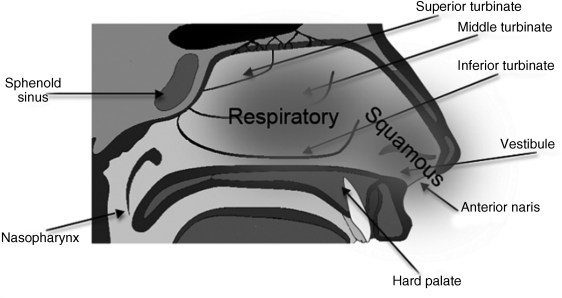
What is the function of nasal mucus?
What is the function of the nasal mucosa? The nasal mucosa plays an important role in mediating immune responses to allergens and infectious particles which enter the nose. It helps prevent allergens and infections from invading the nasal cavity and spreading to other body structures, for example the lungs. Click to see full answer.
What is at the base of the nasal cavity?
The named arteries of the nose are:
- Sphenopalatine artery and greater palatine artery, branches of the maxillary artery.
- Anterior ethmoidal artery and posterior ethmoidal artery, branches of the ophthalmic artery
- Septal branches of the superior labial artery, a branch of the facial artery, which supplies the vestibule of the nasal cavity.
What is normal oral mucosa?
When healthy, the lining of the mouth (oral mucosa) is reddish pink. Sometimes color changes in the mouth are a sign of a bodywide disease. In this regard, what is the oral mucosa? The oral mucosa is the mucous membrane lining the inside of the mouth.
How to relieve swollen nose membranes?
Treating Swollen Nasal Passages
- Natural Nasal Irrigation. Nasal irrigation can help drain your sinuses for quick relief. ...
- Quercetin. Studies have continually demonstrated the antiallergenic activity of quercetin; in particular, quercetin prevents the release of histamine from basophils and mast cells.
- Berberine-Containing Plants. ...
- Remove Food Sensitivities or Allergies. ...

How does the nasal mucosa work?
The nasal mucosa facilitates a relatively high level of drug absorption per unit area. The mucosa is lined with extensive pseudostratified columnar epithelia containing ciliated cells and mucous-secreting goblet cells in the non-olfactory region. Although the volume of the nasal cavity is low (15–20 cm3 ), the surface area for absorption is relatively large (150 cm 2) because of the numerous epithelial microvilli. Moreover, underlining the mucosa is a porous endothelial basement membrane which is richly supplied with blood vessels from both the external and internal carotid arteries [8]. A drug can permeate through the epithelial cell membrane either by the transcellular route, or by the paracellular route. The lipophilicity and size of the drug molecule play important roles in nasal permeation. Lipophilic drugs generally diffuse via the transcellular route, both passively and actively depending on the cell receptors engaged. In contrast, polar drugs with molecular weights below 1000 Da generally pass across the membrane through cell–cell tight junctions, which are less than 10 Å wide; much wider than the equivalent cell–cell tight junctions of the intestinal mucosa. However, since polypeptides are generally large, hydrophilic molecules (> 10 Å in size), only very small amounts can pass the nasal membrane, and this transport generally involves endocytosis [9].
What are the metabolizing enzymes in nasal tissues?
Enzymes known to be present include a variety of CYPs (CYP1A1, 2B1, 2E1, 3A1, 4A1, 2G1), FMOs, carboxylesterases, epoxide hydrolases, glutathione S -transferases, and UDP-glucuronyl transferases. It is of some interest that, despite the low concentrations of nasal CYP enzymes, these have been demonstrated to have greater specific activity toward several substrates than liver CYPs, perhaps as a result of higher ratios of NADPH cytochrome P450 reductase to CYP in the nasal tissues. Nasal CYPs appear to be less inducible than liver isoforms, although they appear to be sensitive to a number of CYP inhibitors.
How does nasal stimulus affect blood flow?
A detailed study of anesthetized dogs performed by James and de Burgh Daly demonstrates the regional blood flow changes that occur upon stimulation of the nasal mucosa with gases or liquids. 2 These authors observed that responses in the arterial pressure varied; in some animals, it was modified and in others it was kept stable. Also, the changes in pressure appeared with concomitant changes to the heart rate. Activation of the nasal reflex reduced blood flow to the extremities by almost 50% as a consequence of an increase of almost 100% in peripheral vascular resistance. Increments between 22% and 47% in vascular resistance were observed in the vertebral, superior mesenteric, splenic, and renal arteries, with the consequent reduction in blood flow. In contrast, no significant changes were observed in the resistance and the vascular flow of the common carotid artery. At the end of the period of apnea, a slight reduction in the levels of blood O 2, as well as a slight increase in the levels of CO 2, was observed. Once the nasal stimulus was suspended, the autonomic reflexes reversed and a transient vasodilatation occurred in limb arteries, along with a hyperventilation of approximately 1 min in length. These mechanisms achieved a compensation of the blood gases to values that existed previous to the reflex’s induction. Changes in the heart rate were blocked with the administration of atropine, vascular effects with β-adrenergic blockers, and all the cardiovascular changes were interrupted with the nicotinic blocker hexamethonium. Moreover, anesthetic blockade of the olfactory mucosa’s nervous terminals prevented activation of the previously mentioned adaptation mechanisms. This study demonstrates that, similarly to what is observed in the DR, the nasopharyngeal reflex is a somatoautonomic reflex, triggered by trigeminal afferents, whose efferent arms are the sympathetic and parasympathetic nerves. The increase in the parasympathetic vagal tone reduces the heart rate and, consequently, cardiac output. By contrast, the increase in the sympathetic tone allows an increase in peripheral vascular resistance (with the exception of the carotid resistance) that maintains similar values of arterial pressure. The differential modification of the vascular flow at a carotid level 14 indicates a possible redistribution of blood flow to the brain and perhaps also to the heart, although coronary blood flow was not studied.
Is the nasal mucosa vascularized?
The nasal mucosa is well vascularized and is capable of rather dramatic variation in blood flow, whether passively as a result of interference with venous return (congestion) or actively because of vasodilation (hyperemia). Congestion of the mucosal vessels is a nonspecific lesion commonly found at necropsy and presumably associated with the circulatory failure preceding death (e.g., heart failure, bloat in ruminants in which the increased intraabdominal pressure causes increased intrathoracic pressure impeding the venous return from the head and neck). Hyperemia of the nasal mucosa is seen in early stages of inflammation, whether caused by irritation (e.g., ammonia and regurgitated feed), viral infections, secondary bacterial infections, toxemia, allergy, or trauma.
Does nasal DNA protect against HIV?
Conversely, many studies in experimental animals and humans have demonstrated that nasal vaccination gives rise to cross-protection against drifted strains ( Brandtzaeg, 2007 ). With an available live attenuated influenza vaccine for intranasal administration (FluMist ® ), good protection was achieved despite the fact that the epidemic strain was not part of the vaccine ( Belshe et al., 2000 ). Also, cross-clade immunity against experimentally applied HIV in mice has been reported after nasal DNA prime followed by nasal peptide boost; the vaccine contained epitopes of clade B, but high and long-lasting serum antibody titers against the neutralizing gp41 ELDKWAS epitopes from both clades A, B, C, and D were observed ( Devito et al., 2004 ).
Is mucormycosis fatal?
Rhinocerebral mucormycosis begins in the upper airways and then spreads to the orbit, face, palate, and brain. Untreated, it is rapidly fatal and is seen in patients with acidosis (particularly uncontrolled diabetes), patients with leukemia, and organ transplant recipients. The initial presentation involves unilateral headache, nasal and sinus congestion/pain, nasal discharge, and fever. There may be periorbital, perinasal swelling, ptosis, proptosis, loss of vision, frontal lobe necrosis and abscess formation, nasal septum or palatal perforation. The disease is rapidly progressive. A diagnosis of pulmonary mucormycosis should be considered in immunocompromised patients presenting with fever and progressive lung infiltrates despite broad-spectrum antibacterial treatment. Gastrointestinal disease is not common and is seen in malnourished infants and children. Necrotic ulcers occur in the stomach, colon, and ileum that perforate causing peritonitis and septic shock. Cutaneous disease is seen in patients with burns. Disseminated disease is usually seen in neutropenic patients with pulmonary infection or via the gastrointestinal tract, burns, or other skin lesions.
What happens when air passes through the nasal mucosa?
As air passes over the nasal mucosa, it is prepared or conditioned to safely pass deeper into the respiratory system. Air passes over the nasal mucosa. The heat radiated from the blood vessels in the lamina propria warms the air to near body temperature. The blood vessels warm the air.
Which glands secrete mucus?
Mucus secreted from the globet cells and seromucosal glands humidify the air.
What is the nasal mucosa?
The nasal mucosa is the lining of the nasal cavity. It moistens air that is coming into the body during inhalation. It is commonly affected during the common cold, and during a cold more mucus than normal is produced; this creates the symptoms of a stuffy, runny nose. The olfactory mucosa is located in the upper nasal cavity and helps us smell. Mucous membranes also line the bronchi of the lungs, where gas exchange takes place. If someone has asthma, their bronchial mucosa can become inflamed, making their bronchi more likely to spasm. This causes a temporary decline in lung functioning.
What are the mucosae of the digestive system?
Mucosae of the Digestive System. The mouth, tongue, esophagus, stomach, and intestines are all lined with mucous membranes. These membranes are referred to as the oral mucosa, esophageal mucosa, gastric mucosa, and intestinal mucosa. Oral mucosa is found in the mouth, and changes in its condition can be signs of vitamin deficiencies, diabetes, ...
What is the membrane of the body?
A mucous membrane , also known as a mucosa (plural: mucosae), is a layer of cells that surrounds body organs and body orifices. It is made from ectodermal tissue. Mucous membranes can contain or secrete mucus, which is a thick fluid that protects the inside of the body from dirt and pathogens such as viruses and bacteria. Many different mucous membranes exist, such as mucous membran es in the respiratory system, digestive system, and reproductive system.
What organs are responsible for odors?
The nasal and olfactory mucosae help odors to break down in the nose so that their particles can be detected and the substance can be smelled. Mucosae are also found in reproductive organs like the vagina; naturally occurring vaginal discharge is produced by the vaginal mucosa to self-clean and keep the vagina moist.
What are the functions of mucous membranes?
In general, the functions of mucous membranes are to protect the body from being infected by viruses and bacteria and to keep the tissues of the body adequately moisturized. Specific mucous membranes have specialized functions.
Where are the oral, gastric, and esophageal mucosae located?
Answer to Question #2. C is correct. The oral, gastric, and esophageal mucosae are found in the mouth, stomach, and esophagus , respectively. They are all part of the digestive system. The preputial mucosa is located on the prepuce (foreskin) of the penis, so it is part of the reproductive system. 3.
Which mucosa is found on the tongue?
The masticatory mucosa provides a firmer surface for chewing, while the specialized mucosa is found on the tongue and contains the taste buds. The esophageal mucosa secretes mucus that protects the esophagus from abrasion by food. The gastric mucosa, found in the stomach, produces mucus, digestive enzymes, and cells that stimulate acid production ...
What is the color of the nasal mucosa?
Nasal mucosa is inspected for color (eg, red or pale), swelling, color and nature of discharge, and (particularly in children) presence of any foreign body.
What is the nose exam?
Examination focuses on the nose and area over the sinuses. The face is inspected for focal erythema over the frontal and maxillary sinuses; these areas are also palpated for tenderness. Nasal mucosa is inspected for color (eg, red or pale), swelling, color and nature of discharge, and (particularly in children) presence of any foreign body.
What causes nasal congestion?
The most common causes (see table Some Causes of Nasal Congestion and Rhinorrhea ) are the following: Viral infections. Allergic reactions . Table. Some Causes of Nasal Congestion and Rhinorrhea. Some Causes of Nasal Congestion and Rhinorrhea. Cause. Suggestive Findings.
What is mucopurulent discharge?
Red mucosa. Sometimes a foul or metallic taste, focal facial pain or headache, and erythema or tenderness over the maxillary or frontal sinus. Clinical evaluation. CT considered in patients with diabetes, immunocompromise, or signs of serious illness. Allergies.
Can nasal congestion be diagnosed?
Symptoms and examination are often enough to suggest a diagnosis (see table Some Causes of Nasal Congestion and Rhinorrhea ).
Can you test for nasal symtoms?
Testing is generally not indicated for acute nasal symptoms unless invasive sinusitis is suspected in a diabetic or immunocompromised patient; these patients usually should undergo CT. If a CSF leak is suspected, a sample of the discharge should be tested for the presence of beta-2 transferrin, which is highly specific for CSF.
How to open nose with head lamp?
Nasal Cavity. Use a head lamp with a nasal speculum to gently open up the nose. Place the index finger in the middle of the speculum. Use your middle and ring finger to support the speculum on each side.
How to check anterior nares?
Ask the patient to tilt their head back (or gently tip the end of the nose up) and inspect the anterior nares; use a pen torch or otoscope if necessary. Check for the same characteristics as listed above.
How to see a patient's nasal cavity?
Nasal cavity. Sit facing the patient with your knees together and to one side of the patient’s knees. Ask the patient to look forward, keeping their head in a neutral position. Carefully elevate the tip of the nose with your thumb, so that the nasal cavity becomes visible.
What is the bone and cartilage in the nose that separates the nasal cavity into the two nostrils?
The nasal septum is the bone and cartilage in the nose that separates the nasal cavity into the two nostrils
What does it mean when your nose is reducing airflow?
Reduced airflow through a particular nostril may indicate the presence of something blocking that air passage, such as a polyp, deviated nasal septum or foreign body.
What is the function of turbinates in the nose?
The turbinates are projections of bone which are covered in nasal mucosa and control airflow through the nose, exposing it to a large surface area of mucosa which both warms and cleans the air prior to it arriving at the lungs.
What is the speculum attached to the nose called?
Further assessment can be performed using an otoscope with a large speculum attached (inserting only the very tip into the nose) or using a nasal speculum (also known as Thudiculum’s speculum) which widens the nasal cavity to allow you to peer in using a light source.
What is the nasal septum?
The nasal septum is the bone and cartilage in the nose that separates the nasal cavity into the two nostrils. The nasal vestibule is the most anterior part of the nasal cavity. It is enclosed by the cartilages of the nose and lined by the same epithelium as the skin.
What is the name of the bleeding from the nose?
Epistaxis . Epistaxis is bleeding from the nose, caused by damage to the blood vessels of the nasal mucosa. Most epistaxis is self-limiting, however, in rare cases, epistaxis can become life-threatening. Epistaxis can be caused by bleeding from anterior or posterior nasal structures.
What should a head, eyes, ears, nose and throat look like?
Documenting a normal exam of the head, eyes, ears, nose and throat should look something along the lines of the following: Head – The head is normocephalic and atraumatic without tenderness, visible or palpable masses, depressions, or scarring. Hair is of normal texture and evenly distributed. Eyes – Visual acuity is intact.
Is the nasal septum patent bilaterally?
The nasal septum is midline. Nares are patent bilaterally. Throat/Mouth – Oral mucosa is pink and moist with good dentition. Tongue normal in appearance without lesions and with good symmetrical movement. No buccal nodules or lesions are noted. The pharynx is normal in appearance without tonsillar swelling or exudates.
Is nystagmus a sign of nystagmus?
No signs of nystagmus. Ears – The pinna, tragus, and ear canal are non-tender and without swelling. The ear canal is clear without discharge. The tympanic membrane is normal in appearance with a good cone of light. Hearing is intact with good acuity to whispered voice. Nose – Nasal mucosa is pink and moist.
