How to examine the ears?
How to examine the ears. The examination requires two hands, one hand to hold the ear and the other to hold the otoscope. ALWAYS REMEMBER that the ear canal is not straight. In order to get the best view of the ear drum, the viewer will have to move both the outer ear and the otoscope up and down or back and forth.
How is a ruptured eardrum diagnosed?
Many times a healthcare provider can diagnose a ruptured eardrum by simply looking in the ear. If looking at your eardrum with an otoscope is not conclusive, your healthcare provider may also do an audiology exam to test your hearing. In addition, they may perform tympanometry, which tests how your eardrums respond to pressure changes.
How do you test for fluid behind the eardrum?
Your doctor may use a pneumatic otoscope, which has a plastic bulb on the end, to blow a small puff of air against your eardrum. Normally, this air will cause your eardrum to move. Your doctor will see little or no movement if you have an infection and fluid buildup behind your eardrum.
How can I get the best view of the ear drum?
In order to get the best view of the ear drum, the viewer will have to move both the outer ear and the otoscope up and down or back and forth. It may take many examinations to become comfortable with the instrument.

How do you examine an eardrum?
A light beam shines through the otoscope into the ear canal. The provider will carefully move the scope in different directions to see the inside of the ear and eardrum. Sometimes, this view may be blocked by earwax. An ear specialist may use a binocular microscope to get a magnified look at the ear.
How do you know your ear drum is damaged?
Symptoms of a perforated eardrumsudden hearing loss – you may find it difficult to hear anything or your hearing may just be slightly muffled.earache or pain in your ear.itching in your ear.fluid leaking from your ear.a high temperature.ringing or buzzing in your ear (tinnitus)
How can I check my eardrum at home?
When checking the ear of a child older than 12 months or an adult, hold the otoscope in one hand and use your free hand to pull the outer ear gently up and back. This straightens the ear canal and improves visualization. In babies younger than 12 months, gently pull the outer ear down and back.
Can you see ear drum with otoscope?
An ear examination can find problems in the ear canal, eardrum, and the middle ear. During an ear examination, a tool called an otoscope is used to look at the outer ear canal and eardrum.
Will eardrum heal itself?
Most ruptured (perforated) eardrums heal without treatment within a few weeks. Your provider may prescribe antibiotic drops if there's evidence of infection. If the tear or hole in the eardrum doesn't heal by itself, treatment will likely involve procedures to close the tear or hole.
Can you still hear with a ruptured eardrum?
Usually, hearing loss is temporary, lasting only until the tear or hole in the eardrum has healed. The size and location of the tear can affect the degree of hearing loss. Middle ear infection (otitis media).
How do you get rid of fluid behind eardrum?
How is a middle ear infection treated?Antibiotics, taken by mouth or as ear drops.Medication for pain.Decongestants, antihistamines, or nasal steroids.For chronic otitis media with effusion, an ear tube (tympanostomy tube) may help (see below)
How do you know if I have fluid in my ear?
Symptoms of fluid buildup may include: Popping, ringing, or a feeling of fullness or pressure in the ear. Trouble hearing. Balance problems and dizziness.
What does fluid in the ear sound like?
Sounds like water in ear and other symptoms The sensation of a muffled ear caused by the presence of water in the ears is common. It is characterized by a rustling noise through the ear canal and gurgling sounds on the eardrum. Noises in the ears can be annoying and can lead to ailments such as insomnia.
Can a doctor see the ear drum?
An ear exam can be done in a doctor's office, a school, or the workplace. For an ear exam, the doctor uses a special tool called an otoscope to look into the ear canal and see the eardrum. Your doctor will gently pull the ear back and slightly up to straighten the ear canal.
What does an infected ear drum look like?
A healthy eardrum looks pinkish-gray. An infection of the middle ear, or an ear with otitis media, looks red, bulging, and there may be clear, yellow, or even greenish hued drainage.
Is an ear examination painful?
The physical examination of the ear using an otoscope usually isn't painful. If you have an ear infection, putting the otoscope into the ear canal may cause mild pain.
How long does a ruptured eardrum take to heal?
However, if the rupture is associated with middle ear infection and fluid in the middle ear, the healing may take up to two months with hearing not completely returning until the entire infection has been resolved. Larger tears may require surgery, which may require up to eight weeks of recovery.
Can your finger reach eardrum?
Inserting an object into the ear. It's important to teach your kids to never stick anything in their ears. This includes fingers, cotton swabs, safety pins and pencils. Any of these can easily rupture the eardrum.
Why is my hearing muffled in one ear?
Ear blockage A common culprit for muffled hearing is excessive ear wax (cerumen). Ear wax can sometimes build up in the ear canal and cause a blockage. This ear wax can dry up and harden over time, increasing the risk of impaction. Impacted ear wax can affect your ability to hear.
How does an ear exam work?
How is an ear examination performed? Your doctor may dim the lights in the exam room to make it easier to see your ear canal and eardrum with an otoscope. An otoscope is a handheld light with a removable plastic tip shaped like a cone that allows the doctor to look inside your ear. Your doctor will gently pull in the following directions ...
How to see the inside of your ear?
They’ll carefully rotate the otoscope in different directions to see the inside of your ear and your eardrum. Your doctor may use a pneumatic otoscope, which has a plastic bulb on the end, to blow a small puff of air against your eardrum. Normally, this air will cause your eardrum to move. Your doctor will see little or no movement ...
What does it mean when your eardrum is red?
If your doctor identifies any of the following in your ear canal or behind your eardrum, you most likely have an ear infection: redness. swelling. amber liquid. pus. If the light doesn’t reflect off of your eardrum, it’s another indication that fluid may have collected behind the eardrum due to an infection.
What is the pain of an ear exam?
chronic ear infections. a punctured eardrum. An ear exam may be slightly uncomfortable or painful if you have an ear infection. Your doctor will stop the exam and remove the otoscope if the pain worsens.
What are the symptoms of ear infection?
Your doctor will perform an ear examination, or otoscopy, if you have: 1 an earache 2 an ear infection 3 hearing loss 4 ringing in your ears 5 any other ear-related symptoms
How to straighten ear canal?
Your doctor will gently pull in the following directions to straighten your ear canal: up. down. forward. back. Then, they’ll place the tip of the otoscope into your ear and shine a light into your ear canal and down to your eardrum. They’ll carefully rotate the otoscope in different directions to see the inside of your ear and your eardrum.
Why do doctors examine your ears?
Your doctor can examine your ear to diagnose an ear infection or to see if treatments for an ear condition are working. Ear infections are common, especially in children.
How to do an ear exam?
Briefly explain to the patient what the examination involves. Approach the examination in a systematic way, starting from the outer parts of the ear before moving to the inner parts of the ear; be prepared to be instructed to move on quickly to certain sections by any examiner.
What is the best way to inspect the outer aspect of the external ear canal?
Inspect the outer aspect of the external ear canal using the otoscope as a light source
How to straighten out a tympanic membrane?
Tympanic Membrane. Hold the otoscope like a pen between thumb and index finger, left hand for left ear and right hand for right ear, resting your little finger on the patient’s cheek – this acts as a pivot. Gently straighten out the ear canal by pulling the external ear superiorly and posteriorly. For a normal tympanic membrane, you should be able ...
How to pivot an otoscope?
Hold the otoscope like a pen between thumb and index finger, left hand for left ear and right hand for right ear, resting your little finger on the patient’s cheek – this acts as a pivot.
Which side of the hearing loss is loudest?
For conductive hearing loss, the sound is loudest on the ipsilateral side to the hearing deficit. For sensorineural hearing loss, the sound is loudest on the contralateral side to the hearing deficit. Completing the Examination. Remember, if you have forgotten something important, you can go back and complete this.
What is the procedure to replace the eardrum?
Tympanoplasty is a more common and also more involved procedure. During a tympanoplasty, a surgeon uses fascia to replace the missing portion of the eardrum. 5
What are the layers of the eardrum?
Anatomy. The eardrum has three layers: the outer layer, inner layer, and middle layer. The middle layer is made of fibers that give the eardrum elasticity and stiffness. Cartilage holds the eardrum in place.
How long does it take for a eardrum to heal?
Most ruptured eardrums heal on their own within a few weeks, though it can take longer. Rarely, ruptured eardrums require surgery to repair. Surgical eardrum repair is performed by an ear, nose, throat (ENT) surgeon under general anesthesia. There are two types of surgical repair: patch myringoplasty and tympanoplasty.
What is the role of the eardrum in the ear?
In addition to helping you hear, the eardrum also acts as a protective barrier, keeping the middle ear free from dirt, debris, and bacteria. If an eardrum becomes perforated or ruptures, the middle ear is vulnerable to infection.
Why does my eardrum tear?
The eardrum is delicate and can rupture or tear. Most often this happens as a result of a middle ear infection (called otitis media). Damage to the eardrum can also occur as a result of trauma from things like: Injury from hitting the eardrum with an object, such as a cotton swab. Loud noises.
What causes a swollen eardrum?
The eardrum is delicate and can rupture or tear. Most often this happens as a result of a middle ear infection (called otitis media). Damage to the eardrum can also occur as a result of trauma from things like: 1 Injury from hitting the eardrum with an object, such as a cotton swab 2 Loud noises 3 Head injury 4 Changes in air pressure 3
What happens when your eardrum ruptures?
Changes in air pressure 3 . When an eardrum ruptures, you may notice hearing loss or muffled hearing, pain in the ear, and/or drainage from the ear. Pain from a ruptured eardrum is often treated with over the counter pain relievers. A warm compress held on the outside of the ear may also offer some relief.
What to do if eardrum is tearing?
With this office procedure, your ENT doctor may apply a chemical to the edges of the tear, which can promote ear drum healing, and then apply a patch over the hole.
What is the procedure to close a hole in the eardrum?
The most common surgical procedure is called tympanoplasty. Your surgeon grafts a patch of your own tissue to close the hole in the eardrum. This procedure is done on an outpatient basis. In an outpatient procedure, you can usually go home the same day unless medical anesthesia conditions require a longer hospital stay.
What is the name of the device that measures the response of the eardrum to slight changes in air pressure?
Tympanometry. A tympanometer uses a device inserted into your ear canal that measures the response of your eardrum to slight changes in air pressure. Certain patterns of response can indicate a perforated eardrum.
How to keep your ear dry?
Keeping your ear dry. Place a waterproof silicone earplug or cotton ball coated with petroleum jelly in your ear when showering or bathing. Refraining from cleaning your ears. Give your eardrum time to heal completely.
How to keep water out of ear when showering?
To keep water out of your ear when showering or bathing, use a moldable, waterproof silicone earplug or put a cotton ball coated with petroleum jelly in your outer ear.
How long does it take for a eardrum to heal?
Most ruptured (perforated) eardrums heal without treatment within a few weeks. Your doctor may prescribe antibiotic drops if there's evidence of infection. If the tear or hole in your eardrum doesn't heal by itself, treatment will likely involve procedures to close the tear or hole. These may include:
What are some events that may be related to your ear problems?
Relevant events that may be related to your ear problems, such as a history of ear infections, recent ear injuries or head traumas, or recent air travel
How to examine the left ear after a otoscope?
After examining the right ear, examine the left ear by holding the otoscope in the left hand and straightening the canal with the right hand.
Which direction is the tympanic membrane visualized?
As the speculum is introduced farther into the canal in a downward and forward direction, the tympanic membrane is visualized. The tympanic membrane should
What are the white plaques on the tympanic membrane?
Healthy tympanic membranes are usually pearly gray. Diseased tympanic membranes may be dull and become red or yellow. Is the eardrum injected? Injection refers to the dilatation of blood vessels, making them more apparent. The blood vessels should be visible only around the perimeter of the membrane. Dense, white plaques on the tympanic membrane may be caused by tympanosclerosis, which is caused by deposition of hyaline material and calcification within the layers of the tympanic membrane. This condition is commonly (in 50% to 60% of cases) secondary to the insertion of ventilation tubes. The classic horseshoe shape of tympanoscle-rosis is seen in the tympanic membrane shown in Figure 11-18. Despite the size of these lesions, they usually do not impair hearing and are rarely of clinical importance. If the lesion extends into the middle ear, however, conductive deafnessmay result.
What happens if the tympanic membrane is perforated?
If the tympanic membrane is perforated, describe the characteristics. Perforation of the tympanic membrane can occur after trauma or infection. The normal position of the tympanic membrane is oblique to the external canal. The superior margin is closer to the examiner's eye.
What is the superior margin of the ear?
The superior margin is closer to the examiner's eye. This is frequently better seen in infants than in adults. In the normal ear, the handle of the malleus attached to the tympanic membrane is the primary landmark. Frequently, the long process of the incus may be seen posterior to the malleus.
What happens to the ossicles of the middle ear?
onto the ossicles of the middle ear. The ossicles may become eroded, with the development of a conductive hearing loss.
Does tympanoscle rosis impair hearing?
The classic horseshoe shape of tympanoscle-rosis is seen in the tympanic membrane shown in Figure 11-18. Despite the size of these lesions, they usually do not impair hearing and are rarely of clinical importance. If the lesion extends into the middle ear, however, conductive deafnessmay result.
How to examine the ear canal?
Point the tip of the otoscope toward the person's nose first. Begin your examination at this angle, which follows the path of the ear canal. Once you have a clear view, gently move the otoscope around at different angles to examine other parts of the eardrum and ear canal walls.
How to record video of inner ear?
Move the speculum around slightly and record video of your inner ear. With the app open and the speculum placed just barely into your ear, you should see video of your inner ear appear on your phone screen. Follow the product instructions for recording video, and move the speculum around slowly to get a full view of your inner ear.
How deep should an otoscope be?
Specula come in different sizes, and the proper size should fit snugly into the outer third (no more than 2 cm (0.79 in) deep) of the person's ear canal.
How deep should a speculum be?
Specula come in different sizes, and the proper size should fit snugly into the outer third (no more than 2 cm (0.79 in) deep) of the person's ear canal. [7]
How far should you insert a speculum?
Insert the speculum no more than 2 cm (0.79 in) into your ear. No matter if you're using a traditional otoscope or a smartphone attachment, never stick the speculum (the pointed part) more than 1–2 cm (0.39–0.79 in) into your ear. Your inner ear is very sensitive, and you definitely don't want to damage your eardrum! [2]
How far do you push a speculum into the ear?
Do not press the speculum into their ear canal with any amount of force. Simply guide the tip 1–2 cm (0.39–0.79 in) in. If it won't go in easily, you likely have too large of a speculum attached.
How to straighten out ear canal?
Lightly pinch their outer ear between your fingers, at either the 10 o'clock (for the right ear) or 2 o'clock (for the left ear) positions. Gently tug their outer ear up and back—this will straighten out the person's ear canal and make it easier for you to get a clear view inside.
What does it mean when your eardrum is red?
The eardrum may have little or no light reflection. You may also see redness bulging, visible amber liquid or bubbles behind the eardrum. There may also be visible hole (s), whitish scars on the surface of the eardrum, wax blockage, and blockage with an object such as a bean or bug.
How to straighten ear canal?
Straighten the ear canal. Use your opposite hand to gently pull the outer ear up and back on patients older than 12 months. Straightening your patient’s ear canal can make it easier to examine the ears. Pull the outer ear down for babies and children younger than 3 years old.
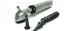
What is an otoscope?
This article has been viewed 33,736 times. An otoscope is a medical instrument used by a doctor to examine the ear. The otoscope magnifies the inside of the ear to detect problems or issues with the outer and middle ears, such as Swimmers ear, earwax build-up, or otitis media. It generally has a magnifying glass, ...
How to hold otoscope?
Handle the otoscope properly. Turn the otoscope’s light on and hold your otoscope “upside down” between your thumb and pointer finger like a pen or pencil. Place the back of your hand along the person’s cheek so that otoscope is steady and braced. While the position may feel awkward at first, it soon will feel natural.
How to treat ear infection?
The ear is a very sensitive organ and can injure easily if improperly examined. Avoid pulling, pushing, or generally being rough with the patient you are examining. This can calm your patient and minimize the risk of injury from sudden movements.
What to ask a patient about pressure?
Ask your patient if the pressure is acceptable to them. For example, “Is the pressure I’m using ok, Mr. Neumaier? Let me know if you have any discomfort.”
How old do you have to be to pull your ear down?
Pull the outer ear down for babies and children younger than 3 years old.