Here are the five basic steps for performing a Gram stain:
- Fix the sample to the slide
- Stain the slide with the primary stain, crystal violet.
- Treat the stain with iodine (a mordant).
- Gently apply a decolorizer on the smear (s), then counterstain with safranin.
- Gram-positive microorganisms such as Staphylococcus aureus appear bluish-purple. Gram-negative microorganisms such as Escherichia coli appear pinkish-red.

How do you prepare a Gram stain?
To prepare a gram stain, fix the bacteria to the slide by passing it through a Bunsen burner flame. Then, flood the sample with several drops of crystal violet dye. The ions will interact with negatively charged bacterial cells and stain them purple. Rinse the crystal violet off and flood the smear with iodine.
Why do we use Gram staining to identify bacteria?
Preparing and interpreting Gram stains Nearly every attempt at identifying an unknown bacterial culture begins with a Gram stain. We start there because the Gram stain differentiates nearly all isolates into one of two major groups, namely Gram-positive or Gram-negative (a small proportion of species stain unpredictably or not at all).
How do you prepare a smear for staining?
We start by preparing a smear on a clean microscope slide, then we heat-fix the cells to the surface and follow with the staining procedure. The objective is to obtain a single layer of cells so that all cells are exposed to the staining reagents.
How do you know if a stain is Gram positive or negative?
Describe the gram reaction of any organisms seen. Gram-positive bacteria stain deep violet to blue and gram-negative bacteria stain pink to red. If your slide is all one color (either pink or blue), then the slide may either have been over or under decolorized.
How to use Gram stain?
Add the sample to the slide. You can use the Gram stain method to help identify bacteria present in medical samples, or bacterial cultures grown in a petri dish. In order for the Gram stain to be useful, add a thin layer of the sample on the stain. A sample under 24 hours old is recommended, as older bacteria may have damaged cell walls that respond less predictably to gram staining.
Who invented the Gram stain?
The Gram stain is named after the Danish scientist Hans Christian Gram (1853 – 1938), who developed the technique in 1882 and published it in 1884 as a technique to discriminate between two types of bacteria with similar clinical symptoms: Streptococcus pneumoniae (also known as the pneumococcus) and Klebsiella pneumoniae bacteria.
What is the color of a Gram stain?
Community Answer. Gram stain stains specific molecules in the cell wall of bacteria. Gram positive bacteria stain purple, gram negative stain pink. Since human cells lack a cell wall, there is nothing to retain the purple stain. When the counter-stain safranin is applied, the cells will be light pink.
How to get rid of Gram positive bacteria on slide?
Tilt the slide and use a wash bottle to squirt a small stream of distilled or tap water over the top of the slide. The water should run down over the surface of the smear, but not be aimed directly at it. Do not rinse excessively, which might remove the stain from Gram positive bacteria .
How to remove crystal violet from slide?
A 1:1 mix of acetone and ethanol is typically used for this critical step, which must be timed carefully. Hold the slide at an angle , then add the decoloriser until no more violet colour is visible in the draining runoff. This typically takes under 10 seconds, or even less time if the decoloriser contains higher concentrations of acetone. Stop immediately, or the decoloriser will remove the crystal violet stain from both gram-positive and negative cells, and the stain will have to be repeated. Immediately rinse off excess decoloriser, using the earlier technique.
What is Gram staining?
Gram staining is a quick procedure used to look for the presence of bacteria in tissue samples and to characterise bacteria as Gram-positive or Gram-negative, based on the chemical and physical properties of their cell walls.
Why do Gram positive bacteria turn purple?
Gram-positive bacteria appear purple, due to the crystal violet trapped within their thick cell walls. Gram-negative bacteria appear pink or red, since the violet washed through the thin cell walls, then the pink counterstain entered them. If the sample is too thick, you may see false positive results.
What color is a Gram positive slide?
Describe the gram reaction of any organisms seen. Gram-positive bacteria stain deep violet to blue and gram-negative bacteria stain pink to red. If your slide is all one color (either pink or blue), then the slide may either have been over or under decolorized.
How to clean decolorizer?
Rinse off the decolorizer with distilled or tap water.
How to get crystal violet off slide?
Place slide on the staining tray. Flood the fixed smear with crystal violet solution (#1) and allow to remain for 1 minute. Rinse off the crystal violet with distilled or tap water. Flood the slide with iodine solution (#2). Allow to remain for one minute.
What is the primary stain used to see decolorized bacteria?
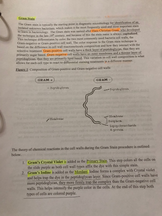
In order to see the decolorized bacteria, a counter stain (Safranine O) is added to exaggerate the contrast with Gram-Negative cells. The crystal violet stain is the primary stain, which stains everything in the smear purple- blue. The Gram's iodine acts as a mordant that causes the crystal violet to penetrate and adhere to the gram-positive cell.
How to fix Gram negative bacteria?
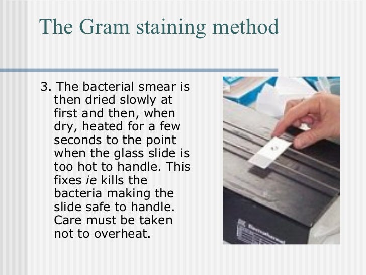
The safranine is the counter-stain that stains everything in the smear that has been decolorized in gram-negative organisms. Apply a smear of bacteria on to a slide. Air dry and then heat fix by passing it through a flame a few times. Make sure you air dry the bacteria before heat fixing.
How to make a bacterial culture?
DIRECTIONS#N#There are six basic steps: 1 Apply a smear of bacteria on to a slide. Air dry and then heat fix by passing it through a flame a few times. Make sure you air dry the bacteria before heat fixing. 2 Add about 5 drops of Hucker’s Crystal Violet to the culture. Let stand for one minute. Bacteria will stain purple. Wash briefly with water and shake off excess. 3 Add about 5 drops of iodine solution to the culture. Let stand for 30 seconds, wash briefly with water and shake off excess. 4 4. Tilt slide and decolorize with solvent (acetone-alcohol solution) until purple color stops running. Be careful not to over-decolorize. Wash immediately (within 5 seconds) with water and shake off excess. 5 Add about 5 drops of Safranine O. Let stand for one minute, wash briefly with water and shake off excess. 6 Examine under microscope at both 400x and 1,000x oil immersion.
What happens when you wash a gram negative with a solvent?
When washed with solvent, the cell pores close becoming less permeable and are able to retain the stain, in this case, purple. Gram-Negative Bacteria have thinner cell walls with more lipids. The solvent dissolves the lipids, which combined with thinner cell walls, washes out or decolorizes the stain. In order to see the decolorized bacteria, ...
How to get rid of purple stains in a bacterial culture?
Make sure you air dry the bacteria before heat fixing. Add about 5 drops of Hucker’s Crystal Violet to the culture. Let stand for one minute. Bacteria will stain purple. Wash briefly with water and shake off excess. Add about 5 drops of iodine solution to the culture. Let stand for 30 seconds, wash briefly with water and shake off excess. 4.
What are the oblong cells in bacteria called?
Slightly oblong bacteria are named ‘coccobacilli’ . You may run across fibrous or filamentous cells similar to fungi, but not quite as wide. These are commonly found in soils. You could find cells with areas of red and purple, often referred to as “Gram-variable”, provided your Gram test was sufficient.
How big are cells in microscopy?
How big are the cells? In microscopy, size variation can be enormous. For instance, microbes are only 10 nanometers or less, while common debris on the slide can be much larger. Even cells taken from your body can be 10 times larger, or more. If you swabbed and stained a sample of your cheek, you may notice a faint pink (cell cytoplasm) along with a light purple (the cell nucleus). These cells are usually bigger than 10 microns in size.
What is the name of the stain that differentiates a culture into two groups?
Nearly every attempt at identifying an unknown bacterial culture begins with a Gram stain. We start there because the Gram stain differentiates nearly all isolates into one of two major groups, namely Gram-positive or Gram-negative (a small proportion of species stain unpredictably or not at all).
How to remove a barely visible amount of material from a culture?
Use a loop or stick to aseptically remove a barely visible amount of material from a culture and stir it into the drop. Try to disperse the culture material completely, so that there are no visible chunks of material, and spread the liquid into a thin layer within the circle. The water should be slightly turbid.
Can you observe Gram stain with a microscope?
We employ bright field microscopy for observing Gram stains. These steps are for your reference only. You will definitely need hands-on training to learn to observe bacteria in a light microscope.
Where do bacteria dry down?
Bacteria often dry down in concentric rings; if you have trouble finding the bacteria look for the rings, which are usually more densely populated than the rest of the smear, around the edges
Can bacteria grow on agar plates?
Sometimes bacteria grown on agar plates adhere strongly to each other andor the agar, making the material very difficult to disperse. With practice, even these "difficult" cultures can be stained and observed.
Is Gram negative culture colorless?
Don't be disappointed if the smear becomes invisible after decolorizing; a Gram negative culture is expected to be colorless until it is counterstained with Gram safranin
What are the prerequisites for a gram staining smear?
Prerequisites: Skill in the use of the microscope, aseptic techniques, experience preparing and gram staining smears, and experience reading smears from cultures.
How to salvage a smear?
If a smear is over-decolorized, it can be salvaged by repeating the Gram staining procedure.