How do I perform a B-scan procedure?
B-scan Pearls 1 • Be sure to direct the patient’s gaze away from the probe and toward the meridian being scanned. 2 • The probe can either be placed on the patient’s closed eyelid or,... 3 • Remember that the top of the displayed image always correlates to the mark on the probe. 4 • Try to keep the probe perpendicular to the tissue being imaged,...
How do you interpret a B scan?
B-Scan Interpretation. When examining the retina on B-scan, conditions such as a retinal tear, retinal detachment and retinoschisis can be clearly differentiated. A retinal tear will appear on longitudinal scans as a “flap.” On occasion, the flap will still be connected to the posterior vitreous surface or membrane.
How do you do an axial scan with a B scan?
The axial scan is obtained by placing the B-scan probe tip directly over the cornea while the patient looks in primary gaze (Figure 3.3) See Clip 3.2.
How do you score an a+ on a B-scan?
Scoring an A+ on a B-Scan 1 Principles of Ultrasonography. B-scan ultrasound uses high frequency soundwaves that are transmitted from a probe/transducer into the eye. 2 Probe Orientations. ... 3 Basic Screening Examination Protocol. ... 4 B-scan Pearls. ... 5 Topographic Evaluation. ... 6 B-Scan Interpretation. ... 7 Know the Norm. ...

What is difference between A-scan and B-scan?
There are two main types of ultrasound used in ophthalmologic practice currently, A-Scan and B-scan. In A-scan, or time-amplitude scan, sound waves are generated at 8 MHz and converted into spikes that correspond with tissue interface zones. In B-scan, or brightness amplitude scan, sound waves are generated at 10 MHz.
Does AB scan hurt?
Your eye is numbed, so you should not have any discomfort. You may be asked to look in different directions to improve the ultrasound image or so it can view different areas of your eye. The gel used with the B-scan may run down your cheek, but you will not feel any discomfort or pain.
Why is B-scan done?
B-scan ultrasonography (USG) is a simple, noninvasive tool for diagnosing lesions of the posterior segment of the eyeball. Common conditions such as cataract, vitreous degeneration, retinal detachment, ocular trauma, choroidal melanoma, and retinoblastoma can be accurately evaluated with this modality.
What is B-scan test?
B scan, or Bright Scan ultrasonography is a diagnostic imaging tool utilized when the view to the back of the eye, or posterior segment is hindered. The posterior segment of the eye is the back two-thirds of the eye and consists of the vitreous, retina, optic nerve, and choroid.
Why would a doctor order an abdominal ultrasound?
It may be ordered to investigate pain, swelling, or other symptoms, and can be the first step in determining the cause for symptoms affecting the soft tissues of the body. For example, in the abdomen it can help check for kidney stones, liver disease, tumours, and the cause of stomach pain or bloating.
What is the most commonly used technique to perform a B-scan?
The most effective method to examine the extent of the retina during a B-scan is to use the limbus-to-fornix technique. To perform this technique, the ultrasonographer should gently glide the probe from the limbus of the eye to the fornix in a sweeping motion to maximize the amount of retina visualized during the scan.
What is A-scan and B-scan in OCT?
In OCT many one-dimensional scans (a-scans) are performed at several depths to create a two-dimensional image (b-scan). Those b-scans, if acquired closely and rapidly, can be translated into a volumetric image (c-scan) of a retina, for example.
What is A-scan B-scan and C scan?
B- scan: If the A- scan - concept is combined with movement of the probe along the surface, a B- scan is the result. It depicts the acoustical side projection of the object, Fig. 4.39. C- scan: Via C- scan are echo amplitudes recorded in relation to probe position.
What is vitreous degeneration?
Vitreous degeneration refers to a change that occurs in the vitreous humor (or vitreous fluid) in the eye, as the vitreous humor changes from a thick vitreous gel to a thin liquid substance. Normally, the vitreous humor is a transparent gel that helps with clarity of vision and maintaining the shape of the eye.
How long does an eye scan take?
In the majority of cases, an eye test will take at least 20 to 30 minutes. Your vision is precious and getting regular check-ups with your optometrist will help to protect and preserve it by helping to monitor your overall eye health.
What is A-scan testing?
Ascan ultrasound biometry, short for amplitude scan, is a routine diagnostic test used by ophthalmologists and optometrists to measure various anatomical dimensions of the eye. This data allows us to select the correct intraocular lens (IOL) for optimal results after Cataract Surgery or Refractive Lens Exchange (RLE).
What are types of scanning?
Here's what you should know.MRI. One of the most common types of scans is a magnetic resonance imaging (MRI) scan. ... X-Ray. X-rays are one of the most common types of scans. ... CT/CAT Scan. Computerized tomography (CT) and computerized axial tomography (CAT) are two names for the same type of scan. ... Ultrasound.
Why did my abdominal ultrasound hurt?
The transducer sends high frequency sound waves through your body. These waves are too high-pitched for the human ear to hear. But the waves echo as they hit a dense object, such as an organ—or a baby. If you're having pain in your abdomen, you may feel slight discomfort during an ultrasound.
How long does an abdominal ultrasound take?
The device sends signals to a computer, which creates images that show how blood flows through the structures in your abdomen. A typical ultrasound exam takes about 30 minutes to complete. It's usually painless. However, you may have some temporary discomfort if the technician presses on an area that is sore or tender.
Is it normal to have stomach pain after ultrasound?
Abdominal ultrasounds are painless, noninvasive imaging tests. You should not feel any aftereffects. Tell your doctor or care team if you have any pain or discomfort after the test. Patients often eat and drink as usual and return to normal activities right after an outpatient ultrasound.
Will a radiographer tell you if something is wrong?
“They aren't doctors, and while they do know how to get around your anatomy, they aren't qualified to diagnose you.” That is true even though the tech likely knows the answer to your question. Imaging techs administer thousands of scans a year.
What are the conditions that B-scans can reveal?
Because B-scans measure the structure and orbit of the eye, they have the potential to reveal details on more conditions, including: vitreous hemorrhage; cancer of the retina, under the retina, and other parts of the eye; damaged tissue or injuries to the orbital socket; any foreign bodies in the eye; swelling; and retinal detachment.
What Conditions May be Diagnosed by an A-Scan or B-Scan?
Generally, they are used in determining the length of the eye for the appropriate replacement lens in cataract surgery. Some tumors may also be found with an A-Scan.
How Do Ultrasounds Work for the Eye?
All ultrasounds are based on how the physics of sound work. Sound is emitted in a parallel, longitudinal pattern (it moves vertically instead of directly across). When the sound contacts an object, it bounces back. When the soundwave returns to the ultrasound probe, it is converted into an electric signal. That electric signal can then be interpreted as an image on a monitor.
What is an A scan?
An A-scan is short for amplitude scan. It is a type of ultrasound biometry that is provides data on the eye’s length. Determining eye length is crucial regarding many common sight disorders, including near-sightedness and far-sightedness. However, for these common conditions, traditional clinician exams are sufficient and far less invasive.
Why is sound imaging important?
To control the depth of the image, ultrasounds change the frequency of the soundwave.
Where is the ultrasound wand placed?
The ultrasound wand will be placed on the front surface of the eye. For a B-scan, the eyelids are closed and a gel is applied prior to the probe’s use. You will likely be asked to move your eyes in multiple directions.
Is an A scan numb?
While A-Scans can be a little uncomfortable in concept, your eyes will be numb and therefore no discomfort should occur.
How to use a B-scan?
Once the patient, personnel and equipment are in the proper orientations, topical anesthetic drops are given to the indicated eye. A methylcellulose-based gel, used as coupling agent, is applied to the B-scan’s probe tip. The patient is instructed to open both eyes and gaze in the direction being imaged. When one of the patient’s eyes is closed, it narrows the contralateral eye and tightens the facial muscles, making the examination more irritating to the patient. When both eyes are open, the contralateral eye is relaxed and can be used to help fixation. The probe is placed directly on the eye. It is possible to obtain B-scan images through closed eyelids. However, it is not recommended in most cases for two reasons. First, the ultrasound waves are attenuated due to the soft eyelid tissue resulting in decreased echo differentiation. Second, it can be difficult to determine the exact position of the B-scan probe on the eye when the lids are closed. In some cases such as the examination of a patient with a ruptured corneal ulcer, or the examination of a small child, the probe should be placed on the eyelids. It is important to note in the examination report that the images were obtained through the eyelids in order to compare subsequent examinations.
What is a B scan?
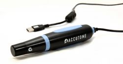
The B-scan probe is a two-dimensional echo display that is used to determine the topographic features of posterior segment pathology including location, shape and extent of lesions (Chapter 2) See Clip 3.1. B-scan probes have a marker along the side of the probe close to the probe tip that indicates the top of the B-scan ultrasound display ( Figure 3.2A ). The transducer inside the B-scan probe oscillates along the plane of the marker only, towards the marker and away from the marker. Therefore, the top of the B-scan display corresponds to the area indicated on the marker and the bottom of the display corresponds to the plane 180° away from the marker. The probe tip corresponds to the white line on the far left side of the B-scan display. The echoes to the right of this line correspond to the ocular structures opposite the probe tip. The further right the ocular structure, the further away is its echo ( Figure 3.2B ).
What muscles are used in ultrasonography?
Unlike fundus photography, where the vasculature of the posterior fundus, the macula and the optic nerve can be used for anatomical reference, ophthalmic ultrasonography is limited to the use of the optic nerve and extraocular muscles.
What is a para axial scan?
Para-axial scans can be helpful in the evaluation of the peripapillary fundus. The para-axial scan images the fundus directly adjacent to the optic nerve. To obtain the scan, the probe tip is placed directly over the cornea as in the axial scan; however, the sound beam is shifted slightly to the peripapillary area of interest. The sound beam is directed through a portion of the crystalline lens in these scans and some sound attenuation, although not as marked as that occurring with the axial scan, occurs resulting in decreased resolution. Para-axial scans are integral in obtaining accurate dimensions of peripapillary mass lesions (Chapter 11).
How to do a longitudinal scan?
The longitudinal scan is obtained by placing the probe marker in the direction of the clock hour to be imaged (Figure 3.4) See Clip 3.3. The transducer located within the probe moves perpendicular to the limbus, sweeping along the radial plane of the fundus located opposite the probe tip. The resulting image shows the fundus along a specific clock hour. The fundus anterior to the equator is located at the top of the B-scan display, the fundus posterior to the equator is imaged centrally and the optic nerve is located at the bottom of the display. In this manner, the longitudinal scan shows the anterior to posterior extent of posterior segment pathology. The longitudinal scan does not usually require the examiner to actively rotate the probe. However, if the desired area to be examined is in the periphery, it can be helpful to place the probe tip closer to the fornix resulting in an image that demonstrates the peripheral fundus at the top of the display with the optic nerve at the far bottom of the display, or completely absent from the display. The longitudinal scans are labeled according to the clock hour imaged anterior to posterior. If the probe is placed at 9 o’clock with the marker towards the pupil, the transducer sweeps along the 3 o’clock plane ( Figure 3.5 ). The resulting image is labeled as a longitudinal scan of 3 o’clock, or L3.
Which scan is the best orientation to evaluate membranes for insertion into the optic disc or adjacent to the optic disc?
Longitudinal scan is the best orientation to evaluate membranes for insertion into the optic disc or adjacent to the optic disc (Chapter 10). It is also essential in the localization of small fundus abnormalities such as a retinal tear or a focal tractional retinal detachment as well as for the evaluation of the macula described later in this chapter.
Where is the optic nerve located on a B scan?
The fundus anterior to the equator is located at the top of the B-scan display, the fundus posterior to the equator is imaged centrally and the optic nerve is located at the bottom of the display. In this manner, the longitudinal scan shows the anterior to posterior extent of posterior segment pathology.
How to scan a bladder?
Press the Scan button a) Press the probe button to start ultra sound scanning to locate the bladder b) Make sure the ultrasound bladder image is the biggest and centered. c) When you find the ideal ultrasound bladder image, press the probe button again. Pad Scan Bladder Scanner will start calculation automatically. d) When you hear a 'beep', the calculation is finished. The result of urine volume will be displayed.
What is a pad scan?
A Pad Scan bladder scanner features new 3D sector probe and real-time ultrasound imaging algorithms that measures the urinary bladder volume and post-void residual (PVR) quickly, safely, automatically and non-invasive. It is your ideal assistant to meet today Point-Of-Care challenges.
How to examine the retina during a B scan?
The most effective method to examine the extent of the retina during a B-scan is to use the limbus-to-fornix technique. To perform this technique, the ultrasonographer should gently glide the probe from the limbus of the eye to the fornix in a sweeping motion to maximize the amount of retina visualized during the scan.
Where should the area of interest be placed in an ultrasound?
Thus, the area of interest should be placed along the equatorial line of the image. In ocular ultrasound, the retina will appear on the right hand side of the image; this is where any pathology should be focused. The denser the tissue, the brighter (hyperechoic) it will appear and vice versa.
How to get an ultrasound image of the eye?
Ultrasound images can be obtained through the patient's eyelids ( as depicted in this tutorial) or with the probe directly on the surface of the eye with appropriate topical anesthesia. Begin with the gain on high. The patient should look in the direction of the quadrant to be evaluated.
Where to place the probe in the T3 quadrant?
For the T3 quadrant of the patient's right eye, instruct the patient to look left. Place the probe on the temporal limbus (L). After obtaining an image of the retina and optic nerve, gently sweep the probe to the fornix (F) to complete evaluation of this quadrant. To view the T3 quadrant of the left eye, the patient should still gaze to the left, but the probe will be placed at the medial limbus, with the marker oriented superiorly.
Where to place a probe on a globe?
Place your probe on the superior aspect of the globe with the marker aimed nasally. Again, begin at the limbus (L)). Ensure you have an image of the retina and optic nerve before sweeping the probe toward the superior fornix (F). Repeat if necessary, centering any pathology.
Where to place a probe for optic nerve?
Figure 3: Ask the patient to look up. Place your probe on the inferior aspect of the globe with the marker oriented nasally. Begin at the limbus (L) and locate the optic nerve shadow, both to orient yourself and assure you are imaging the posterior segment. Slowly sweep your probe toward the inferior fornix (F) until visualization of the T12 quadrant is complete. Repeat if necessary. Remember to center any pathology along the equatorial plane of the image for the best resolution.
How many maneuvers can you do to examine the globe?
One can examine the entire globe in just five maneuvers, i.e. four dynamic quadrant views and one more static slice through the macula and optic disc, also known as longitudinal macula (LMAC). The quadrants views are designated T12, T3, T6, and T9. These numbered quadrants correspond to a clock face superimposed on the eye. For example, T12 is a view through the superior quadrant of the eye, T3 the nasal quadrant of the right eye (temporal quadrant of the left eye), and so on (Figure 1) (2).
What Are A & B Scans?
A scan is short for amplitude scan, while B scan refers to the brightness scan. Both are ultrasound tests performed on the eye by an ophthalmologist. These tests measure various data about the eyes. An ophthalmologist will perform an A or B scan in order to get a better understanding of the condition of your eyes and assess whether you may have an injury, disease, or condition of the eye. After all, monitoring our eye health is very important, so it is best to catch these ailments as early as possible.
What is the purpose of an A scan?
An ophthalmologist uses an A scan to determine the length of the eye through ultrasound technology. The length of the eye is most often used to determine whether a patient has any sight disorders, such as cataracts. On the other hand, an ophthalmologist will use a B scan to diagnose other issues by comparing different parts of the eye, ...
Why do ophthalmologists do A or B scans?
An ophthalmologist will perform an A or B scan in order to get a better understanding of the condition of your eyes and assess whether you may have an injury, disease, or condition of the eye. After all, monitoring our eye health is very important, so it is best to catch these ailments as early as possible.
Why do we run A/B tests?
As you might guess, we run many A/B tests to increase engagement and drive conversions across our platform. Here are five examples of A/B tests to inspire your own experiments.
What is A/B testing?
A/B testing, also known as split testing, is a marketing experiment wherein you split your audience to test a number of variations of a campaign and determine which performs better. In other words, you can show version A of a piece of marketing content to one half of your audience, and version B to another.
What are the independent variables of the A/B test?
The independent variable of this A/B test was the inclusion and type of TOC module in blog posts, and the dependent variables were conversion rate on content offer form submissions and clicks on the CTA inside the TOC module.
How many recipients are needed for a 50/50 A/B test?
It'll let you do a 50/50 A/B test of any sample size — although all other sample splits require a list of at least 1,000 recipients. If you're testing something that doesn't have a finite audience, like a web page, then how long you keep your test running will directly affect your sample size.
Why is A/B testing important?
Above all, though, these tests are valuable to a business because they're low in cost but high in reward.
How much money do you lose if your A/B test fails?
If the test fails, of course, you lost $192 — but now you can make your next A/B test even more educated. If that second test succeeds in doubling your blog's conversion rate, you ultimately spent $284 to potentially double your company's revenue.

Principles of Ultrasonography
Probe Orientations
- The B-scan can be used in three different orientations for the eye: transversal, longitudinal and axial. In a transverse scan, the probe is oriented tangential to the limbus with the probe marker pointing superiorly for vertical scans and oblique scans, or nasally for horizontal scans. You designate the meridian being scanned by the clock hour in the center of the scan. A transverse s…
B-Scan Pearls
- • Be sure to direct the patient’s gaze away from the probe and toward the meridian being scanned. • The probe can either be placed on the patient’s closed eyelid or, for better resolution and certain globe position, place the probe directly on the conjunctiva. • Remember that the top of the displayed image always correlates to the mark on the probe. • Try to keep the probe perpendicul…
Topographic Evaluation
- Topographic evaluation is performed once a lesion is found; this allows for detailed analysis regarding various aspects of the lesion. Typically, a standard sequence of steps is followed in this process. The transverse orientation is classically done first. The probe is placed on the opposite side of the globe as the lesion and moved from limbus to fornix. Following this pattern, sound be…
B-Scan Interpretation
- • Vitreous.When examining the vitreous cavity on a B-scan, common conditions, such as a posterior vitreous detachment, can be evaluated and easily differentiated from more serious abnormalities such as a vitreous hemorrhage. In a young, healthy eye, the vitreous is mostly echolucent. When vitreous degeneration occurs, the cholesterol crystals within the liquefied vitr…
Know The Norm
- It is vital to have a good grasp of what a normal ultrasound looks like. Not only are there minor differences in normal findings from patient to patient, but variation can exist within the same eye, due to the heterogenous nature of a normal eye. It is highly recommended that you gain as much experience as possible examining a normal eye to better evaluate patients with suspected eye di…