- Place the slide on a flat surface.
- Use tweezers or a forceps to place the sample on the slide.
- Place the coverslip on top of the sample. In some cases, it's okay to view the sample without a coverslip, as long as care is taken not to bump the sample into the microscope lens. ...
How to observe plant cells under microscope?
Cut a thin section of stem or leaf which you want to observe. Place it on a slide and put a small amount of colouring agent. Place the slide under the microscope. Focus the lens. Now you can see the plant cell.
How do you prepare a specimen for a microscope?
Then, using tweezers, place the specimen on the center of the slide. Grab a cover slip, making sure to hold it on the edges, and place it on top of the specimen on the slide. Lower the prepared slide onto the microscope stage, positioning it in such a way that the sample is clearly visible through the eyepiece.
How do you make a wet mount on a microscope?
To prepare a wet mount using a flat slide or a depression slide: Place a drop of fluid in the middle of the slide (e.g., water, glycerin, immersion oil, or a liquid sample). If viewing a sample not already in the liquid, use tweezers to position the specimen within the drop.
How to prepare a specimen slide?
How the specimen slide must be prepared largely depends on its properties, such as whether it’s organic or inorganic, live or fixed, wet or dry, thick or thin, large or small, and so on. In fact, these things determine not only how the specimen preparation will go, but also the type of microscope that is most suited for viewing the specimen.

How do you make a slide for a plant cell microscope?
0:081:58DIY Plant Microscope Slides - YouTubeYouTubeStart of suggested clipEnd of suggested clipHere are the materials you will need microscope slides alcohol wipes cover slips water a pipette andMoreHere are the materials you will need microscope slides alcohol wipes cover slips water a pipette and plants. Making the slides wipe the slides with the alcohol wipes. Do the same with the coverslips.
How do you observe plant cells under a microscope?
1:502:51Plant cells under the microscope - YouTubeYouTubeStart of suggested clipEnd of suggested clipInside by removing a thin piece of onion tissue just one cell thick and placing it onto a slide it'sMoreInside by removing a thin piece of onion tissue just one cell thick and placing it onto a slide it's ready to view under a light microscope. The cell walls are easily visible.
How do you prepare and sample a plant cell using a microscope?
1:143:44How to use a Microscope | Cells | Biology | FuseSchool - YouTubeYouTubeStart of suggested clipEnd of suggested clipYou place a slide containing the specimen onto the stage. And secure it using the clips. You firstMoreYou place a slide containing the specimen onto the stage. And secure it using the clips. You first choose the lowest objective lens by turning around the nose.
How do you prepare a slide to view a plant epidermis under a microscope?
Plant cellsPeel a thin, transparent layer of epidermal cells from the inside of an onion.Place cells on a microscope slide.Add a drop of water or iodine (a chemical stain).Lower a coverslip onto the onion cells using forceps or a mounted needle. This needs to be done gently to prevent trapping air bubbles.
How do you observe the plant cell?
A number of stains can be used, including iodine solutions (iodine-potassium iodide, Lugol solution, Gram iodine), crystal violet, toluidine blue, and methylene blue. After 1 minute, rinse away the stain with tap water, add a coverslip, and observe the cells. Nuclei will be evident.
How can u see a plant cell?
Plant cells can be distinguished from most other cells by the presence of chloroplasts, which are also found in certain algae.
How do you make a plant cell slide?
0:383:27Preparing animal and plant cells slides - YouTubeYouTubeStart of suggested clipEnd of suggested clipThree or four minutes before taking a thin glass coverslip. And then very gently place the coverslipMoreThree or four minutes before taking a thin glass coverslip. And then very gently place the coverslip over the top of the cells.
How do you prepare a microscope slide from an animal cell?
First, to prepare an animal cell slide, start by pouring 30 mL of distilled water into a beaker. Then, use a glass dropper to place 2 - 3 drops of the water onto the center of a clean microscope slide. Next, take a clean toothpick and scrape the inside of your cheek to gather cells.
How will you prepare an onion slide on a plant cell?
0:336:50Onion Skin Epidermal Cells: How to Prepare a Wet Mount Microscope ...YouTubeStart of suggested clipEnd of suggested clipI need to break my onion layer towards the shiny side and then very gently peel the two pieces apartMoreI need to break my onion layer towards the shiny side and then very gently peel the two pieces apart. There's a thin transparent layer of cells holding these two pieces of the onion layer.
How do you make a microscope slide to look at onion cells?
Peel a thin layer of onion (the epidermis) off the cut onion. STEP 2 - Place the layer of onion epidermis carefully on the glass slide, and cover with a cover slip. STEP 3 - Stain the layer of onion with food colouring. STEP 4 - View your onion cells.
How do you use a microscope step by step?
1:264:23BIOLOGY 10 - Basic Microscope Setup and Use - YouTubeYouTubeStart of suggested clipEnd of suggested clipLens. Start with a low-power objective lens 4x. And the stage in its highest position. Then lookMoreLens. Start with a low-power objective lens 4x. And the stage in its highest position. Then look through the ocular lens. And focus on the specimen.
How can you tell the difference between a plant and animal cell under a microscope?
Plant cells have a rigid cell wall that surrounds the cell membrane. Animal cells do not have a cell wall. When looking under a microscope, the cell wall is an easy way to distinguish plant cells.
Can you see a plant cell with a light microscope?
The light microscope works by creating a magnified image of the object. Organelles that can be seen and observed under a light microscope include the nucleus, cytoplasm, cell membrane, chloroplasts, and cell wall. The chloroplasts and cell walls are only present in plant cells.
How to prepare a specimen for a microscope?
Prepare the specimen by thinly slicing the portion you wish to view. Darker and more opaque specimens need to be sliced as finely as possible. Get a clean piece of a microscope slide, and hold it carefully on its edges. Then, using tweezers, place the specimen on the center of the slide. Grab a cover slip, making sure to hold it on the edges, ...
How to adjust microscope slide?
Carefully lower the prepared slide onto the microscope stage, and slowly adjust the position of the slide until the specimen is right beneath the aperture.
How to collect liquid specimens?
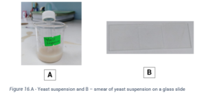
Use a pipette to collect a small amount of the liquid specimen. Take a piece of microscope slide, and place a drop of the specimen on to the slide. Using a second piece of a glass slide, smear the specimen around the slide by dragging the smearing slide across the first.
What type of microscope does a specimen have to be prepared before it can be viewed?
For many types of microscopes, especially light or bright field microscopes, some form of specimen sample preparation must be done before the specimen can be viewed under the microscope. Take for example a compound light microscope. It makes use of microscope slides to mount the specimen on, before loading it onto the microscope for viewing.
What is a flat slide?
Flat slides. The most basic type of microscope slides are tiny rectangular pieces of clear glass or plastic made of soda lime or borosilicate, which measure at around 1 by 3 inches, and are roughly 1 millimeter thick. Flat slides need to be covered with a cover slip when using.
What is a cover slip for a microscope?
When working with flat slides, it’s a must to use a cover slip to protect the specimen from moving, leaking, getting contaminated, and coming into contact with the objective lens of the microscope. It’s a small piece typically made of clear square borosilicate or silicate glass.
What is the easiest slide to make?
Perhaps the most common and the easiest type of microscope slide preparation is making a dry mount slide, since it essentially just involves placing the specimen on the slide. This is a technique used for most inorganic specimens and dead matter.
What is a microscope slide?
Microscope slides are pieces of transparent glass or plastic that support a sample so that they can be viewed using a light microscope. There are different types of microscopes and also different types of samples, so there is more than one way to prepare a microscope slide. The method used to prepare a slide depends on the nature of the specimen.
What is the best way to test a slide?
Place a drop of fluid in the middle of the slide (e.g., water, glycerin, immersion oil, or a liquid sample).
How to draw liquid into a line?
Place a small drop of a liquid sample onto the slide. Take a second clean slide. Hold it at an angle to the first slide. Use the edge of this slide to touch the drop. Capillary action will draw the liquid into a line where the flat edge of the second slide touches the first slide.
What is dry mount slide?
Dry mount slides can consist of a sample placed on a slide or else a sample covered with a coverslip. For a low power microscope, such as a dissection scope, the size of the object isn't critical, since its surface will be examined. For a compound microscope, the sample needs to be very thin and as flat as possible.
What organisms need more space than what forms between a coverslip and a flat slide?
Some organisms (like Paramecium) need more space than what forms between a coverslip and a flat slide. Adding a couple of strands of cotton from tissue or swab or else adding tiny bits of broken coverslip will add space and "corral" the organisms.
What are some good slides for food?
Many common foods and objects make fascinating subjects for slides. Wet mount slides are best for food. Dry mount slides are good for dry chemicals. Examples of appropriate subjects include:
How to stop evaporation of a slide?
As the liquid evaporates from the edges of the slide, living samples may die. One way to retard evaporation is to use a toothpick to coat the edges of the coverslip with a thin rim of petrol eum jelly before dropping the coverslip over the sample. Press gently on the coverslip to remove air bubbles and seal the slide.
What microscopes can we see inside cells?
Light and electron microscope s allow us to see inside cells. Plant, animal and bacterial cells have smaller components each with a specific function.
What is the purpose of a coverslip?
A small square or circle of thin glass called a coverslip is placed over the specimen. It prevents the slide from drying out when it's being examined. Iodine stain can be used to stain plant cells to make the internal structures more visible.

