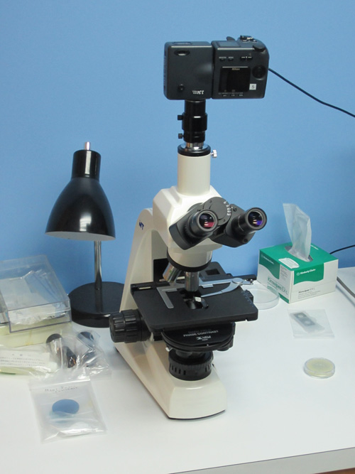
- Place a brightly stained specimen on the stage and rotate the 10x phase contrast objective into the optical pathway in brightfield illumination mode. ...
- Remove the stained specimen and place a phase specimen on the microscope stage.
How do you adjust a phase contrast microscope?
1:298:15Adjusting Phase - YouTubeYouTubeStart of suggested clipEnd of suggested clipNow we typically would use a centering telescope this is a lens assembly that basically fits intoMoreNow we typically would use a centering telescope this is a lens assembly that basically fits into one of the eyepiece slots. So we pull out the eyepiece. And we simply put this in its place.
How do you set up a DIC on a microscope?
Remove an eyepiece and look down the microscope tube. Turn the adjustment knob on the DIC slider until a black band extends diagonally across the objective back focal plane. Replace the eyepiece, and make any final adjustment of the slider to give best DIC contrast.
How can you tell if your microscope is set for phase contrast illumination?
0:092:40Phase contrast microscope - YouTubeYouTubeStart of suggested clipEnd of suggested clipIf these two waves meet at a point there will be a destructive interference as a result of which theMoreIf these two waves meet at a point there will be a destructive interference as a result of which the amplitude decreases. The decrease in amplitude in turn decreases the intensity.
What is the purpose of using the phase contrast setting on the microscope?
Phase contrast is a light microscopy technique used to enhance the contrast of images of transparent and colourless specimens. It enables visualisation of cells and cell components that would be difficult to see using an ordinary light microscope.
How do I align my DIC?
Objective DIC Prism Alignment - Install the objective prism by either inserting the slider or a prism confined to a fixed mount (for systems utilizing de Sénarmont bias retardation). Once the prism is in position, examine the objective rear focal plane once again with the phase telescope or Bertrand lens.
What is the difference between phase contrast and DIC?
In both cases, contrast in the images obtained from DIC is largely dependent upon the orientation of the specimen with respect to the shear axis of the microscope, while the phase contrast image features are independent of specimen rotation around the microscope optical axis.
Why is green light used in phase contrast microscopy?
Most of the microscope manufacturers provide a green interference or absorption filter with their auxiliary phase contrast kits, because the filter will produce monochromatic light having the same wavelength used for the original calibration of the objective phase plates.
What is the magnification of phase contrast microscope?
Phase contrast is preferable to bright field microscopy when high magnifications (400x, 1000x) are needed and the specimen is colorless or the details so fine that color does not show up well.
How do you take good pictures under a phase contrast microscope?
Thus, in order to achieve high-quality phase-contrast images, the correct phase plate and condenser annulus pair must be used and the condenser annulus must be properly centered such that the image of the annulus corresponds exactly with the position of the phase ring.
What is the principle of phase contrast microscopy?
The phase contrast microscopy is based on the principle that small phase changes in the light rays, induced by differences in the thickness and refractive index of the different parts of an object, can be transformed into differences in brightness or light intensity.
Why is oil used in 100X objective?
While we want light to refract differently between the specimen and the medium, we do not want to lose any light rays, as this would decrease the resolution of the image. By placing immersion oil between the glass slide and the oil immersion lens (100X), the light rays at the highest magnification can be retained.
What type of microscopy is phase contrast microscopy?
optical microscopy techniquePhase-contrast microscopy (PCM) is an optical microscopy technique that converts phase shifts in light passing through a transparent specimen to brightness changes in the image. Phase shifts themselves are invisible, but become visible when shown as brightness variations.
How does a DIC microscope work?
DIC microscopy is a light microscopic technique based on an interference principle involving two coherent beams of light (from the same small light source) and image contrast achieved with gradients in optical path. It produces clear optical sections of thick transparent specimens and a 3D shadowed image.
How do you use DIC?
Aligning Differential Interference ContrastFocus on a blank sample plate using either a 4x or 10x objective.Move the DIC slider with the U-AN analyser into the light path. ... If using a trinocular head, remove one eyepiece and view the sample directly down the trinocular head.a.More items...
When would you use DIC microscopy?
DIC is used for imaging live and unstained biological samples, such as a smear from a tissue culture or individual water borne single-celled organisms. Its resolution and clarity in conditions such as this are unrivaled among standard optical microscopy techniques.
What is DIC slider?
Differential interference contrast (DIC) optical components can be installed on virtually any brightfield transmitted, reflected, or inverted microscope, provided the instrument is able to accept polarizing filters and the specially designed condenser and objective prisms (together with the housings) necessary to ...
How to arrange condenser annular plates?
The condenser annular plates should also be sequentially arranged, starting with the annulus designed for the lower magnification objectives and proceeding up to the one designed for the highest magnification. In general, the entire magnification range can be covered with three or four individual annuli. If a universal condenser is employed, position the lowest magnification annulus (for example, PhL or Ph1) to the right of the brightfield slot, and the other plates in sequential order in the neighboring slots. Often, the specimen must be examined in brightfield either before or after phase contrast observation, so this arrangement will provide an easy work flow.
How does a microscope align with an objective phase plate?
By matching the condenser annulus to the objective phase plate, the microscope can be aligned to superimpose illuminating light rays passed through the annulus onto the objective phase ring to achieve phase contrast illumination.
What is phase contrast annulus?
The phase contrast annuli ( annulus is the Latin term for ring, with the plural being annuli) utilized in the substage condenser on an upright microscope, or within the condenser turret on the illumination pillar of an inverted microscope, must be specifically matched to a particular objective equipped with a corresponding phase plate. For example, a 20x objective and a 100x objective, both of which contain a phase plate near the diffraction plane (rear focal plane), will require condenser annuli having different diameters that correspond to the objective magnification and numerical aperture. By matching the condenser annulus to the objective phase plate, the microscope can be aligned to superimpose illuminating light rays passed through the annulus onto the objective phase ring to achieve phase contrast illumination.
What is annulus insert?
Condenser annulus inserts are circular aluminum plates having a stamped ring of varying dimensions in the center (as illustrated in Figures 1 and 2 ). After the stamping operation, annular disk units are anodized and dyed with a flat-black pigment to absorb stray light and ensure that illuminating light rays passing through the annulus follow a defined pathway. The central light stop, which varies in size depending upon the target objective aperture and magnification range, is positioned in the center of the plate and secured by three slim tabs spaced at 120-degree intervals. The annulus plate is either pressed or glued into a circular frame (also anodized and dyed black), which is fitted into a round opening in the condenser turret.
What type of plate is used in a condenser?
Modern universal condenser system turrets (see Figure 2) usually contain between five and eight open slot positions that can be fitted with annular phase contrast plates, DIC Nomarski (Wollaston) prism plates, or darkfield light stop plates. The basic construction of these plates is similar. Darkfield light stops are manufactured in a manner similar to phase annular diaphragms, but the annulus is generally wider and has a larger diameter to enable highly oblique light wavefronts to pass through. In contrast, DIC prisms are cut into circular slabs that are glued into an anodized frame and inserted into the turret.
How to focus a Bertrand lens?
In a similar manner, a majority of the Bertrand lenses in modern microscopes are equipped with a focusing thumbwheel that enables the operator to focus the Bertrand-lens-eyepiece combination on the phase plate in the objective. When utilizing a matched set of objectives (parfocal, with the same optical correction and tube length), the rear focal plane position should remain essentially the same as magnification is changed, thus reducing the necessity to refocus the phase telescope or Bertrand lens.
Where is the aperture diaphragm located in the focal plane?
In order to adapt a standard Abbe, achromatic, or aplanatic substage condenser for phase contrast observations, a mechanism must be established for placing the annular diaphragm in or very near the condenser front focal (aperture) plane. Several condenser models are fabricated with the aperture iris diaphragm (always located in the focal plane) positioned at the base of the condenser, adjacent to slots designed to accept phase plates or darkfield light stops. Other condensers have the aperture plane located in the central region of the housing, which is relatively inaccessible to auxiliary components, such as a phase annulus.
How does a tungsten halogen lamp work?
Light from a tungsten-halogen lamp goes through the condenser annulus in the substage condenser before it reaches the specimen. This allows the specimen to be illuminated by parallel light that has been defocused. Some of the light that passes through the specimen will not be diffracted (bright yellow in the picture).
Why is phase contrast used in microscopy?
As phase contrast microscopy does not require cells to be killed, fixed or stained, the technique enables living cells, usually in culture, to be visualised in their natural state. This means biological processes can be seen and recorded at high contrast and specimen detail can be observed. Fluorescence staining can be used in combination with phase contrast to further improve the visualisation of samples.
What are the limitations of phase contrast?
Limitations of phase contrast 1 Phase contrast images often have halos surrounding the outline of details that have a high phase shift. These halos are optical artefacts and can make it hard to see the boundaries of details. 2 The resolution of phase images can be reduced due to the phase annuli limiting the numerical aperture of the system. 3 Phase contrast doesn’t work well with thick specimens as these can appear distorted.
Why are phase contrast images reduced?
The resolution of phase images can be reduced due to the phase annuli limiting the numerical aperture of the system.
How does phase plate affect background light?
The phase plate then changes the background light’s speed by ¼ wavelength. When the light is focused on the image plane, the diffracted and background light will cause destructive or constructive interference, which changes the brightness of the areas that contain the sample in comparison to the background light. Often the background is also dimmed by 60 to 90% by a grey filter ring.
How does the light phase change?
The difference of the light phase is increased by slowing down (or advancing) the background light by a ¼ wavelength, with a phase plate just before the image plane. When the light is focused on the image plane, the diffracted and background light cause destructive (or constructive) interference which decreases (or increases) ...
What is an unstained object?
Unstained specimens that do not absorb light are known as phase objects. This is because they slightly change the phase of light that is diffracted by them; the light is usually phase-shifted by about ¼ wavelength compared to the background light.
How to use phase contrast on a microscope?
In cases where the target specimen has only minimal optical path differences (and may be difficult to visualize), align the microscope with a specimen known to produce high contrast in phase contrast mode. Rotate the condenser turret until the appropriate annulus is positioned in the optical pathway ( Ph1 or the equivalent for the 10x objective). Check to ensure that the condenser annulus color code or inscription matches that of the objective. Examine the position of the condenser aperture diaphragm lever and move it to the widest iris position (condensers designed for phase contrast may do this automatically).
How many annuli can a condenser have?
In general, the entire magnification range can be covered with three or four individual annuli. If a universal condenser is employed, position the lowest magnification annulus (for example, PhL or Ph1) to the right of the brightfield slot, and the other plates in sequential order in the neighboring slots.
What happens if the image of the annulus does not fit within the dark circle of the phase plate?
If the image of the annulus does not fit within the dark circle of the phase plate, then either the condenser is out of focus (and Köhler illumination is not established), or the phase telescope (or Bertrand lens) is not focused on the objective rear focal plane.
How to arrange condenser annular plates?
The condenser annular plates should also be sequentially arranged, starting with the annulus designed for the lower magnification objectives and proceeding up to the one designed for the highest magnification. In general, the entire magnification range can be covered with three or four individual annuli. If a universal condenser is employed, position the lowest magnification annulus (for example, PhL or Ph1) to the right of the brightfield slot, and the other plates in sequential order in the neighboring slots. Often, the specimen must be examined in brightfield either before or after phase contrast observation, so this arrangement will provide an easy work flow.
What happens when the annulus is properly centered?
When the condenser annulus is properly centered, the ring of light passing through the condenser will be attenuated by the neutral density material applied to the objective phase plate, reducing the intensity of the annulus image.
Why is it important to align a phase contrast microscope?
Careful alignment of the phase contrast microscope is essential in order to produce the maximum contrast effect without introducing artifacts that degrade specimen appearance or lead to erroneous interpretation. This interactive tutorial examines variations in how specimens appear through the eyepieces (at different magnifications) when the condenser annulus is shifted into and out of alignment with the phase plate in the objective.
What happens if a microscope is out of phase contrast?
Once the microscope has been aligned for phase contrast, it will generally hold its centration for a considerable number of objective/annuli changes, but should be checked periodically to ensure proper alignment. If the microscope starts to slip out of alignment, the images appearing in the eyepieces (or on a computer monitor) will appear increasingly more like those observed with brightfield illumination.
How to achieve phase contrast?
In order to achieve a phase contrast image your microscope must be set-up with the proper phase contrast components. These components must be properly aligned and centered. Each objective must be centered to the corresponding phase annulus. In order to center for phase contrast please follow the instructions below.
What are some examples of a system that is not centered and aligned for phase contrast?
Images a & c are an example of a system that is not centered and aligned for phase contrast. Image e shows a perfectly centered system. The segmented circle of light falls directly into the black ring.
Do you need to recenter a microscope?
Once your microscope is fully centered for phase contrast it should hold its position. There really should be no need to re-center unless the system is tampered with or it is a written protocol. You should check for proper centration periodically by inserting your phase centering telescope.
What happens when direct undeviated light and diffracted light proceed to the image plane?
Now, when the direct undeviated light and the diffracted light proceed to the image plane, they are 1/2 wavelength out of phase with each other. The diffracted and direct light can now interfere destructively so that the details of the specimen appear dark against a lighter background (just as they do for an absorbing or amplitude specimen). This is a description of what takes place in positive or dark phase contrast.
What is an unstained object?
Unstained specimens that do not absorb light are called phase objects because they slightly alter the phase of the light diffracted by the specimen, usually by retarding such light approximately 1/4 wavelength as compared to the undeviated direct light passing through or around the specimen unaffected. Unfortunately, our eyes as well as camera ...
Why is a green filter used in phase images?
Also, because the speeding up or slowing down of the direct light is calculated on a 1/4 wavelength of green light, the phase image will appear best when a green filter is placed in the light path (a green interference filter is preferable). Such a green filter also helps achromatic objectives produce their best images, since achromats are spherically corrected for green light.
Why are unstained specimens called phase objects?
Unstained specimens that do not absorb light are called phase objects because they slightly alter the phase ...
How does a phase plate work?
To speed up the direct undeviated zeroth order light, a phase plate is installed with a ring shaped phase shifter attached to it at the rear focal plane of the objective. The narrow area of the phase plate is optically thinner than the rest of the plate. As a result, undeviated light passing through the phase ring travels a shorter distance in traversing the glass of the objective than does the diffracted light.
Why is phase microscopy important?
Phase microscopy continues to be a widely used and important tool, particularly for the microscopist studying living and/or unstained material, such as cells and tissues in culture. The method is also currently used simultaneously with reflected light fluorescence to reveal areas of a specimen that do not fluoresce.
What are the limitations of phase contrast?
There are some limitations of phase contrast microscopy: 1 Phase images are usually surrounded by halos around the outlines of details. Such halos are optical artifacts, which sometimes obscure the boundaries of details. 2 The phase annuli do limit the working numerical aperture of the optical system to a certain degree, thus reducing resolution. 3 Phase contrast does not work well with thick specimens because of shifts in phase occur from areas slightly below or slightly above the plane that is in focus. Such phase shifts confuse the image and distort image detail. 4 Phase images appear gray if white light is used and green if a green filter is used. In the past, many microscopists restricted their film to black and white when performing photomicrography on phase specimens. Today, many color films reproduce black, white, and grayscales very effectively, especially the tungsten-balanced transparency films from Fuji, Kodak, and Agfa.
How to set up a microscope for phase optics?
To set up your microscope for phase optics, you first set it at BF and focus on the specimen. Adjust the height of the condenser for optimum image quality. Next, set the condenser turret to the phase setting for that particular lens and remove the specimen.
What is phase contrast?
Phase contrast is a method used in microscopy and developed in the early 20th century by Frits Zernike. Zernike discovered that if you speed up the direct light path, you can cause destructive interference patterns in the viewed image. These patterns make details in the image appear darker against a light background.
What are the long adjustment screws on a phase condenser?
The long adjustment screws on the phase condenser push in to engage set screws for proper alignment of the phase ring.
How do you align a telescope?
When looking through the telescope, you will see two rings. They may or may not be concentric. By turning centering adjustment screws on the condenser, you align the rings so that they become concentric.
When did Zernike win the Nobel Prize?
This phase contrast technique proved to be such an advancement in microscopy that Zernike was awarded the Nobel prize (physics) in 1953. See more on Frits Zernike biography. Cheek cells captured with regular brightfield objectives. Notice the air bubbles at three locations, some cells are visible on the left.
What is BF on a microscope?
This allows you to use the phase objectives as standard brightfield lenses. Not all phase contrast microscopes are the same but generally they rely on similar techniques to set up the system for optimum results.
What are the controls that stick out from both sides on the back of a condenser?
The controls that stick out from both sides on the back are for centering the condenser.
History Of Phase Contrast Microscopy
The discovery of phase contrast microscopy was made by a Dutch physicist, Frits Zernike in 1938.
Basic Components Of Phase Contrast Microscopy
To obtain the perfect enlarged image of the biological samples, the inverted phase contrast microscope uses some basic components. These components work in harmony to give out the perfect high contrasted image of the specimens.
Limitations of Phase Contrast Microscopy
Phase Annuli or rings limit the aperture to some extent, which decreases the resolution of the specimen’s images.
.JPG)