What do you need to do before putting a slide on a stage?
Do you need glasses for a microscope?
Do you clean the glass of a lamp housing?

How do you use a microtome?
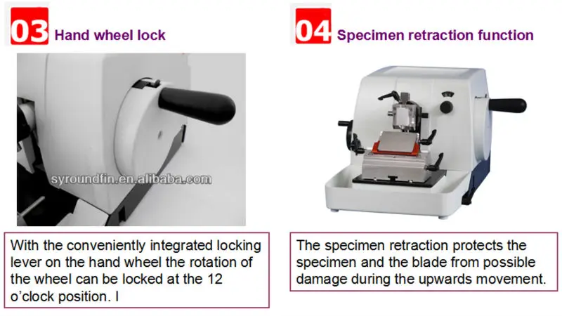
2:314:47MICROTOME TUTORIAL - YouTubeYouTubeStart of suggested clipEnd of suggested clipUnlock the wheel on the right release the finger guard and turn the wheel swiftly. Until you haveMoreUnlock the wheel on the right release the finger guard and turn the wheel swiftly. Until you have begun to cut into the sample.
How do I reset my microtome?
Specimen advance can be reset by turning back crank on left side of microtome clockwise until stop, then counterclockwise a quarter turn. While doing this, place pressure on specimen holder to make sure that advance resets.
What are the 3 major parts of a microtome?
There are different microtomes, but they all consist of three main parts: Base (microtome body) Knife attachment and blade. Material or tissue holder.
What are the steps in tissue processing?
Tissue processing is the technique by which fixed tissues are made suitable for embedding within a supportive medium such as paraffin, and consists of three sequential steps: dehydration, clearing, and infiltration.
What are common problems during Microtomy?
Large holes, missing tissue, or mushy sectionsTissue not embedded flat; will not show during sectioning.Tissue not processed properly and will not form a section (especially if center is raw)Under-processed portion of tissue bursts on contact with warm water.Block faced too aggressively.
What are the four types of microtome?
Types of microtomeRotary microtome. The rotary microtome is often referred to as the “Minot” after its inventor. ... Base sledge microtome. With the sledge microtome, the specimen is held stationary and the knife slides across the top of the specimen during sectioning. ... Sliding microtome. ... Ultra microtome.
Which blade is used in microtome?
Microtomes use steel, glass or diamond blades depending upon the specimen being sliced and the desired thickness of the sections being cut. Steel blades are used to prepare histological sections of animal or plant tissues for light microscopy.
What is the most commonly used microtome?
Rotary Mictrotome -Rotary Mictrotome - It is most commonly used microtome. This device operates with a staged rotary action such that the actual cutting is part of the rotary motion. In a rotary microtome, the knife is typically fixed in a horizontal position.
What is the simplest type of microtome?
Rocking microtome is the simplest type and is used for cutting unembedded tissues. Cryostat is used to cut under-hydrated tissues in a frozen state. Both statement is false. Sliding microtome has two types; small and large sliding microtome.
What are the 4 steps done at the tissue processor?
Overview of the steps in tissue processing for paraffin sectionsObtaining a fresh specimen. Fresh tissue specimens will come from various sources. ... Fixation. The specimen is placed in a liquid fixing agent (fixative) such as formaldehyde solution (formalin). ... Dehydration. ... Clearing. ... Wax infiltration. ... Embedding or blocking out.
What is the first step in tissue processing?
The technique of getting fixed tissue into paraffin is called tissue processing. The main steps in this process are dehydration and clearing. Wet fixed tissues (in aqueous solutions) cannot be directly infiltrated with paraffin. First, the water from the tissues must be removed by dehydration.
What is the most important step in tissue processing?
FIXATION. Fixation of tissues is the most crucial step in the preparation of tissue for observation in the transmission electron microscope. Fixation consists of two steps: cessation of normal life functions in the tissue (killing) and stabilization of the structure of the tissue (preservation).
What is the maintenance of microtome?
Cleaning of the Microtome • The rotary when must be locked and blade removed from the holder before cleaning. Ensure that the lock is properly engaged. Always wear gloves when cleaning the microtome. Use a disinfectant that is effective against possible infectious agents.
How do you clean microtome?
Either discard the knife directly in a sharps container or if re-using it, place in a container of disinfectant to soak. Ensure complete contact time, then use forceps and a handled brush to remove residue and scrub clean, followed by water rinse.
How do you fix histopathology tissue?
Fixation The specimen is placed in a liquid fixing agent (fixative) such as formaldehyde solution (formalin). This will slowly penetrate the tissue causing chemical and physical changes that will harden and preserve the tissue and protect it against subsequent processing steps.
How do you maintain a damaged microtome knife?
A. Your microtome knife has been coated with an oil mixture to prevent rust and corrosion when not in use. B. Before using your knife, take a lint-free facial tissue saturated in either zylene, benzene or acetone to remove the protective oil coating on the knife.
What do you need to do before putting a slide on a stage?
Before putting a slide on the stage - turn on the illumination & set the light to a comfortable level. IT IS NOT NECESSARY to use the light at maximum - to do so can be very tiring to the eyes.
Do you need glasses for a microscope?
If you wear glasses for mild near- or far-sightedness, you generally won’t need them while looking through the scope – the microscope can defocus or overfocus the image as needed. However, if you have astigmatism or a heavy prescription, you may find it necessary to wear glasses –just keep in mind that the oculars may scratch your lenses, particularly if they are plastic.
Do you clean the glass of a lamp housing?
The glass of the Lamp Housing MUST be clean
What is the eyepiece of a microscope?
The eyepiece is the portion that you will look into to view your specimen. Simple compound microscopes will only have one eyepiece while more complex microscopes will have a binocular eyepiece. Here are the components: The stage is a platform where you will place your slides for viewing.
How to keep a microscope manual?
Keep your microscope manual nearby. Read it carefully, if you want to see instructions on how to handle your specific model. The manual will also have instructions on maintenance and cleaning if those things are necessary .
How to clean a microscope?
Clear your surface of any debris that could potentially harm your microscope. Clean the area with a surface cleaner and lint-free rag, if necessary. Make sure the table is located near an electrical outlet. Carry the microscope below the base and on the arm.
How to get the lowest magnification?
Start focusing on the lowest power objective. You may have two or three different rotating objective lenses that you can switch into place to magnify the object. You should start with the 4x objective and increase until it is focused. Usually, the 4x (sometime 3.5x) objective is the standard for the lowest magnification on a basic microscope.
How many testimonials does wikihow have?
wikiHow marks an article as reader-approved once it receives enough positive feedback. This article has 12 testimonials from our readers, earning it our reader-approved status.
How to see two images at the same time?
If you see two images when you look through the eyepieces, you need to continue to adjust the distance. Move the eyepieces closer together or further apart until you see a single circle of light. Remove your glasses, if you wear them. You can use the microscope's settings to focus the object according to your sight.
What is a microscope?
A microscope is a device that magnifies an image, allowing you to see small structures in detail. Although they come in a variety of sizes, microscopes for home and school use generally have similar parts: a base, an eyepiece, a lens and a stage. Learning the basics of using a microscope will protect the equipment and provide you ...
How to set up a digital microscope?
To set up your digital microscope, begin by installing the required software and connecting your device via the included USB cable. Attach the microscope body to the stand and choose your preferred magnification and lighting. Adjust the focus wheel until the image on the screen is sharp and in focus. Before you get started, you’ll need ...
How to use a microscope stand?
If you are using a microscope stand, place it on a hard, flat surface. Make sure that it is close enough to your laptop for the USB cable to reach. When you connect the cable, the image captured by the microscope camera should automatically appear on your screen.
What is the best way to see detail on a microscope?
If your microscope comes with LED lighting, you usually want to set this to the maximum. As a rule of thumb, the brighter the lighting, the more detail is visible.
What happens if you change your magnification?
If you change your magnification midway through your examination, the image will become blurred and you will have to refocus again. At this point, your digital microscope is set up and ready to go. Depending on the software you are using, there may be additional options to play with such as time-lapse or video capture.
How long does it take for a phone to recognize a camera?
If you have any issues with your phone recognizing the device, unplug your USB and then wait a few seconds before reinserting. It can occasionally take up to a minute for the cam image to appear on the screen.
Where is the snap button on a microscope?
Finding your way around your software should be moderately straightforward but if you need a quick way to capture your microscopic image, the “snap” button on the side of your microscope should do the trick.
Can I connect my microscope to my phone?
These days, most digital microscopes give you the option of connecting your device to your mobile phone. Although most USB microscopes are not compatible with iOS, your device may have WiFi options to enable iPhone connection. If you are connecting to an android, you will need an OTG cable.
What do you need to do before putting a slide on a stage?
Before putting a slide on the stage - turn on the illumination & set the light to a comfortable level. IT IS NOT NECESSARY to use the light at maximum - to do so can be very tiring to the eyes.
Do you need glasses for a microscope?
If you wear glasses for mild near- or far-sightedness, you generally won’t need them while looking through the scope – the microscope can defocus or overfocus the image as needed. However, if you have astigmatism or a heavy prescription, you may find it necessary to wear glasses –just keep in mind that the oculars may scratch your lenses, particularly if they are plastic.
Do you clean the glass of a lamp housing?
The glass of the Lamp Housing MUST be clean
