To test cranial nerve III (oculomotor nerve), IV (trochlear), VI (abducens): Have the patient follow your pen light by moving it 12-14 inches from the patient’s face in the six cardinal fields of gaze (start in the midline) Watch for any nystagmus (involuntary movements of the eye)
How do you do a normal response cranial nerve assessment?
Cranial Nerve Assessment Normal Response Documentation; Hold a penlight 1 ft. in front of the client’s eyes. Ask the client to follow the movements of the penlight with the eyes only. Move the penlight through the six cardinal fields of gaze. Both eyes coordinated, move in unison with parallel alignment. Both eyes move in coordination.
What are the components of a cranial nerve exam?
One component of the examination (III) uses the pupillary light reflex to assess the status of the oculomotor nerve. The cranial nerve exam is a type of neurological examination. It is used to identify problems with the cranial nerves by physical examination. It has nine components.
How do you test the three nerves of the eye?
These three nerves are tested together as the control movement of the eye. This is done by asking the patient to keep their head perfectly still directly in front of you, you should draw two large joining H’s in front of them using your finger and ask them to follow your finger with their eyes.
What equipment is required for a cranial nerve examination?
The following equipment is required for a Cranial Nerve Examination: 1.) Olfactory Nerve (I) The olfactory is a sensory nerve, and damage in the nasal epithelium or the basal gangliamight impair the ability to discriminate different smells.
How do you test cranial nerve III IV VI?
Cranial Nerve III, IV, and VI – Oculomotor, Trochlear, AbducensTest eye movement by using a penlight. Stand 1 foot in front of the patient and ask them to follow the direction of the penlight with only their eyes. ... Test bilateral pupils to ensure they are equally round and reactive to light and accommodation .
How do you examine trochlear nerve?
This muscle depresses and abducts the eyeball when working independently. The extraocular muscles, on the other hand, synergistically move the eye. As a result, the trochlear nerve is tested by having the patient look 'down and in,' as the superior oblique contributes the most to this motion.
How do you test for trochlear nerve palsy?
Measure the patient's vertical misalignment while in the upright position. Then, measure again while the patient is supine. A decrease of more than 50% in supine is a positive result. The vertical misalignment caused by skew deviation depends on head position, whereas it would not change in trochlear nerve palsy.
What is cranial nerve IV?
The trochlear nerve is the fourth cranial nerve (CN IV) and one of the ocular motor nerves that controls eye movement. The trochlear nerve, while the smallest of the cranial nerves, has the longest intracranial course as it is the only nerve to have a dorsal exit from the brainstem.
What is CN IV palsy?
Fourth nerve palsy means that a certain muscle in your eye is paralyzed. It is caused by disease or injury to the fourth cranial nerve. In children, it is most often present at birth (congenital). In adults, it is most often caused by injury. Many cases of fourth nerve palsy are idiopathic.
What does cranial nerve 4 palsy look like?
Fourth cranial nerve palsy may affect one or both eyes. Because the superior oblique muscle is paretic, the eyes do not adduct normally. Patients see double images, one above and slightly to the side of the other; thus, going down stairs, which requires looking down and inward, is difficult.
How do you fix 4th nerve palsy?
Eye muscle surgery is generally recommended as the treatment for fourth nerve palsy in children and adults. Following corrective eye muscle surgery for fourth nerve palsy, the associated abnormal head tilt usually disappears.
Which part of the H is testing the trochlear nerve?
Another way to examine the trochlear nerve is with the 'Bielchowsky head tilt test. ' When a trochlear nerve palsy is suspected, the diplopia can be abolished by tilting the head toward the shoulder of the unaffected side.
What does trochlear nerve palsy look like?
Patients with trochlear nerve palsy complain of double vision vertically (vertical diplopia) or the images being tilted or rotated (torsional diplopia). The diplopia is binocular and may worsen or improve in different gazes.
Does an MRI show cranial nerve damage?
Cranial nerve dysfunctions may be the result of pathological processes of the cranial nerve itself or be related to tumors, inflammation, infectious processes, or traumatic injuries of adjacent structures. Magnetic resonance imaging (MRI) is considered the gold standard in the study of the cranial nerves.
What would happen if cranial nerve 4 was damaged?
Effects of a Cranial Nerve 4 Palsy Inability to move the eye down and in toward the nose. Double vision (because the two eyes are not pointed in the same direction). This double vision is vertical and torsional (tilted) Tilting of the head to compensate for the double vision.
How do you evaluate the 5th cranial nerve?
3:074:02Trigeminal Nerve | Cranial Nerve V Assessment - YouTubeYouTubeStart of suggested clipEnd of suggested clipFunction. And what I want you to do is I want you to bite down for me okay. And what you want to doMoreFunction. And what I want you to do is I want you to bite down for me okay. And what you want to do is take your hands and you're gonna feel the masseter muscle in the temporal muscle. And you should
Which part of the H is testing the trochlear nerve?
Another way to examine the trochlear nerve is with the 'Bielchowsky head tilt test. ' When a trochlear nerve palsy is suspected, the diplopia can be abolished by tilting the head toward the shoulder of the unaffected side.
How do you evaluate the 5th cranial nerve?
5th Cranial nerve For the 5th (trigeminal) nerve, the 3 sensory divisions (ophthalmic, maxillary, mandibular) are evaluated by using a pinprick to test facial sensation and by brushing a wisp of cotton against the lower or lateral cornea to evaluate the corneal reflex.
Which nerve is tested to examine the nerve of the neck?
CN IX – Glossopharyngeal nerve In a face-to-face consultation, an unilateral lesion in the glossopharyngeal nerve can manifest as loss of the ipsilateral gag reflex that is triggered with a tongue depressor touching gently the back of the throat on one side (tests CN IX and X together).
Which way does the trochlear nerve move the eye?
In each eye, the superior oblique muscle functions as the trochlea. The trochlear nerve innervates this muscle to lift the eyes so you can look down. The nerve also enables you to move your eyes toward your nose or away from it.
Which cranial nerves are evaluated together?
The 9th (glossopharyngeal) and 10th (vagus) cranial nerves are usually evaluated together. Whether the palate elevates symmetrically when the patient says "ah" is noted. If one side is paretic, the uvula is lifted away from the paretic side.
How to test trigeminal nerve?
For the 5th (trigeminal) nerve, the 3 sensory divisions (ophthalmic, maxillary, mandibular) are evaluated by using a pinprick to test facial sensation and by brushing a wisp of cotton against the lower or lateral cornea to evaluate the corneal reflex. If facial sensation is lost, the angle of the jaw should be examined; sparing of this area (innervated by spinal root C2) suggests a trigeminal deficit. A weak blink due to facial weakness (eg, 7th cranial nerve paralysis) should be distinguished from depressed or absent corneal sensation, which is common in contact lens wearers. A patient with facial weakness feels the cotton wisp normally on both sides, even though blink is decreased.
What nerves are used to test for symmetry of movement?
For the 3rd (ocolomotor), 4th (trochlear), and 6th (abducens) cranial nerves , eyes are observed for symmetry of movement, globe position, asymmetry or droop of the eyelids (ptosis), and twitches or flutters of globes or lids. Extraocular movements controlled by these nerves are tested by asking the patient to follow a moving target (eg, examiner’s finger, penlight) to all 4 quadrants (including across the midline) and toward the tip of the nose; this test can detect nystagmus and palsies of ocular muscles. Brief fine amplitude nystagmus at end-lateral gaze is normal.
What nerve is evaluated for hemifacial weakness?
The 7th (facial) cranial nerve is evaluated by checking for hemifacial weakness. Asymmetry of facial movements is often more obvious during spontaneous conversation, especially when the patient smiles or, if obtunded, grimaces at a noxious stimulus; on the weakened side, the nasolabial fold is depressed and the palpebral fissure is widened. If the patient has only lower facial weakness (ie, furrowing of the forehead and eye closure are preserved), etiology of 7th nerve weakness is central rather than peripheral.
What is the 2nd cranial nerve?
For the 2nd (optic) cranial nerve, visual acuity is tested using a Snellen chart for distance vision or a handheld chart for near vision; each eye is assessed individually , with the other eye covered.
What is the function of the 1st cranial nerve?
Smell, a function of the 1st (olfactory) cranial nerve, is usually evaluated only after head trauma or when lesions of the anterior fossa (eg, meningioma) are suspected or patients report abnormal smell or taste.
What type of plate is used to test color perception?
Color perception is tested using standard pseudoisochromatic Ishihara or Hardy-Rand-Ritter plates that have numbers or figures embedded in a field of specifically colored dots.
Where does CN IV enter the brain?
After leaving the brainstem, CN IV enters the superior cistern. It turns laterally, then anteriorly as it wraps around the cerebral peduncles. It travels anterosuperiorly toward the middle cranial fossa, where it enters the lateral wall of the cavernous sinus. Within the sinus, CN IV is inferiorly related to the oculomotor nerve (CN III) and superiorly related to the ophthalmic division of the trigeminal nerve (CN V1). It emerges from the cavernous sinus through the superior orbital fissure. Subsequently, it passes lateral to the common tendinous ring (also called the common annular tendon and the annulus of Zinn) and continues superiorly, where it pierces and innervates the superior oblique muscle .
Which cranial nerve has the longest intracranial course?
Another interesting point is that CN IV has the longest intracranial course of all the cranial nerves. These two facts are two prominent distinguishing features of this cranial nerve.
What is the abducens nerve?
The abducens nerve (CN VI) is also a paired cranial nerve whose fibers innervate the lateral rectus muscle (another extraocular muscle). Therefore the abducens nerve function is to move the eyeball laterally on the horizontal plane. The nuclei can be found in the inferodorsal aspect of the pons, beneath the facial colliculus of the fourth ventricle .
Why do I have CN IV palsies?
This is usually because the symptoms of CN IV or CN VI palsies can be caused by disorders of other extraocular muscles or their associated nerves. Therefore they are carried out as either part of a cranial nerve examination or specific examination of the eyes where other nerves are tested as well.
What nerve is responsible for the movement of the eyeball downward?
The trochlear nerve (CN IV) is a paired cranial nerve that is responsible for innervating the superior oblique muscle. As a result, it causes the eyeball to move downward and inward. The nucleus of CN IV is located in the periaqueductal grey matter of the inferior part of the midbrain.
How far should you sit in front of a patient for an extraocular exam?
A proper introduction and informed consent are required before beginning the examination. Ask the patient to sit up if possible. The practitioner should then sit directly in front of the patient at a distance of roughly 1 yard (1 meter).
Which nerve supplies the extraocular muscles?
While the oculomotor nerve supplies most of the extraocular muscles, the trochlear and abducens nerves each supply their own muscle. The bulk of the article will address the clinical examination of the trochlear and abducens nerves.
Which nerve controls the position of the eyeballs?
Cranial nerv es III (CNIII) (oculomotor), IV (trochlear), and VI (abducens) control the position of the eyeballs; CNIII influences the position of the eyelids and the size of the pupils. In addition to their value in localizing lesions, these three oculomotor nerves (sensory function is limited to proprioception) can reveal subtle changes in general ...
What nerves are involved in oculomotor activity?
Motor activity affecting the direction of gaze, the position of the eyelids, and the size of the pupils are served by cranial nerves III, IV, and VI. Unusual oculomotor activity is often encountered in psychiatric patients and can be quite informative. Evaluation techniques include casual observation and simple tests that require no equipment in addition to the sophisticated methods used in specialty clinics and research labs. This article reviews pupil size, extraocular movements, nystagmus, lid retraction, lid lag, and ptosis. Beyond screening for diseases and localizing lesions, these tests yield useful information about the individual’s higher cortical function, extrapyramidal motor functioning, and toxic/pharmacologic state.
How to test pupil size?
Pupils. Because of factors such as ambient light, arousal, and attention, subtle variations in pupil size are difficult to interpret in clinical practice. Unusually large and small pupils and anisocoria might be noted with simple observation. Testing the light response should be done in a dimly lit room. The patient is asked to focus on some distant object, even if the view of the object is obstructed. A bright light is passed over the patient’s eyes, shining it into each eye for about one second before shifting to the other eye. The normal response is for both eyes to constrict promptly and equally and to dilate slightly during the shift from one eye to the other. In normal light, during extraocular motor testing, observe the pupils’ response during convergence. The normal response is bilateral constriction. It should be noted that in specialized labs pupilometry can reveal the extent of a subject’s interest or effort, and can reveal much about autonomic nervous function.25–28
Which part of the optic tract is responsible for sending efferent projections through the ciliary ganglion?
The optic tract synapses in the pretectal nucleus, which projects equally to the Edinger-Westphal nucleus (part of CNIII) on both sides. The Edinger-Westphal nucleus sends efferent projections through CNIII to the ciliary ganglion, then to the pupil.
Where is the nucleus of CNIII?
The nucleus is in the midbrain.
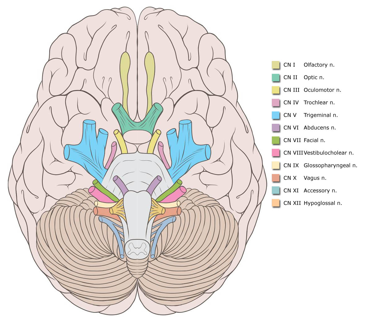
How many cranial nerves are there?
Listed below is a chart of the 12 cranial nerves, the assessment technique used, if the response elicited is normal, and how to document it.
How many cranial nerves are there in the nervous system?
Assessment of the cranial nerves provides insightful and vital information about the patient’s nervous system. There are 12 cranial nerves that are often forgotten by nurses, so with that in mind, here’s a free assessment form that you can use!
How to test light sensation?
(same as above) (same as above) To test deep sensation, use alternating blunt and sharp ends of an object. Determine sensation to warm and cold object by asking client to identify warmth and coldness. (same as above)
What reflex should a client have to respond to light and deep sensation?
While the client looks upward, lightly touch the lateral sclera of eye to elicit blink reflex. Client should have a (+) corneal reflex, able to respond to light and deep sensation and able to differentiate hot from cold. Client was able to elicit corneal reflex, sensitive to pain stimuli and distinguish hot from cold.
What is the purpose of cranial nerve exam?
The cranial nerve exam is a type of neurological examination. It is used to identify problems with the cranial nerves by physical examination. It has nine components. Each test is designed to assess the status of one or more of the twelve cranial nerves (I-XII).
Which nerve is used to perform the Corneal Reflex test?
Corneal reflex is conducted along with the facial nerve section of the test. Note the sensory innervation of the cornea is provided by the trigeminal nerve while the motor innervation for blinking the eye is provided by the facial nerve .-. Muscles of mastication ( temporalis, masseter) should be inspected for atrophy.
What muscles should be inspected for atrophy?
Muscles of mastication ( temporalis, masseter) should be inspected for atrophy. Palpate the temporalis and masseter as the patient clenches the jaw. The pterygoids can be tested by asking the patient to keep the mouth open against resistance, and move from side to side against resistance.
How to test hearing?
Hearing is tested by whispering numbers in one ear as patient covers the other and ask the patient to repeat the numbers. Alternatively, have patient close their eyes and say "left" or "right" depending on the side from which they hear the sound. Vigorously rub fingers together in one ear at a time to produce rustling sound. Conduct the Rinne test and Weber test.
How are visual fields assessed?
Visual fields are assessed by asking the patient to cover one eye while the examiner tests the opposite eye. The examiner wiggles the finger in each of the four quadrants and asks the patient to state when the finger is seen in the periphery.
What nerve innervates the mandible?
Be careful not to test the mandibular division too laterally, as the mandible is innervated by the great auricular nerve (C2 and C3). A common mistake is to use a stroking motion, which will trigger pain and temperature nerves. Instead, a point stimulus should be applied.
What nerve is used to test smell?
I: Olfactory nerve. Sense of smell. Smell is tested in each nostril separately by placing stimuli under one nostril and occluding the opposing nostril. The stimuli used should be non-irritating and identifiable. Some example stimuli include cinnamon, cloves, and toothpaste.
What is the clinical presentation of acquired fourth nerve palsy?
Clinical presentation of acquired fourth nerve palsy is similar to that of congenital palsy. Patients may present most commonly with diplopia but can also present with blurry vision or a minor vision problem when looking down like reading a book or going down the stairs. The diplopia presented in trochlear nerve palsy is either vertical or diagonal and is worse with a downward gaze. Compensation for the nerve palsy usually includes a head tilt to the opposing side and tucking in the chin, so the affected eye’s pupil can move up and extort, instead of downwards and intort. During clinical examination, the eyes will display hypertropia with the affected eye being slightly elevated relative to the other normal eye. Under cover, the affected eye will show an upward drift relative to the other eye. [15]
Which cranial nerve controls the movement of the eye?
The trochlear nerve is the fourth cranial nerve (CN IV) and one of the ocular motor nerves that controls eye movement. The trochlear nerve, while the smallest of the cranial nerves, has the longest intracranial course as it is the only nerve to have a dorsal exit from the brainstem. It originates in the midbrain and extends laterally and anteriorly to the superior oblique muscle. [1]
What causes a 4th nerve palsy?
The most common cause of an isolated fourth nerve palsy is congenital. [5][6][7][8] These patients may commonly present with eye deviation and complain of diplopia and postural head changes. This characteristic head tilt is towards the unaffected side to compensate for a lack of intorsion from the superior oblique muscle. Congenital trochlear nerve palsies are almost always unilateral. These nerve palsies in children can be initially mistaken for torticollis because of the head tilt many of these children display. [9][10] Most commonly, this can be corrected surgically or with prism. A more conservative approach for minor deviations involves patching of one eye that can alleviate diplopia. However, if patients defer or fail initial therapy, surgical approaches include tucking of the superior oblique tendon or inferior oblique weakening.[11] Interestingly, there is evidence to suggest that congenital superior oblique palsy is more common in young males. [12]
What is the trochlear nerve?
Moreover, the trochlear nerve is a somatic efferent (motor) nerve, and along with oculomotor (III) and abducens (VI) nuclei, it is responsible for eye movement. Through its innervation of the superior oblique, the trochlear nerve controls the abduction and intorsion of the eye. [1]
Why is the trochlear nerve so fragile?
Because of its fragility and extensive intracranial course, the trochlear nerve is especially vulnerable to trauma compared to most cranial nerves. Thus, the most common cause of an acquired defect of the trochlear nerve is trauma.[13] Traumatic trochlear nerve palsies are associated with motor vehicle accidents and boxing, as they involve rapid deceleration of the head. Because the trochlear nerve is so fragile, this can occur in minor head injuries that do not involve loss of consciousness or skull fracture. Shearing forces can result in disruption particularly at the superior orbital fissure, where the trochlear nerve enters the orbit.
Where does the trigeminal nerve enter the cavernous sinus?
It enters the cavernous sinus where it runs anteriorly above the abducens nerve and ophthalmic branch of the trigeminal nerve. Here in the cavernous sinus, a few sympathetic fibers join the trochlear nerve with the possibility of some sensory fibers from the trigeminal nerve. The nerve then enters the orbit through the superior orbital fissure and continues to extend anteriorly to the superior oblique muscle. [1][3]The superior orbital fissure is also the nerve pathway for cranial nerves III, VI, and V and is vulnerable to shearing forces in the setting of trauma.
Which nerve is a homologous of the ventral roots of the spinal nerves?
The trochlear nerve, as well as the abducens (VI), hypoglossal (XII), and oculomotor (III) nerves, is a homolog of the ventral roots of the spinal nerves . The somatic efferent columns of the brainstem give rise to these cranial nerves. The muscles that they innervate are derived from the cranial (preoptic and occipital) myotomes in early skeletal muscle development.