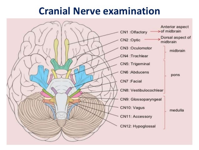
Cranial nerve examination. The cranial nerve exam is a type of neurological examination. It is used to identify problems with the cranial nerves by physical examination. It has nine components. Each test is designed to assess the status of one or more of the twelve cranial nerves (I-XII).
How to conduct a cranial nerve examination?
• Ask patient to turn head to one side and push against examiners hand or ask to flex head against resistance, palpate and evaluate strength of sternocleidomastoid muscle. • Evaluate both right and left side, compare for symmetry. CRANIAL NERVES 39 40.
How can therapists evaluate cranial nerves?
Therapists can evaluate cranial nerves at an initial consultation which provides the therapist a window into the patient’s neurological status and the location of the insult. It makes reading the neurologist’s consult clear and the therapist’s objectives more to the point.
How do you test hypoglossal cranial nerve?
The hypoglossal nerve communicates with several other nerves as well, including:
- Vagus nerve
- Sympathetic trunk
- Cervical plexus
- Lingual branch of the trigeminal nerve
How to assess cranial nerves?
- Have patient stand 20 feet from chart
- First the patient will cover the right eye, then left eye, and lastly read the chart with both eyes.
- Covering the right eye first, have the patient recite the lowest line they can read with ease.
- Repeat this with the left eye and then both eyes.

What nerve is used for the 7th cranial nerve test?
Seventh Cranial Nerve Test: It is a mixed nerve. The facial nerve nucleus is situated in the pons, lateral to that of the abducent nerve. It receives the taste fibers from anterior two thirds of tongue through lingual chorda tympani nerve.
What nerves are used to test motor parts?
Test For Motor Part: All the muscles of face and scalp are supplied by facial nerves except levator palpebral superioris which is supplied by occulomotor nerve. It also supplies to buccinator, stapedius and styloid muscles. Look for facial expression, furrows over forehead, nasolabial fold, angle of the mouth and width of the palperbral fissure.
Where do the facial nerves go?
The facial nerve proper and intermediate nerve lie in the cerebellopontine angle with the sixth and eighth cranial nerves. The seventh, intermediate, and eighth nerves enter the internal auditory meatus. The facial and intermediate nerves then enter the facial canal of the petrous portion of the temporal bone.
What is the motor portion of the facial nerve?
Definition. The motor portion, or the facial nerve proper, supplies all the facial musculature. The principal muscles are the frontalis, orbicularis oculi, buccinator, orbicularis oris, platysma, the posterior belly of the digastric, and the stapedius muscle. In nuclear or infranuclear ("peripheral") lesions, there is a partial to complete facial ...
How to tell if you have facial weakness?
Look for asymmetry about the mouth. The most subtle signs of mild facial weakness are the blink reflex and incomplete lid closure. Observe the blink reflex during conversation, or tap gently on the glabella with your index finger or reflex hammer in an attempt to bring out a mild asymmetry of blink.
Which nerve is involved in the corneal reflex?
The facial nucleus participates in the corneal reflex. Corneal pain and temperature fibers go through the ophthalmic division of the fifth cranial nerve to the spinal nucleus of the fifth and thence to the ipsilateral seventh nucleus, causing the eyelid to blink.
Where do the cortical fibers of the facial nerve originate?
The cortical fibers of the facial nerve proper originate from the lower third of the motor strip. They course in the genu of the internal capsule and the middle third of the cerebral peduncle, supplying the seventh nucleus in the lower pons.
Which part of the facial nerve supplies the anterior two-thirds of the tongue and the submandibular
The chorda tympani supplies the anterior two-thirds of the tongue and the submandibular and sublingual glands. The motor part of the facial nerve leaves the stylomastoid foramen and supplies the facial musculature. A major part of the nerve forms a plexus within the parotid gland.
Which cranial nerves are evaluated together?
The 9th (glossopharyngeal) and 10th (vagus) cranial nerves are usually evaluated together. Whether the palate elevates symmetrically when the patient says "ah" is noted. If one side is paretic, the uvula is lifted away from the paretic side.
What is the function of the 1st cranial nerve?
Smell, a function of the 1st (olfactory) cranial nerve, is usually evaluated only after head trauma or when lesions of the anterior fossa (eg, meningioma) are suspected or patients report abnormal smell or taste.
What is the slow component of vestibular nystagmus?
Vestibular nystagmus has 2 components: A slow component caused by vestibular input. A quick, corrective component that causes movement in the opposite direction (called beating) The direction of the nystagmus is defined by the direction of the quick component because it is easier to see.
What nerve is evaluated by asking the patient to extend the tongue and inspecting it for atrophy, fasciculations,
The 12th (hypoglossal) cranial nerve is evaluated by asking the patient to extend the tongue and inspecting it for atrophy, fasciculations, and weakness (deviation is toward the side of a lesion).
What is the 2nd cranial nerve?
For the 2nd (optic) cranial nerve, visual acuity is tested using a Snellen chart for distance vision or a handheld chart for near vision; each eye is assessed individually , with the other eye covered.
How to test for taste in the anterior two thirds of the tongue?
Taste in the anterior two thirds of the tongue can be tested with sweet, sour, salty, and bitter solutions applied with a cotton swab first on one side of the tongue, then on the other. Hyperacusis, indicating weakness of the stapedius muscle, may be detected with a vibrating tuning fork held next to the ear.
Is nystagmus peripheral or central?
If nystagmus is absent with visual fixation but present with Frenzel lenses, it is probably peripheral. If nystagmus changes direction (eg, from one side to the other when, for example, when the direction of gaze changes), it is probably central. However, absence of this finding does not exclude central causes.
Where are the 7th cranial nerves located?
The two 7th Cranial Nerves (CN VII) are located on either side of the brainstem, at the top of the medulla. They are mixed cranial nerves with BOTH sensory and motor function. CN VII controls the face and is mainly FACE MOVEMENT with some face sensation. See Diagram.
What is the function of the 7th cranial nerve?
What is the function of Cranial Nerve VII? The main function of each of the two 7th cranial nerves is facial movement on the same side (ipsilateral). Left sided forehead wrinkle, left eyelid closure, and movement of the left half of the face is stimulated by the left 7th cranial nerve. Lacrimation (tearing) and salivation are also stimulated by ...
How to tell if CN VII is damaged?
Because of the close proximity to CN VIII, balance and hearing should also be assessed when the CN VII is damaged.
Which nerve stimulates salivation?
Lacrimation (tearing) and salivation are also stimulated by the 7th cranial nerve. The 7th cranial nerve also has some sensory component including the sense of taste (anterior 2/3 of the tongue).
What is CN VII?
CN VII disorders can be associated with dry mouth or choking (impa ired salivation or loss of sensation to anterior portion of tongue), dry eyes/corneal injury or associated symptoms involving the adjacent CN VIII (hearing and balance). Unconscious Patient.
Why wouldn't cranial nerves work?
Why wouldn’t a cranial nerve “work ”? In many neuro diseases, the neurons that supply a particular nerve is damaged, which makes the nerve not function properly. For example, in multiple sclerosis the myelin sheath of the neurons in the central nervous system are damaged, which leads to some sensory and motor problems.
What nerve causes blurry vision?
Therefore, you can assess this nerve (cranial nerve II) for any type of abnormalities.
How far can a patient see with normal vision?
This means the patient can see at 20 feet what a person with normal vision can see at 30 feet.
Which nerve is responsible for mastication?
Cranial Nerve V. To test Cranial Nerve V…..trigeminal nerve: This nerve is responsible for many functions and mastication is one of them. Have the patient bite down and feel the masseter muscle and temporal muscle. Then have the patient try to open the mouth against resistance.
How many cranial nerves are there in the nervous system?
Assessment of the cranial nerves provides insightful and vital information about the patient’s nervous system. There are 12 cranial nerves that are often forgotten by nurses, so with that in mind, here’s a free assessment form that you can use!
How to test light sensation?
(same as above) (same as above) To test deep sensation, use alternating blunt and sharp ends of an object. Determine sensation to warm and cold object by asking client to identify warmth and coldness. (same as above)
What reflex should a client have to respond to light and deep sensation?
While the client looks upward, lightly touch the lateral sclera of eye to elicit blink reflex. Client should have a (+) corneal reflex, able to respond to light and deep sensation and able to differentiate hot from cold. Client was able to elicit corneal reflex, sensitive to pain stimuli and distinguish hot from cold.
Which cranial nerve innervates the forehead?
This is because a portion of the VII cranial nerve nucleus innervating the forehead receives input from both cerebral hemispheres. The portion of the VII cranial nerve nucleus innervating the mid and lower face does not have this dual cortical input.
How to test visual acuity?
First test visual acuity by using a pocket visual acuity chart. Perform this part of the examination in a well lit room and make certain that if the patient wears glasses, they are wearing them during the exam. Hold the chart 14 inches from the patient's face, and ask the patient to cover one of their eyes completely with their hand and read the lowest line on the chart possible. Have them repeat the test covering the opposite eye. If the patient has difficulty reading a selected line, ask them to read the one above. Note the visual acuity for each eye.
What is anisocoria test?
Anisocoria is a neurological term indicating that one pupil is larger than another.
How to test for a lesion of the afferent pathway?
To test for a lesion of the afferent pathway one must perform a "swinging light test". To interpret this test one must understand that the level of pupillary constriction is directly related to the total "perceived" illumination the brain appreciates from both eyes.
Which cranial nerve is affected by anisocoria?
As an aside, the parasympathetics run with the III cranial nerve and are usually affected with an abnormal III cranial nerve. Anisocoria can only be produced if the efferent pathway of the pupillary light reflex is disrupted. A lesion of the afferent pathway along the II cranial does not yield anisocoria.
Which nerve innervates the levator palpebrae?
The III cranial nerve also innervates a much larger muscle that elevates the eye lid: the levator palpebrae. Thus, disruption of either will cause ptosis. The ptosis from a III nerve palsy is of greater severity than the ptosis due to a lesion of the sympathetic pathway, due to the size of the muscles innervated.
What is XII in the nervous system?
XII - Movement and protrusion of tongue. Lesions of the nervous system above the spinal cord are often classified as peripheral or central in location. Peripheral lesions are lesions of the cranial nerve nuclei, the cranial nerves or the neuromuscular junctions.