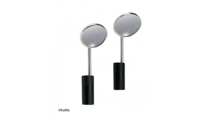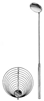
Using the Laryngeal Mirror The tongue is held while a small, slightly warmed mirror is introduced into the mouth. The mirror should not be excessively warm and should avoid contact with the tongue. The patient is asked to breathe normally through the mouth. The mirror should be pushed upward against the uvula and positioned in the oropharynx.
What is a laryngeal mirror and how does it work?
The Laryngeal Mirror provides thermal-tactile stimulation during swallowing therapy. It helps trigger a swallow by stimulating the anterior pillars of faces. The Mirror can be sterilized by steam, hot air or antiseptic solutions.
How do you sterilize a laryngeal mirror?
The Mirror can be sterilized by steam, hot air or antiseptic solutions. The Laryngeal Mirror provides thermal-tactile stimulation during swallowing therapy. It helps trigger a swallow by stimulating the anterior pillars of faces. The Mirror can be sterilized by steam, hot air or antiseptic solutions.
How is laryngoscopy used to examine the larynx?
Mirror Laryngoscopy The first and oldest (having been available for over a century) method of examining the larynx involves inserting a small angled mirror, such as a dentist might use, into the back of the mouth. This mirror deflects a beam of light down onto the vocal folds and reflects the image of the vocal cords back up to the examiner.
What is the surtex® laryngeal mirror?
The SURTEX® Laryngeal Mirror assists otolaryngologists to view and take biopsies from the upper airways. This includes structures like the posterior tongue, the posterior pharyngeal wall and the larynx in pharyngeal examinations.
How to visualize the larynx?
How to determine extent of resection of uvula?
Why isn't the Curettage of the tissue in the Fossa of Rosenmüller done?
What is surface electromyography?
See 1 more
About this website

Why should a laryngeal mirror be warmed before use?
Warm the mirror over an alcohol lamp or with warm water to prevent fogging. The patient should be sitting upright with a straight back, leaning slightly toward you with chin pointing upward (“sniffing position”). Sit to the patient's side, and be higher than the patient.
How do you keep a laryngeal mirror from fogging up?
Warm the mirror with warm water (about body temperature) to prevent fogging (check to make sure mirror is not too hot). Alternatively, coat the mirror with antifogging solution or alcohol. Wrap the patient's tongue with gauze and grasp it with your nondominant hand.
What is a mirror laryngoscopy?
Mirror (indirect) laryngoscopy is viewing of the pharynx and larynx using a small, curved mirror. Mirror laryngoscopy is typically done to evaluate symptoms in the pharynx and larynx. (See also Evaluation of the Patient with Nasal and Pharyngeal Symptoms.
How do you find your throat in a mirror?
Indirect laryngoscopy. Your doctor uses a small mirror and a light to look into your throat. The mirror is on a long handle, like the kind a dentist often uses, and it's placed against the roof of your mouth. The doctor shines a light into your mouth to see the image in the mirror.
Can you see your vocal cords in the mirror?
You may not be able to see this in a mirror, but through a laryngoscopy, we can see the muscles on the inside of your throat straining when you speak or sing.
How do you sterilize a laryngeal mirror?
Sklar Laryngeal Mirrors can be processed in Pre-Vacuum Steam (wrapped configuration) for 5 min. hold @ 134°C / 273°F, total 25-35 min. NOTE: Make sure autoclave chambers are cleaned regularly and as recommended by the manufacturer.
What can a laryngoscopy detect?
This test can be used to look for the causes of symptoms in the throat or voice box (such as trouble swallowing or breathing, voice changes, bad breath, or a cough or throat pain that won't go away). Laryngoscopy can also be used to get a better look at an abnormal area seen on an imaging test (such as a CT scan).
How do I get my voice back in minutes?
Inhale steam from a bowl of hot water or a hot shower. Rest your voice as much as possible. Avoid talking or singing too loudly or for too long. If you need to speak before large groups, try to use a microphone or megaphone.
How does an ENT check your throat?
To look at the back of the throat, larynx and vocal cords, the doctor wears a head mirror with a bright light hooked to it and inserts a thin, flexible scope (laryngoscope) into your nose and gently moves it down your throat.
How do you take a throat pic?
1:122:28send a photo of your throat to your doctor - YouTubeYouTubeStart of suggested clipEnd of suggested clipAnd make sure you tap on the screen in the middle of the throat. Area so the phone autofocus takes aMoreAnd make sure you tap on the screen in the middle of the throat. Area so the phone autofocus takes a good image of the middle part of the throat. Then simply tap the button to take the photograph.
Can an endoscopy detect throat problems?
Your doctor may use a special lighted scope (endoscope) to get a close look at your throat during a procedure called endoscopy. A camera at the end of the endoscope transmits images to a video screen that your doctor watches for signs of abnormalities in your throat.
Can you see your epiglottis in the mirror?
The epiglottis is an anatomical cartilage flap that has a leaf like shape. It is located in the throat between the tongue and the larynx but can be seen popping just above the tongue when you open the mouth and say ah in front of a lighted mirror.
What is used for preventing fogging in indirect Laryngoscopic examination?
Many methods are described in literature to prevent fogging over the mirror during the examination. Authors have used hydrogen peroxide 0.75% w/v solution as a defogging agent during indirect laryngoscopy examination and found it safe and cost effective.
Do dental mirrors fog up?
Fog develops on dental mirrors as well, making visibility in the mouth difficult. When patients inhale and exhale, the breath may form condensation, fogging the mirror and distorting tooth and tissue images.
How do you clean a fog free mirror?
The cleaning instructions for fog-free mirrors from Home Decorators Collection say to use “a generous amount” of window cleaner, and they mention Windex, Zep Streak-Free Glass Cleaner and HDX Glass Cleaner.
Why does my mirror have a film on it?
A simple reason may be because of an accumulation of dirt and lack of maintenance. Another reason could be desilvering - mirrors are made of glass with a silver backing, over time the mirror may begin to develop black spots. This is called desilvering and it usually happens due to moisture.
Laryngeal mirror examination
Laryngeal mirror examination is a time-honored method for visualizing the interior of the larynx and pharynx, and especially the vocal folds. This method was originally described in the 19th century by famed singing teacher Manuel Garcia.An angled “dental” mirror is held against the soft palate and over the base of the tongue, and illuminated, typically by head mirror or headlight.
Amazon.com: laryngeal mirror
Set of 2 LARYNGEAL BOILABLE Hygiene Dental Mirrors with Handle #000#00 (DDP Quality)
A Medical Equipment List of Must-Haves for Your New Exam Room - CME Corp
About CME: CME Corp is the nation’s premier source for healthcare equipment, turnkey logistics, and biomedical services, representing 2 million+ products from more than 2,000 manufacturers. With two corporate offices and 35+ service centers, our mission is to help healthcare facilities nationwide reduce the cost of the equipment they purchase, make their equipment specification, delivery ...
What is mirror laryngoscopy?
Mirror (indirect) laryngoscopy is viewing of the pharynx and larynx using a small, curved mirror. Mirror laryngoscopy is typically done to evaluate symptoms in the pharynx and larynx. (See also Evaluation of the Patient with Nasal and Pharyngeal Symptoms and Overview of Laryngeal Disorders .)
How to rotate a mirror?
Rotate the mirror from side to side with thumb and forefinger to bring lateral structures into view.
What to do if you gagging your oropharynx?
If gagging occurs, remove the mirror and spray the posterior oropharynx with a topical anesthetic.
How to stop gagging in a patient?
Instruct the patient to breathe deeply through the mouth, to help prevent gagging.
What is the failure to align the light source as closely as possible with line of sight?
Failure to align the light source as closely as possible with line of sight. Failure to warm the mirror, because a cold mirror will fog. Failure to maintain hold of the patient's tongue to keep it retracted. Allowing the patient to lean back, which will prevent full visualization.
How to stop a patient from slipping?
Wrap the patient's tongue with gauze and grasp it with your nondominant hand. The gauze will prevent the tongue from slipping and protect it from injury by the lower incisor teeth. Gently pull on the tongue. Instruct the patient to breathe deeply through the mouth, to help prevent gagging.
Can laryngopharynx be stimulated?
In such cases, stimulation of the laryngopharynx may further compromise the airway. If laryngoscopy is essential, it should be done in the controlled setting of an operating room with a person skilled at difficult airway management (including surgical techniques) present.
What is a laryngeal mirror used for?from alimed.com
One-piece laryngeal mirror can be used for dysphagia evaluation, pre-feeding intra-oral structure inspection, or speech training
How to visualize the larynx?from sciencedirect.com
Following a thorough history and head and neck examination, including subjective evaluation of the voice, visualization of the larynx in the dysphonic patient is of paramount importance. Classically this has been accomplished with an angled laryngeal mirror and light directed by a semispherical reflector, which has been the standard of laryngeal examination for over 100 years. 19 Although mirror laryngoscopy remains an important part of the head and neck examination, modern comprehensive evaluation of the larynx and interventions demand greater resolution, a larger field of view, and the ability to record and play back logged examinations. Advances in optics led to the development of an array of rigid telescopes, flexible fiberoptic endoscopes, and flexible distal chip endoscopes that provide high resolution and magnification and can be attached to video recording devices for examination storage and review. Two studies prospectively compared the use of laryngeal mirror and rigid endoscopy with regard to patient tolerance and accuracy of diagnosis. Barker and Dort successfully performed rigid endoscopy using a 90-degree telescope in 83% of patients compared with only 52% with laryngeal mirror examination; direct laryngoscopy confirmed no false-negative rigid endoscopy evaluations. 20 In 2009, Dunkelbarger et al. found rigid laryngoscopy using a 30-degree endoscope to be more comfortable and preferred by a majority of patients (80%) when compared with mirror examination, provided greater detail to the clinician, and was helpful to 84% of patients to see the examinations on the monitor. 21 Direct comparison of mirror examination and flexible laryngoscopy has not been reported. Studies comparing flexible and rigid laryngoscopy are varied with regard to patient demographics, study design, and outcome measures. Overall, flexible transnasal laryngoscopy is well tolerated by the majority of adult patients with minimal morbidity. 22 However, although flexible laryngoscopy was tolerated by all participants in a study of 35 pediatric patients, 80% of children preferred rigid transoral examinations because of less perceived pain and irritation. 23
How to determine extent of resection of uvula?from sciencedirect.com
The extent of reflection of the uvula and distal palate and the extent of resection of the uvular tip are then determined by grasping the uvula with medium-length forceps and reflecting it cephalad toward the junction of the hard and soft palate while simultaneously examining the retropalatal airway diameter with a number 5 laryngeal mirror. The uvula is retracted sufficiently to create a crease between the intervening mucosal edges. Standard UPPP principles are used to determine the extent of shortening desired. Because the patient is awake and able to phonate, the palatal dimple point is easily identified; VPI is more likely if the palate is shortened much beyond this point. Though varying significantly between patients, the final position of the repositioned uvular tip is generally 5–10 mm from the hard–soft palate junction.
How to administer anesthesia to the uvula?from sciencedirect.com
Topical local anesthetic is applied to the entire soft palate and uvula, and additional anesthesia to the nasal surface of the palate can be obtained by spraying the nasal cavities with a 1:1 mixture of tetracaine hydrochlor ide and phenylephrine hydrochloride. After allowing sufficient time for the topical anesthetic to take effect, the surgical site is infiltrated with 2–4 ml of injectable anesthetic with adrenaline. It is important not to distort the tissue or create blebs of submucosal anesthetic by injecting too much solution or by injecting too superficially. In addition to making the patient more comfortable during the procedure, meticulous injection of anesthetic results in enough vasoconstriction that the surgical field is surprisingly bloodless; any oozing can easily be controlled with a battery-operated ophthalmic cautery unit. Electrosurgical cautery should not be necessary except when the procedure is performed under general anesthesia with concurrent tonsillectomy.
Why isn't the Curettage of the tissue in the Fossa of Rosenmüller done?from sciencedirect.com
Curettage of the tissue in the fossa of Rosenmüller is not done because it may lead to scar tissue formation and contracture that might result in stenosis or Eustachian tube reflux or both. Direct injury to the Eustachian tube also is avoided.
What is surface electromyography?from sciencedirect.com
Surface electromyography has been used to retrain brainstem stroke patients with chronic dysphagia to eat safely by mouth 19 and also has been demonstrated to be useful in providing biofeedback for relaxing high tone in laryngeal musculature, which allows an improved swallow response. 40
How to examine the larynx?
The first and oldest (having been available for over a century) method of examining the larynx involves inserting a small angled mirror, such as a dentist might use, into the back of the mouth. This mirror deflects a beam of light down onto the vocal folds and reflects the image of the vocal cords back up to the examiner.
Where is the mic used in stroboscopy?
In stroboscopy, a microphone, usually applied to the skin of the neck overlying the larynx, registers the frequency of the voice. This is connected to a strobe light, which flashes just slightly out of sync with the frequency, offering a video image of the vibration of the vocal fold, known as the mucosal wave.
How to check vocal folds?
An endoscope measuring less than 4mm in diameter is inserted through one nostril and guided through the nose to the back of the throat, until it lies just above the larynx. The flexible fiberoptic laryngoscope is the workhorse endoscope in ear, nose and throat medicine – no otolaryngologist’s office is without one. Its advantages include ease of use and general availability, and the capability to examine the larynx during a variety of tasks - such as swallowing, connected speech, and singing. This is very important in the evaluation of many neurologic disorders such as spasmodic dysphonia and vocal fold paralysis. However, flexible endoscope optics are generally inferior to that of a rigid endoscope. The view can appear grainy or pixilated, and may not permit precise differentiation among masses of the vocal cords.
Why is stroboscopy the best method to evaluate masses or irregularities of the vocal fold?
Because this vibration is the source of sound, stroboscopy is the best method to evaluate masses or irregularities of the vocal fold. However, stroboscopy is technically more difficult than simple endoscopy, and interpreting the examination is not always straightforward.
What is stroboscopic view of vocal folds?
Stroboscopic view of the vocal folds - the stroboscopic light allows us to see vocal fold vibration.
What is the term for an examination of the internal structures of the larynx, including the vocal folds,?
An examination of the internal structures of the larynx, including the vocal folds, is called laryngoscopy . There are three principal ways to perform laryngoscopy, reviewed below.
Can you magnify vocal folds?
However, it is not possible to magnify the view or record the examination. For these reasons, and because it requires a certain skill and lightness of touch, it is not often used today.
How to visualize the larynx?
Following a thorough history and head and neck examination, including subjective evaluation of the voice, visualization of the larynx in the dysphonic patient is of paramount importance. Classically this has been accomplished with an angled laryngeal mirror and light directed by a semispherical reflector, which has been the standard of laryngeal examination for over 100 years. 19 Although mirror laryngoscopy remains an important part of the head and neck examination, modern comprehensive evaluation of the larynx and interventions demand greater resolution, a larger field of view, and the ability to record and play back logged examinations. Advances in optics led to the development of an array of rigid telescopes, flexible fiberoptic endoscopes, and flexible distal chip endoscopes that provide high resolution and magnification and can be attached to video recording devices for examination storage and review. Two studies prospectively compared the use of laryngeal mirror and rigid endoscopy with regard to patient tolerance and accuracy of diagnosis. Barker and Dort successfully performed rigid endoscopy using a 90-degree telescope in 83% of patients compared with only 52% with laryngeal mirror examination; direct laryngoscopy confirmed no false-negative rigid endoscopy evaluations. 20 In 2009, Dunkelbarger et al. found rigid laryngoscopy using a 30-degree endoscope to be more comfortable and preferred by a majority of patients (80%) when compared with mirror examination, provided greater detail to the clinician, and was helpful to 84% of patients to see the examinations on the monitor. 21 Direct comparison of mirror examination and flexible laryngoscopy has not been reported. Studies comparing flexible and rigid laryngoscopy are varied with regard to patient demographics, study design, and outcome measures. Overall, flexible transnasal laryngoscopy is well tolerated by the majority of adult patients with minimal morbidity. 22 However, although flexible laryngoscopy was tolerated by all participants in a study of 35 pediatric patients, 80% of children preferred rigid transoral examinations because of less perceived pain and irritation. 23
How to determine extent of resection of uvula?
The extent of reflection of the uvula and distal palate and the extent of resection of the uvular tip are then determined by grasping the uvula with medium-length forceps and reflecting it cephalad toward the junction of the hard and soft palate while simultaneously examining the retropalatal airway diameter with a number 5 laryngeal mirror. The uvula is retracted sufficiently to create a crease between the intervening mucosal edges. Standard UPPP principles are used to determine the extent of shortening desired. Because the patient is awake and able to phonate, the palatal dimple point is easily identified; VPI is more likely if the palate is shortened much beyond this point. Though varying significantly between patients, the final position of the repositioned uvular tip is generally 5–10 mm from the hard–soft palate junction.
Why isn't the Curettage of the tissue in the Fossa of Rosenmüller done?
Curettage of the tissue in the fossa of Rosenmüller is not done because it may lead to scar tissue formation and contracture that might result in stenosis or Eustachian tube reflux or both. Direct injury to the Eustachian tube also is avoided.
What is surface electromyography?
Surface electromyography has been used to retrain brainstem stroke patients with chronic dysphagia to eat safely by mouth 19 and also has been demonstrated to be useful in providing biofeedback for relaxing high tone in laryngeal musculature, which allows an improved swallow response. 40
