How do you use Köhler illumination?
- You will need a specimen to perform this, so grab a slide. ...
- Switch on the microscope.
- Use the 10x objective lens (20x if necessary). ...
- Place your slide on the microscope stage. ...
- Close down the condenser diaphragm (rotate it fully anti-clockwise).
- Focus on your specimen using transmitted light.
- Close down the field aperture (field diaphragm), you should see an octagon shaped aperture appear (or if it is really badly out of focus, the entire image will get darker). Close the aperture until it occupies about 2/3 of the field of view.
How to set up Koehler illumination 3 minutes?
How to set up Koehler Illumination 3 minutes to optimize your image quality Field diaphragm (FD) Condenser Aperture diaphragm (AD) Specimen Objective Step 1: Fully open the FD and AD, then adjust brightness using the illumination intensity control Step 2:Focus on specimen Step 4:Bring the FD into focus by ad- justing the height of the condenser
How do you use a Köhler illumination switch?
Steps in Establishing Köhler Illumination Switch on the light source and hold a small sheet of paper directly above the luminous field diaphragm collector lens in the base of the microscope (Figure 2(a)). Next, open the field diaphragm to its widest position (fully open) by turning the lever or knob.
What is Köhler illumination?
Köhler Illumination. Illumination of the specimen is the most important variable in achieving high-quality images in microscopy and critical photomicrography or digital imaging. Köhler illumination was first introduced in 1893 by August Köhler of the Carl Zeiss corporation as a method of providing the optimum specimen illumination.
How do I set up my microscope for Köhler illumination?
Now that the basic components of the microscope are set, the instrument is ready to be configured for Köhler illumination. The first step is to narrow the size of the field diaphragm and translate the condenser up and down via the adjustment knob until you see a sharp image of the edges from field diaphragm leaves (Figure 5 (a)).

What is Köhler illumination and how is it performed?
Koehler Illumination is a process that provides optimum contrast and resolution by focusing and centring the light path and spreading it evenly over the field of view. Sophisticated and well-equipped microscopes fail to yield quality images because of incorrect use of the light source.
Why do you use Köhler illumination?
In practice, Köhler illumination is used in most microscopes, and a specialized form of critical illumination is used in confocal microscopes. Köhler illumination provides a uniformly illuminated, bright field of view, which is important when using an uneven light source, like a coiled tungsten filament.
What is the most critical step in Koehler illumination?
The aperture diaphragm is opened and closed with either a swinging arm, a lever, or by rotating a collar on the condenser housing. It should be noted that correct adjustment of the substage condenser is probably the most critical aspect of achieving proper Köhler illumination.
What is the first objective used when setting up for Köhler illumination?
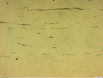
10 Simple Steps To Köhler Illumination 1) You will need a specimen to perform this, so grab a slide. Any regular H&E stained tissue section is ideal. 2) Switch on the microscope. 3) Use the 10x objective lens (20x if necessary).
How do I set up Kohler illumination?
0:374:08Set up Köhler Illumination on your ZEISS Axio Imager - YouTubeYouTubeStart of suggested clipEnd of suggested clipOne switch on the imager via the power supply and power button to adjust the lamp brightness to aMoreOne switch on the imager via the power supply and power button to adjust the lamp brightness to a comfortable level by turning the rotary dial clockwise.
Does Kohler illumination increase magnification?
Resolution and contrast, however, are largely dependent on specimen illumination. Optical illumination for most specimens is called Kohler Illumination. Once a given selection of optics is in place, Kohler illumination cannot affect magnification, but it does affect resolution and contrast.
How do you set up a light microscope for Köhler illumination?
1:583:44Setting up a compound microscope for Kohler illumination - YouTubeYouTubeStart of suggested clipEnd of suggested clipUntil it fills about 7/8 of the field of you put the ocular back firmly the microscope is now setupMoreUntil it fills about 7/8 of the field of you put the ocular back firmly the microscope is now setup for color illumination. If you want to use a higher power objective rotate the nosepiece.
How do you get critical illumination?
0:3112:47How to achieve Kohler illumination - YouTubeYouTubeStart of suggested clipEnd of suggested clipTo begin we place the specimen onto the stage. And bring it into focus. And that's essential as theMoreTo begin we place the specimen onto the stage. And bring it into focus. And that's essential as the starting. Point. We then fully close down the field aperture.
What is illumination microscope?
The illumination system of the standard optical microscope is designed to transmit light through a translucent object for viewing. In a modern microscope it consists of a light source, such as an electric lamp or a light-emitting diode, and a lens system forming the condenser.
What is critical illumination microscope?
Critical illumination or Nelsonian illumination is a method of specimen illumination used for transmitted and reflected light (trans- and epi-illuminated) optical microscopy. Critical illumination focuses an image of a light source on to the specimen for bright illumination.
Technical requirements
The substage condenser must be capable of being focused up and down and must be fitted with an aperture iris diaphragm that can be opened and closed by a lever or knob.
A quick guide - 6 steps to Koehler Illumination
1) Place a thin sample on the stage and focus on it using a 4x or 10x objective.
Sign up to receive our latest news
Find out about Scientifica's latest product releases, company news, and developments through a range of news articles, customer interviews and product demonstration videos.
What is Kohler illumination?
Optical microscopes generate the magnified images through the interaction of visible lights and the specimens. Illumination of the specimen is the most important variable in achieving high-quality images in microscopy. However, the light source (i.e., a halogen lamp) used for our microscopes is not homogeneous.
Is my microscope compatible with Kohler illumination?
Köhler illumination requires several optical components to function. Most regular compound microscopes with the Abbe condenser and Iris diaphragm should be able to do so.
How to set up Kohler Illumination?
a. You may need to establish Kohler illumination each time you change the objective.
Introduction
In microscopy, we aim to produce a sharply-defined, magnified image of our sample in our detector or our eyes.
How Is An Image Formed With A Lens?
At its simplest, a microscope may be seen as a series of simple (aberration/defect-free) lenses. The behavior of a single lens is illustrated in Fig.1.
The Optics Underlying Köhler Illumination
The predecessor to Köhler illumination was critical illumination. This method used two lenses as discussed above to produce a magnified image of the light source at the sample plane. This provided effective illumination, however, an image of the light source was superimposed over the sample.
Illumination Control Using Köhler Illumination
Köhler’s optical design also allows for the control of: the illumination field of view, the power delivered to the sample, and the effective illumination numerical aperture (NA).
Setting Up Köhler Illumination On A Modern Microscope
Most microscopes can be used in either transmission/trans-fluorescence mode, where the light travels through from the light source through the sample to the eye or camera, or in reflectance/epi-fluorescence mode where the light delivered through the objective follows the same path back before being deviated off to the eye or camera.
Common Problems In Setting Up Köhler Illumination
Setting up Köhler illumination becomes second nature with practice. One common condition can confound most beginners and even some experts is the adjustment of the aperture diaphragm.
Summary
Köhler illumination is critical to removing illumination source structure from the image of the sample. The correct application of Köhler illumination provides the best possible contrast in both brightfield, phase contrast, and fluorescence techniques.
A Brief History
In the 1893 paper entitled "Ein neues Beleuchtungsverfahren für mikrophotographische Zwecke" which was published in Zeitschrift für wissenschaftliche Mikroskopie und für mikroskopische Technik (“A New System of Illumination for Photomicrographic Purposes” in the Journal of Scientific Microscopy and Microscopic Technique) he set out the main problems microscopists faced at the time.
An Important Principle
Even today, his consistent illumination technique is still widely used and forms the basis of many techniques including confocal, phase contrast and differential interference contrast ( DIC) microscopy. Once you know how to set up a microscope for correct Koehler illumination, it should become second nature and will transform your imaging work.
The Field Diaphragm
The ‘field diaphragm’ is usually located in the base (in an upright microscope) of the instrument as part of the microscope body (Figure 1). Some older (or simpler) microscopes may just have an ‘on/off’ switch for the light source and there may be no field diaphragm to control the intensity of light from the source.
The Sub-Stage Condenser
Directly under the stage in an upright microscope (or above the stage but in front of the light source/field diaphragm in an inverted microscope), is the sub-stage condenser (Figure 1).
Five Steps for Koehler Illumination
Before you start your imaging session always check to see that the lenses, eyepieces, objectives are clean. The cleaning of microscope lenses is covered in another article, but you should always use lens cleaning tissue and a recommended solvent. Place the slide on the stage, and turn on the lamp to its lowest setting.
The History and Creation of Kohler Illumination
Kohler Illumination was named after its creator, August Koehler (1866-1948), who had produced this new method in 1893 when he was 27 years old. Previous to his method, scientists were viewing their specimens with light that was provided through a complex setup of mirrors and gas lamps.
A More Thorough Explanation of Kohler Illumination
Without proper lighting and illumination, it can be impossible to properly view and examine your specimen. Kohler knew how important this was and had created the method which most scientists now use while examining a specimen.
How to Set Up Your Own Microscope Using The Kohler Illumination Method
Setting up Kohler Illumination on your own microscope is actually a very quick thing to do. While the first time may take a little longer and be a little confusing, after you have set it up once, it will be easy to repeat the process each time you use a microscope.
Why Kohler Illumination is Important
After you have learned how to set up a microscope using the Kohler Illumination Method, it should become second nature for you. Like buckling your seatbelt as soon as you get in a car, you should immediately set up a microscope to these standards. It only takes about a minute to set up and greatly enhances the visibility of your specimen.