What results do you get with a Gram stain?
These may include:
- itchy bump that turns into a sore with black center
- nausea
- vomiting
- abdominal pain
- coughing up blood
- high fever
What are four possible results of a Gram stain?
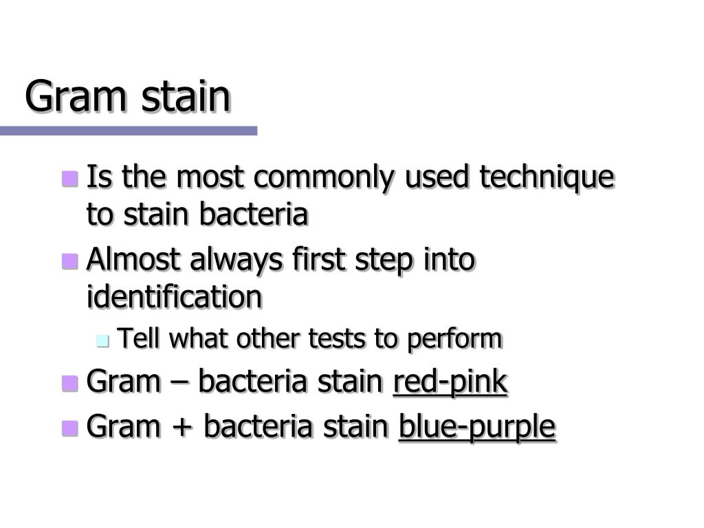
The staining results of gram stain are as follows:
- Gram-positive will be dark-purple color
- Gram-negative will be pale to dark red color
- Yeasts will be dark purple color
- Epithelial cells will be pale red color
What are the steps in Gram staining?
Gram Staining - Step 1. Flood heat-fixed emulsion with Crystal Violet, let stand for 1 minute, rinse with water. Gram Staining - Step 2. Add iodine solution, let stand for 1 minute, rinse with water. Gram Staining - Step 3. Decolorize with alcohol solution holding slide on slight angle, add until run off is clear (20-30 sec), rinse with water.
How long should a Gram stain take?
The gram stain procedure
- Prepare smear from specimen, air dry, heat fix.
- Add crystal violet (purple), wait 30 seconds.
- Rinse with water.
- Add Iodine (Iodine is a mordant; Mordant allows the crystal violet to stick to the cell wall of a gram positive bacteria. ...
- Decolorize with alcohol until slide runs clear
- Rinse with water.
- Counter stain with Safranin (red) and wait 30 seconds.

What information should included in a Gram stain report?
This typically includes: Whether the bacteria are Gram-positive (purple) or Gram-negative (pink) Shape — round (cocci) or rods (bacilli) Size, relative quantity, and/or arrangement of the bacteria, if relevant.
How do you write Gram-positive?
'Gram' should be capitalized and not hyphenated when used as Gram stain; gram negative and gram positive should be lowercase and only hyphenated when used as a unit modifier. Note: present as two words unless being used to modify a noun.
What is Gram staining and explain its procedure?
The Gram stain is a differential method of staining used to assign bacteria to one of two groups (gram-positive and gram-negative) based on the properties of their cell walls. It is also known as Gram staining or Gram's method.
What is the practical significance of Gram staining procedure?
This test differentiate the bacteria into Gram Positive and Gram Negative Bacteria, which helps in the classification and differentiations of microorganisms.
What can you conclude about the Gram stained specimen?
What can you conclude about the pictured bacteria? If a Gram stain was performed on this bacterium within 24 hrs of a fresh culture, it would most likely be Gram-positive. 1)If a Gram stain was performed on this bacterium within 24 hrs of a fresh culture, it would most likely be Gram-positive.
Why is the G in Gram stain capitalized?
The word “Gram” is always to be capitalized, as it is the name of Hans Christian Gram, who invented this method, and published it in 1884.
What are the 4 steps of gram staining?
The Gram staining process includes four basic steps, including:Applying a primary stain (crystal violet).Adding a mordant (Gram's iodine).Rapid decolorization with ethanol, acetone or a mixture of both.Counterstaining with safranin.
Which is the correct order of steps in gram staining?
The performance of the Gram Stain on any sample requires 4 basic steps that include applying a primary stain (crystal violet) to a heat-fixed smear, followed by the addition of a mordant (Gram's Iodine), rapid decolorization with alcohol, acetone, or a mixture of alcohol and acetone and lastly, counterstaining with ...
Which step in gram staining is most important?
The thickness of the smear used in the Gram stain will affect the result of the stain. The step that is most crucial in effecting the outcome of the stain is the decolorizing step.
Which stain is used in gram staining?
[1] Often the first test performed, gram staining involves the use of crystal violet or methylene blue as the primary color. [2] The term for organisms that retain the primary color and appear purple-brown under a microscope is Gram-positive organisms.
How do you remember Gram-positive and negative bacteria?
2:1511:23Mnemonics for gram positive and gram negative bacteria - YouTubeYouTubeStart of suggested clipEnd of suggested clipAnd coke i haz CC. So it's a gram negative coke i.MoreAnd coke i haz CC. So it's a gram negative coke i.
Why is Gram-positive purple?
gram stain test Gram-positive bacteria remain purple because they have a single thick cell wall that is not easily penetrated by the solvent; gram-negative bacteria, however, are decolorized because they have cell walls with much thinner layers that allow removal of the dye by the solvent.
What are the steps in a Gram stain and the possible results?
Steps of Gram Staining Application of mordant: The iodine solution (mordant) is added to form a crystal violet-iodine (CV-I) complex; all cells continue to appear blue. Decolorization step: The decolorization step distinguishes gram-positive from gram-negative cells.
What is Gram staining PDF?
It is a differential staining method of differentiating bacterial species into two large groups (Gram-positive and Gram-negative) based on the chemical and physical properties of their cell walls.
What is the procedure of acid fast staining?
Some of the sample is placed on a glass slide, stained, and heated. The cells in the sample hold onto the dye. The slide is then washed with an acid solution and a different stain is applied. Bacteria that hold onto the first dye are considered "acid-fast" because they resist the acid wash.
What are the steps of Gram staining quizlet?
Steps of gram staining technique:Apply primary stain (crystal violet). All bacteria are stained purple by this basic dye.Apply mordant (Gram's iodine). ... Apply decolorizing agent (ethyl alcohol or ethyl alcohol acetone). ... Apply secondary stain or counterstain (safranin).
What is Gram staining?
Gram Staining is the common, important, and most used differential staining techniques in microbiology, which was introduced by Danish Bacteriologist Hans Christian Gram in 1884. This test differentiate the bacteria into Gram Positive and Gram Negative Bacteria, which helps in the classification and differentiations of microorganisms.
How to clean a gram of iodine?
Flood the gram’s iodine for 1 minute and wash with water. Then ,wash with 95% alcohol or acetone for about 10-20 seconds and rinse with water. Add safranin for about 1 minute and wash with water. Air dry, Blot dry and Observe under Microscope. Interpretation.
What happens to the cell wall of Gram negative bacteria?
When they are exposed to alcohol, decolorizer dissolves the lipids in the cell walls, which allows the crystal violet-iodine complex ...
What is the primary stain for Gram staining?
The Gram stain involves staining bacteria, fixing the color with a mordant, decolorizing the cells, and applying a counterstain. The primary stain ( crystal violet) binds to peptidoglycan, coloring cells purple. Both gram-positive and gram-negative ...
What is Gram staining?
The Gram stain is a differential method of staining used to assign bacteria to one of two groups (gram-positive and gram-negative) based on the properties of their cell walls. It is also known as Gram staining or Gram's method.
What is the primary stain for Gram positive bacteria?
The primary stain ( crystal violet) binds to peptidoglycan, coloring cells purple. Both gram-positive and gram-negative cells have peptidoglycan in their cell walls, so initially, all bacteria stain violet. Gram's iodine ( iodine and potassium iodide) is applied as a mordant or fixative. Gram-positive cells form a crystal violet-iodine complex.
How to get a crystal violet stain off a slide?
If too little heat is applied, the bacteria will wash off the slide during staining. Use a dropper to apply the primary stain (crystal violet) to the slide and allow it to sit for 1 minute. Gently rinse the slide with water no longer than 5 seconds to remove excess stain.
Why is Gram stain important?
Because the bacteria are colored, not only is their Gram stain group identified, but their shape, size, and clumping pattern may be observed . This makes the Gram stain a valuable diagnostic tool for a medical clinic or lab.
How does Gram stain work?
How the Gram Stain Works 1 The primary stain ( crystal violet) binds to peptidoglycan, coloring cells purple. Both gram-positive and gram-negative cells have peptidoglycan in their cell walls, so initially, all bacteria stain violet. 2 Gram's iodine ( iodine and potassium iodide) is applied as a mordant or fixative. Gram-positive cells form a crystal violet-iodine complex. 3 Alcohol or acetone is used to decolorize the cells. Gram-negative bacteria have much less peptidoglycan in their cell walls, so this step essentially renders them colorless, while only some of the color is removed from gram-positive cells, which have more peptidoglycan (60-90% of the cell wall). The thick cell wall of gram-positive cells is dehydrated by the decolorizing step, causing them to shrink and trapping the stain-iodine complex inside. 4 After the decolorizing step, a counterstain is applied (usually safranin, but sometimes fuchsine) to color the bacteria pink. Both gram-positive and gram-negative bacteria pick up the pink stain, but it is not visible over the darker purple of the gram-positive bacteria. If the staining procedure is performed correctly, gram-positive bacteria will be purple, while gram-negative bacteria will be pink.
What is the process of dehydrating gram positive cells?
The thick cell wall of gram-positive cells is dehydrated by the decolorizing step, causing them to shrink and trapping the stain-iodine complex inside. After the decolorizing step, a counterstain is applied (usually safranin, but sometimes fuchsine) to color the bacteria pink.
When was Gram staining first used?
Last updated on May 30th, 2021. Gram staining method, the most important procedure in Microbiology, was developed by Danish physician Hans Christian Gram in 1884. Gram staining is still the cornerstone of bacterial identification and taxonomic division. This differential staining procedure separates most bacteria into two groups on the basis ...
What is the best way to check for staining reactions?
Always check new batches of stain and reagents for correct staining reactions using a smear containing known Gram-positive and Gram-negative organisms.
What color are Gram negative bacteria?
The gram-negative bacteria appear colorless and gram-positive bacteria remain blue. Application of counterstain (safranin): The red dye safranin stains the decolorized gram-negative cells red/pink; the gram-positive bacteria remain blue. Find information and process for the Preparation of Gram Staining Regent.
What is the difference between Gram positive and Gram negative bacteria?
The differences in cell wall composition of Gram-positive and Gram-negative bacteria account for the Gram staining differences. Gram-positive cell wall contains a thick layer of peptidoglycan with numerous teichoic acid cross-linking which resists the decolorization.
What is the decolorizing agent in bacteria?
The decolorizing agent, (ethanol or an ethanol and acetone solution), interacts with the lipids of the membranes of both gram-positive and gram-negative bacteria.
How long to flood cells with crystal violet stain?
Flood air-dried, heat-fixed smear of cells for 1 minute with crystal violet staining reagent. Please note that the quality of the smear (too heavy or too light cell concentration) will affect the Gram Stain results.
What is the solvent that extracts the blue dye complex from the lipid-rich, thin-walled gram?
The organic solvent such as acetone or ethanol extracts the blue dye complex from the lipid-rich, thin-walled gram-negative bacteria to a greater degree than from the lipid-poor, thick-walled, gram-positive bacteria. The gram-negative bacteria appear colorless and gram-positive bacteria remain blue.
Overview
A Gram stain is a laboratory test that checks for bacteria at the site of a suspected infection or in certain bodily fluids. A medical laboratory scientist processes the Gram stain, which gives relatively quick results, so healthcare providers can know if bacteria are present, and, if so, the general type (s).
Test Details
You don’t need to do anything special to prepare for a Gram stain test.
Results and Follow-Up
Gram stain test results reveal one of two categories: a negative Gram stain or a positive Gram stain. This is not to be confused with gram-negative bacteria or gram-positive bacteria.

Steps of Gram Staining
Principle of Gram Stain
- The differences in Gram-positive and Gram-negative bacteria cell wall composition account for the Gram staining differences. Gram-positive cell wall contains a thick layer of peptidoglycanwith numerous teichoic acid cross-linking, which resists decolorization. In aqueous solutions, crystal violet dissociates into CV+ and Cl – ions that penetrate through Gram-positive and Gram-negativ…
Procedure of Gram Staining
- Smear Preparation
Fix material on a slide with methanol or heat. If the slide is heat fixed, allow it to cool to the touch before applying the stain. - Gram Staining Procedure
After performing a gram stain, thetechnicianshould first determine whether the Gram stain is adequate. In an appropriately stained biological specimen, the nuclei of neutrophils are red. If the nuclei are blue, the decolorization is insufficient.
Results
- Gram-negative bacteria will stain pink/red and
- Gram-positive bacteria will stain blue/purple.
Limitations
- The sensitivity of the Gram stain procedure is low. Sometimes, you may fail to see the organism in Gram Stain smear, but the same clinical specimen may yield organisms when cultured. To be visible on a slide, organisms that stain by the Gram method must be present in about 104 to 105organisms per milliliter of centrifuged fluid. Gram staining technique is not recommended fo…
Quality Control
- Always check new batches of stain and reagents for correct staining reactions using a smear containing known Gram-positive and Gram-negative organisms.
Variations in Gram Reaction
- Various factors influence the results of Gram staining. Sometimes the result might be entirely different than you have anticipated. 1. Gram-positive bacteria may lose their ability to retain crystal violet and stain Gram negatively for the following reasons: 1.1. cell wall damage of bacteria due to antibiotic therapy or excessive heat fixation of the smear. 1.2. over- decolorizatio…