How the Microscope Forms Images In the optical microscope, when light from an illumination source passes through the condenser and then through the specimen, some of the light passes both around and through the specimen undisturbed in its path. This light is called direct, undeviated, or non-diffracted light, and represents the background light.
How does a microscope magnify an image?
The closer the object is to the lens, the larger the magnified image becomes. A light source directs a certain amount of light to pass through the specimen, then through the lens. For a better image resolution, the size and intensity of the light can be modified through the microscope’s condenser and diaphragm.
How does a light microscope work?
The Principle behind How a Light Microscope Works? A light microscope uses incident light and a sequence of lenses to create a magnified image. There are two lenses; an objective lens and an eyepiece. The third type of lens called the condenser lens focuses the light rays, and then the objective lens and eyepiece magnify the specimen.
What is a simple microscope?
A simple microscope is essentially a magnifying glass made of a single convex lens with a short focal length, which magnifies the object through angular magnification, thus producing an erect virtual image of the object near the lens. It’s the most elementary form of microscopy, dating back as far as the 14th century.
What is the modern theory of image formation in microscope?
The modern theory of image formation in the microscope was founded in 1873 by the German physicist Ernst Abbe. The starting point for the Abbe theory is that objects in the focal plane of the microscope are illuminated by convergent light from a condenser. The convergent light from the source can be considered as a collection ...

Does a microscope form a real image?
Two convex lenses can form a microscope. The objective lens is positioned close to the object to be viewed. It forms an upside-down and magnified image called a real image because the light rays actually pass through the place where the image lies.
How does a compound microscope form an image?
The compound microscope, in its simplest form is a system of two converging lenses used to look at very small objects at short distances. The lens closest to the object, called the Objective, is used to enlarge and invert the object into a 'real' image.
How is an image being formed?
An image is formed because light emanates from an object in a variety of directions. Some of this light (which we represent by rays) reaches the mirror and reflects off the mirror according to the law of reflection.
What image is formed by simple microscope?
A simple microscope forms a magnified, real and erect image.
How does a compound microscope work?
A compound light microscope contains two sets of lens which increases magnification. Normally light bounces off an object in a straight line. In a microscope the lens causes the light waves to bend in toward each other forming a "cone" of light which focuses on the next lens.
How does a compound microscope magnify an object?
The image of an object is magnified through at least one lens in the microscope. This lens bends light toward the eye and makes an object appear larger than it actually is.
What does a compound microscope do?
Compound Microscopes Typically, a compound microscope is used for viewing samples at high magnification (40 - 1000x), which is achieved by the combined effect of two sets of lenses: the ocular lens (in the eyepiece) and the objective lenses (close to the sample).
What can you see with a compound microscope?
Compound microscopes are designed to view specimens that are transparent -- they have been stained and affixed to a slide. Stereoscopes are able to view non-transparent objects at much lower magnifications than compound microscopes. The lower magnification of a stereoscope is not a shortcoming but a design decision.
Who developed the concept of phase microscopy?
Abbe was a pioneer in developing these concepts to explain image formation of absorbing or so-called amplitude specimens. In the 1930's, F. Zernike, a Dutch physicist, extended these principles when he devised and explained phase microscopy.
Who developed the concept of image formation?
This concept of image formation was largely developed by Ernst Abbe, the famous German microscopist and optics theoretician of the 19th century.
What color is the central spot of the aperture diaphragm?
If you close down the condenser diaphragm most of the way, you will see a bright white central spot of light which is the image of the aperture diaphragm. To the right and left of the central spot, you will see a series of spectra, each colored blue on the part closest to the central spot and colored red on the part of the spectrum farthest ...
Why is the aperture diaphragm fainter?
The fainter colored diffracted images of the aperture diaphragm are caused by light deviated or diffracted, spread out in fan shape , at each of the openings of the line grating (Figure 4 (b)). The blue wavelengths are diffracted at a lesser angle than the green wavelengths, which are at a lesser angle than the red wavelengths.
How many spectra can be seen on a 10x objective?
Note that the spaces between the colored spectra appear dark. If you examine the grating with the 10x objective, you will observe that only one pair of spectra can be seen, one to the left of the central spot, one to the right.
What happens when light is projected across the image plane?
The light diffracted by the specimen is brought to focus at various localized places on the same image plane, as illustrated in Figure 2; and there the diffracted light causes destructive interference, and reduces intensity resulting in more or less dark areas. These patterns of light and dark are what we recognize as an image of the specimen. Since our eyes are sensitive to variations in brightness, the image then becomes a more or less faithful reconstitution of the original specimen.
Why do colored spectra disappear when the grating is removed?
Since the colored spectra disappear when the grating is removed, it can be assumed that it is the specimen itself that is affecting the light passing through , thus producing the colored spectra. Further, if you close down the aperture diaphragm, you will observe that objectives of higher numerical aperture "grasp" more of these colored spectra than do objectives of lower numerical aperture. The crucial importance of these two statements for understanding image formation will become clear in the ensuing paragraphs.
What is the body of a microscope?
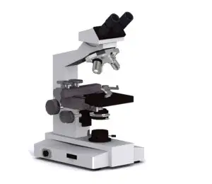
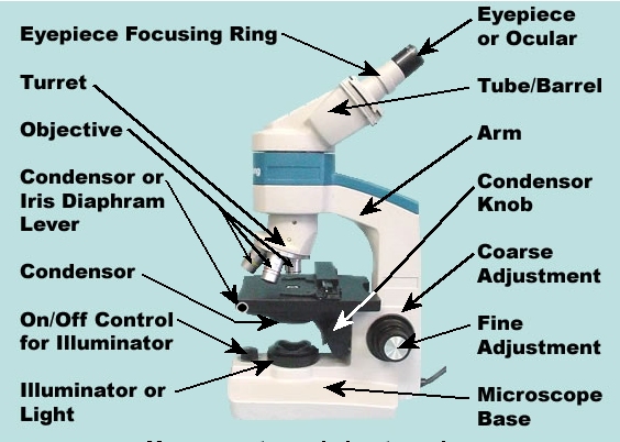
Body – the microscope’s body is typically a metal stand with an arm that supports the lens tube connecting the eyepiece to the lens, and connects it to the base, which is a heavy plate that supports the weight of the microscope and keeps it upright.
What are the components of a microscope?
Simple microscopes are actually made up of several mechanical and optical components. The mechanical components are what supports the microscope, holds the specimen, and controls some functions of the microscope. On the other hand, the optical components are what works on enlarging the image of the specimen, as well as allowing ...
What are the parts of a simple microscope?
Simple microscopes are actually made up of several mechanical and op tical components. The mechanical components are what supports the microscope, holds the specimen, and controls some functions of the microscope.
What is a simple microscope used for?
While simple microscopes are, well, simple imaging devices, these microscopes still have a variety of uses and applications, from scientific studies like biology, to more practical activities such as jewelry and watchmaking, and in looking at books, cloths, stamps, and engravings.
What is the purpose of an eyepiece in a microscope?
Eyepiece – on the first simple microscopes, the lens also serves as the eyepiece, but on some modern models, the microscope has a separate optical lens to view the specimen image. Eyepieces with their own magnification are used on compound microscopes.
What is the lens of a microscope called?
It’s the most elementary form of microscopy, dating back as far as the 14th century. The lens of a simple microscope is interchangeably referred to as a loupe, and is typically used as an eyepiece for other types of simple magnification devices such as compound microscopes, telescopes, and reading glasses.
How much magnification does a simple microscope have?
Since a simple microscope only makes use of one objective lens, its magnification capability is greatly limited. In fact, most simple microscopes only have a 10x magnification power.
How to find the total magnification of a microscope?
The total magnification of a microscope is obtained by multiplying the magnification power of objective lenses and the eyepiece. A light microscope usually has 40x to 100x magnification power.
How many lenses are in a light microscope?
A light microscope uses incident light and a sequence of lenses to create a magnified image. There are two lenses; an objective lens and an eyepiece.
What is the objective power of a lens?
Objective lenses are four in number. They are of 4x, 10x, 40x, and 100x magnification power. The lens with 100x objective power is also known as an oil immersion lens.
Why is the objective lens upside down?
The objective lens forms an upside-down/inverted and a real image because it is close to the object. The ocular lens or the eyepiece then further magnifies the image.
How does the ocular lens work?
In other words, the ocular lens functions further to magnify the image created by the objective lens.
What is the most used microscope?
Light microscopes are the most used microscopes. You will find them in every school, hospital, research center, etc. They use light and a number of lenses to magnify objects. If you own a microscope or are planning to have one, you should know how to use it and it works as this will help you yield maximum advantages from your microscope.
What is the magnification of an eyepiece?
The eyepiece, also known as an ocular lens, is present at the top of the body tube. Its magnification power is usually 10x but ranges from 5x to 30x.
