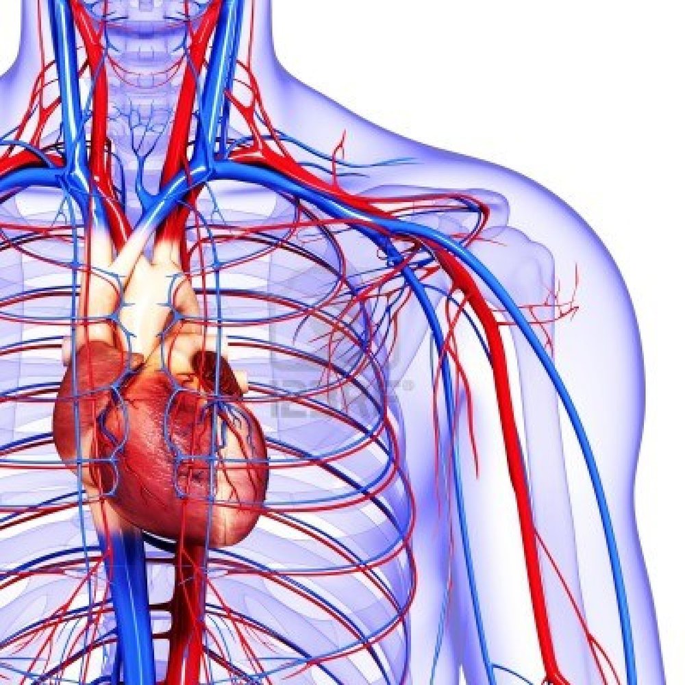
With each heartbeat, the heart sends blood throughout our bodies, carrying oxygen to every cell. After delivering the oxygen, the blood returns to the heart. The heart then sends the blood to the lungs to pick up more oxygen.
How does the cardiovascular system deliver nutrients and oxygen?
How does the cardiovascular system deliver nutrients and oxygen to all cells in the body via the flow of blood? Select all correct answers. (The arteries carry blood away from the heart; the veins carry it back to the heart.) (In the systemic circulation, the left ventricle pumps oxygen-rich blood into the aorta.)
How does the cardiovascular system work?
Your cardiovascular system, which is made up of your heart and blood vessels, is a crucial part of your body. When your cardiovascular system is working right, the cells in your body get a continuous supply of oxygen and nutrients from your blood. Blood vessels also remove carbon dioxide and other waste.
How is oxygen absorbed and excreted from the body?
During inhalation, air enters the lungs and oxygen is absorbed through the air sacs into the bloodstream. This oxygen-rich blood is pumped through the heart into the arterial circulation. In the capillaries, oxygen diffuses out of the blood and into the cells of the body's organs and tissues.
What is the relationship between blood flow and oxygen supply?
Since the convective supply of oxygen depends directly on blood flow, the regulation of tissue oxygenation depends critically on the regulation of blood flow.

How does the cardiovascular system deliver oxygen and nutrients?
Blood moves through the circulatory system as a result of being pumped out by the heart. Blood leaving the heart through the arteries is saturated with oxygen. The arteries break down into smaller and smaller branches to bring oxygen and other nutrients to the cells of the body's tissues and organs.
How does the cardiovascular system transport oxygen and carbon dioxide?
The heart, blood and blood vessels work together to service the cells of the body. Using the network of arteries, veins and capillaries, blood carries carbon dioxide to the lungs (for exhalation) and picks up oxygen. From the small intestine, the blood gathers food nutrients and delivers them to every cell.
How does the heart distribute oxygen?
The right ventricle pumps the oxygen-poor blood to the lungs through the pulmonary valve. The left atrium receives oxygen-rich blood from the lungs and pumps it to the left ventricle through the mitral valve. The left ventricle pumps the oxygen-rich blood through the aortic valve out to the rest of the body.
How does blood flow through the heart step by step?
Blood comes into the right atrium from the body, moves into the right ventricle and is pushed into the pulmonary arteries in the lungs. After picking up oxygen, the blood travels back to the heart through the pulmonary veins into the left atrium, to the left ventricle and out to the body's tissues through the aorta.
How circulatory system works step by step?
The circulatory system begins in your right atrium, the upper right-hand chamber of your heart. Blood moves from the right side of your heart through your lungs to get rid of carbon dioxide and pick up oxygen, and then returns to the left side of your heart, ending up in the left ventricle.
How does the cardiac muscle of the heart wall get its oxygen?
Coronary arteries supply blood to the heart muscle. Like all other tissues in the body, the heart muscle needs oxygen-rich blood to function. Also, oxygen-depleted blood must be carried away. The coronary arteries wrap around the outside of the heart.
What carries oxygenated blood to the heart?
The oxygenated blood is brought back to the heart by the pulmonary veins which enter the left atrium. From the left atrium blood flows into the left ventricle. The left ventricle pumps the blood to the aorta which will distribute the oxygenated blood to all parts of the body.
What are the 7 steps of the circulatory system?
Blood flows through the heart in the following order: 1) body –> 2) inferior/superior vena cava –> 3) right atrium –> 4) tricuspid valve –> 5) right ventricle –> 6) pulmonary arteries –> 7) lungs –> 8) pulmonary veins –> 9) left atrium –> 10) mitral or bicuspid valve –> 11) left ventricle –> 12) aortic valve –> 13) ...
How is carbon dioxide primarily transported in the cardiovascular system?
There are three means by which carbon dioxide is transported in the bloodstream from peripheral tissues and back to the lungs: (1) dissolved gas, (2) bicarbonate, and (3) carbaminohemoglobin bound to hemoglobin (and other proteins).
What transports oxygen and carbon dioxide to and from body cells?
One of the key functions of blood is transport. Blood vessels are like networks of roads where deliveries and waste removal take place. Oxygen, nutrients and hormones are delivered around the body in the blood and carbon dioxide and other waste products are removed.
What exchanges oxygen and carbon dioxide between blood and air?
respiratory systemThe lungs and respiratory system allow us to breathe. They bring oxygen into our bodies (called inspiration, or inhalation) and send carbon dioxide out (called expiration, or exhalation). This exchange of oxygen and carbon dioxide is called respiration.
What is the ability of the cardiovascular system of the body to supply energy?
Cardiorespiratory endurance refers to the ability of the heart and lungs to deliver oxygen to working muscles during continuous physical activity, which is an important indicator of physical health.
Is This an Emergency?
If you are experiencing serious medical symptoms, please see the National Library of Medicine’s list of signs you need emergency medical attention or call 911. If you think you may have COVID-19, use the CDC’s Coronavirus Self-Checker .
What is the role of the cardiovascular system in the delivery of hormones?
The cardiovascular system serves as the transportation connection between the endocrine glands and the organs or tissues they control via hormones.
How does carbon dioxide get into the lungs?
At the same time, carbon dioxide -- a waste product produced by cells -- is absorbed into the blood and transported to the lungs through the venous circulation. When this oxygen-poor blood reaches the lungs, carbon dioxide diffuses through the air sacs and is then exhaled. This cycle occurs with every breath.
What is the role of capillaries in the cardiovascular system?
The arterial circulation delivers blood from the heart to the body , and the venous circulation carries it back to the heart. Capillaries are tiny blood vessels at the interface of the arterial and venous circulation where exchange of substances between the blood and body tissues occurs. The cardiovascular system serves several major functions ...
What is the heart?
The heart is the core of the cardiovascular system. Image Credit: EpicStockMedia/iStock/Getty Images. The cardiovascular system, also known as the circulatory system, includes the heart, arteries, veins, capillaries and blood. The heart functions as the pump that moves blood through the body.
What is the function of the circulatory system?
The circulatory system also carries chemical messengers that attract cells to heal tissues that have been damaged due to injury or disease.
How does the cardiovascular system regulate body temperature?
If body temperature begins to rise, blood vessels close to the body surface dilate, increasing in size. This allows the body to rid itself excess heat through the skin. Conversely, if body temperature drops, surface blood vessels constrict to conserve body heat. The cardiovascular system works in concert with the body's sweating mechanism as the primary regulators of body temperature.
Which part of the heart is responsible for the flow of blood from the heart to the aorta?
The arteries carry blood away from the heart; the veins carry it back to the heart. In the systemic circulation, the left ventricle pumps oxygen-rich blood into the aorta.Veins contain a series of one-way valves that close to prevent the back flow of circulating blood.Blood, which is low in oxygen, is collected in veins and travels to the right atrium and into the right ventricle.
Which artery channels oxygen-poor blood from the right ventricle into the lungs?
The inferior and superior vena cava bring oxygen-poor blood from the body into the right atrium. The pulmonary artery channels oxygen-poor blood from the right ventricle into the lungs, where oxygen enters the bloodstream. The pulmonary veins bring oxygen-rich blood to the left atrium.
Which system delivers oxygen to all cells in the body?
The blood circulatory system (cardiovascular system) delivers nutrients and oxygen to all cells in the body. It consists of the heart and the blood vessels running through the entire body. ... In the systemic circulation, the left ventricle pumps oxygen-rich blood into the main artery (aorta).
Which part of the body transports blood away from the heart?
The arteries carry blood away from the heart; the veins carry it back to the heart. ... The blood travels from the main artery to larger and smaller arteries and into the capillary network. There the blood drops off oxygen, nutrients and other important substances and picks up carbon dioxide and waste products.
Which ventricle pumps oxygen-rich blood into the aorta?
2: (In the systemic circulation, the left ventric le pumps oxygen-rich blood into the aorta.)
How can the composition of ISF be maintained near its desired value?
An organism is faced with the following problem: How can the composition of ISF be maintained near its desired value? The solution of this problem is to introduce a circulatory system which continuously refreshes the ISF by putting it in intimate contact with “fresh, reconditioned” fluid (i.e., arterial blood). The circulating blood must be brought close to the cells (<10 μm) since nutrient and metabolic waste exchange takes place by passive diffusion, a transport mechanism which is most efficient over short distances. Thus, the cardiovascular system uses bulk flow (convection) to reduce the effective distance between the pumping action of the heart and the various parts of an organism.
What are some examples of local blood flow control?
Examples of local blood flow control processes are autoregulation, reactive hyperemia and active (or functional) hyperemia. The term “autoregulation” in this context refers to the tendency for organ blood flow to remain constant in the face of local changes in arterial or perfusion pressure. Autoregulation is observed in virtually every vascular bed. It is most pronounced in the brain and kidney and is prominent in the heart, skeletal muscle, intestine and liver. Recall that flow (Q) equals perfusion pressure (ΔP= difference between inflow arterial pressure and outflow venous pressure, Pa− Pv) divided by vascular resistance (R) so that, as ΔPrises through the autoregulatory range (Pa≈ 80–160 mm Hg in brain and kidney), Rmust increase to maintain constant flow. Reactive hyperemia refers to the elevated blood flow observed in an organ when flow is restored following a period of circulatory arrest (i.e., occlusion of the blood supply). Hyperemia is literally an excess of blood in a region. The magnitude of the hyperemia is related both to the duration of the occlusion period and to the pre-occlusion blood flow. Active (or functional) hyperemia refers to the increase in blood flow which accompanies an increase in the metabolic activity of an organ or tissue. It has been described in skeletal and cardiac muscle, brain, intestine, stomach, salivary glands, kidney and adipose tissue. The name of the hyperemia depends upon the specific function of the tissue (e.g., contraction hyperemia for muscle or secretory hyperemia for various glands). Each one of these examples of local regulatory processes can be linked to the regulation of tissue oxygenation.
Why is tissue oxygenation important?
However, the maintenance of tissue oxygenation is such an important feature for survival of the organism that it seems necessary that some mechanisms must exist to ensure an adequate oxygen supply to all cells of the organism.
How does the cardiovascular system regulate oxygenation?
The cardiovascular system controls blood flow to individual organs (1) by maintaining the input pressure to each organ within narrow limits by the mechanisms designed to regulate arterial pressure and (2) by allowing each organ to adjust its vascular resistance (R) to blood flow to an appropriate value. The cardiac output (CO) is distributed among the various organs according to their respective resistances so that flow (Q) in an organ is given by:
How are the systemic and pulmonary circulation connected?
The systemic circulation and pulmonary circulation are connected in series through the four chambers of the heart, so that all the blood that is pumped from the left ventricle into the systemic organs eventually makes its way back to the right ventricle from where it is pumped into the lungs. The systemic organs (tissues) are connected in parallel, and the following statements are consequences of this parallel architecture: (1) the stroke volume ejected from the left ventricle is divided among the various organs, and a given volume of blood passes through only one organ before entering the venous outflow of the organ; (2) the arterial blood entering each organ has the same composition; (3) the blood pressure at the entrance to each organ is the same; and (4) the blood flow to each organ can be controlled independently (local regulation of blood flow).
What is NCBI bookshelf?
NCBI Bookshelf. A service of the National Library of Medicine, National Institutes of Health.
What are the three mechanisms of TPR?
There are three major mechanisms that control the function of the cardiovascular system: local, neural and humoral. They can work independently of each other, but there are also interactions among them. The local mechanisms are intrinsic to a tissue and will be described in more detail below. The neural mechanisms involve the central nervous system and rely primarily on the release of norepinephrine from the sympathetic nerve endings of the autonomic nervous system. Finally, the humoral mechanisms rely on circulating vasoactive hormones, such as angiotensin II and epinephrine. It is important to recognize that the vasoregulation occurs in the resistance vessels. In the context of the regulation of tissue oxygenation, it is most appropriate to focus on the mechanisms that control blood flow at a local level.
Why do muscles produce more carbon dioxide?
During exercise the muscles need more oxygen in order to contract and they produce more carbon dioxide as a waste product. To meet this increased demand by the muscles, the following happens: Breathing depth (tidal volume) and rate (frequency) increase – this gets more oxygen into the lungs and removes more carbon dioxide out of the lungs. ...
How does the cardiorespiratory system work?
Cardio-respiratory system and exercise. The cardio-respiratory system works together to get oxygen to the working muscles and remove carbon dioxide from the body. During exercise the muscles need more oxygen in order to contract and they produce more carbon dioxide as a waste product.
What happens to the tidal volume when you exercise?
The graph shows that as a person goes from rest to exercise, their tidal volume increases. Heart rate increases – this increases the rate that oxygen is transported from the blood to the working muscles and carbon dioxide is transported from the working muscles to the lungs.
Which system transports oxygen from the air we breathe, through a system of tubes, into our lungs and then?
The respiratory system transports oxygen from the air we breathe, through a system of tubes, into our lungs and then diffuses it into the bloodstream, whilst carbon dioxide makes the opposite journey.
What is the heart rate of a person at 8 minutes?
This graph indicates the following: the person's resting heart rate is around 60 bpm. at 8 minutes, just before taking part in exercise their heart rate increases – this is called the anticipatory increase in heart rate which occurs when a person starts to think about taking part in exercise.
How long does it take for your heart rate to increase?
at 10 minutes the person starts to take part in exercise and there is a steep increase in heart rate which reaches 145 bpm at 13 minutes. the heart rate remains high during exercise. when the person stops taking part in exercise the heart rate decreases. previous.
