What is the catalytic triad?
The catalytic triad provides a paradigm for the structural and chemical features of enzymes that allow them to facilitate a difficult reaction.
What is the role of the catalytic triad in serine protease?
The catalytic triad and its role in the serine protease mechanism. The catalytic triad provides a paradigm for the structural and chemical features of enzymes that allow them to facilitate a difficult reaction. The reaction in this case is hydrolysis of a peptide bond, which -...
What is the catalytic triad of TEV protease?
The enzyme TEV protease contains an example of a catalytic triad of residues (red) in its active site. The triad consists of an aspartate ( acid ), histidine ( base) and serine ( nucleophile ).
What is an example of a triad enzyme?
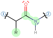
Examples of triads. The range of amino acid residues used in different combinations in different enzymes to make up a catalytic triad for hydrolysis. On the left are the nucleophile, base and acid triad members. On the right are different substrates with the cleaved bond indicated by a pair of scissors.

How does the Triad in chymotrypsin work?
Chymotrypsin contains a collection of three amino acids called the catalytic triad. This triad consists of serine-195, histidine-57 and aspartate-102. These amino acids work together to carry out the catalytic function of breaking peptide bonds.
Is the catalytic triad the active site?
These enzymes are found in prokaryotic and eukaryotic cells and all use a common set of three amino acids in the active site called a catalytic triad (Figure 4.53). It consists of aspartic acid, histidine, and serine.
What does histidine do in the catalytic triad?
The histidine residue forces serine into a position that facilitates nucleophilic attack later on through the process of catalysis by approximation. In the presence of a substrate, a chain reaction occurs.
Where is the catalytic triad in the trypsin?
The catalytic triad; Asp 102, His 57, and Ser 195, shown here in yellow, is positioned near the substrate. The catalytically active histidine and serine side chains are even near an amide bond in UB-THR 10, just like the amide bond broken in peptide hydrolysis.
What is the catalytic triad in serine protease?
The triad is located in the active site of the enzyme, where catalysis occurs, and is preserved in all superfamilies of serine protease enzymes. The triad is a coordinated structure consisting of three amino acids: His 57, Ser 195 (hence the name "serine protease") and Asp 102.
What is the mechanism of chymotrypsin action?
More specifically, chymotrypsin operates through a particular type of ping-pong mechanism called covalent hydrolysis. This means that the enzyme first forms a covalent bond with the target substrate, displacing the more stable moiety into solution. This enzyme-substrate complex is called the enzyme intermediate.
How does the catalytic triad function to lower the pKa of serine?
- The catalytic serine has a decreased pKa, due to the hydrogen-bond network created in the active site. This alteration in pKa causes Ser to donate a proton at physiological pH.
Why is histidine important in the function of enzymes?
Histidine is required for synthesis of proteins. It plays particularly important roles in the active site of enzymes, such as serine proteases (e.g., trypsin) where it is a member of the catalytic triad. Excess histidine may be converted to trans-urocanate by histidine ammonia lyase (histidase) in liver and skin.
What is the catalytic triad in chymotrypsin?
0:1414:51Mechanism of Chymotrypsin and Catalytic Triad - YouTubeYouTubeStart of suggested clipEnd of suggested clipAnd this collection of three amino acids is known as the catalytic triad. So it's the catalyticMoreAnd this collection of three amino acids is known as the catalytic triad. So it's the catalytic triad inside the active site of climate trypsin that essentially catalyzes the cleavage of peptide bonds
How does trypsin break down proteins?
Trypsin is an enzyme in the first section of the small intestine that starts the digestion of protein molecules by cutting these long chains of amino acids into smaller pieces. It is a serine protease from the PA clan superfamily, found in the digestive system of many vertebrates, where it hydrolyzes proteins.
What breaks down trypsin?
Once trypsinogen moves from the pancreas to the small intestine, it is cleaved and released as the active enzyme trypsin. Trypsin breaks down protein into peptides through a hydrolysis reaction. Hydrolysis occurs when a bond is broken in the presence of water.
Why does trypsin cleave after lysine?
Trypsin cleaves the peptide bond between the carboxyl group of arginine or the carboxyl group of lysine and the amino group of the adjacent amino acid. The rate of cleavage occurs more slowly when the lysine and arginine residues are adjacent to acidic amino acids in the sequence or cystine.
What are the roles of His Ser and Asp in chymotrypsin catalyzed reaction?
The His-57 role is to position the serine residue and polarize the hydroxyl group so it can be deprotonated to the alkoxide ion. In the presence of the substrate, this accepts a proton by acting as a base. Asp-102 orients the His-57 and stabilizes it through hydrogen bonding and electrostatics.
Why is histidine found in active sites?
Message: Jithesh, Histidine is found so often in active sites of enzymes because the imidazole ring at the end of this amino acid s side chain can perform many different roles in catalysis. As you correctly suggested, the pKa of about 6 is one important factor.
Which of the following amino acids can act as a general acid only in an active site with an extensive salt bridging hydrogen bond network?
Which of the following amino acids can act as a general acid only in an active site with an extensive salt bridging/hydrogen bond network? - Serine is a polar neutral amino acid.
What amino acids are catalytic?
Using MACiE as a knowledge base, we have seen that the top 10 most catalytic residues are histidine, aspartate, glutamate, lysine, cysteine, arginine, serine, threonine, tyrosine and tryptophan.
What is the catalytic triad of RTX?
A catalytic triad consisting of D-587, H-653, and C-698 ( that has first been identified on the Vibrio cholerae RTX toxin) is also present on TcdB [36].
Where is the catalytic triad located in the CP structure?
The catalytic triad is located at the interface of the two subdomains in the CP structure. The catalytic triad residues Ser, His, and Asp are located far apart in the sequence; however, they come closer in the tertiary structure to form the active site of CP (Fig. 5.6 ). In CVCP, the residue Ile227 is located near the active site and has been proposed to play an important role in the catalytic action of the protease. The PCR-generated mutation I227K results in the loss of proteolytic activity of CVCP ( Thomas et al., 2010 ). This might be due to the formation of a salt bridge between active-site residue Asp161 and Lys227. This additional salt bridge would disrupt the interactions within the active-site residues. Hence, even a single–amino acid change can lead to a complete loss of enzyme function. The residue Ser218 side chain is flexible in the active form, while it gets stabilized in native AVCP with bound Trp267 ( Aggarwal et al., 2012, 2014) ( Fig. 5.6 B). The active-site residue Asp166 is exposed to the solvent in both inactive and active AVCP.
Where is the oxyanion hole located in the catalytic triad?
In the immediate vicinity of catalytic triad residues at the base of the active center gorge, just above the active serine, is located the electrophilic oxyanion hole, which has the capacity to attract carbonyl oxygen of ACh and other substrates as well as phosphyl oxygen of covalent, OP inhibitors. Three backbone nitrogens well conserved throughout the cholinesterase family (Table 2) form the oxyanion hole, lending their amide protons for the interaction. In oxyanion holes of proteases, only two donor residues are involved. Site-directed mutagenesis of AChEs and use of substrates and inhibitors specific for interaction with either AChE or BuChE helped delineate specific locations of additional ligand binding sites within the enzyme gorge ( Shafferman et al., 1992a; Vellom et al., 1993; Ordentlich et al., 1993; Radić et al., 1993 ). The acyl pocket and the choline binding site are located next to the active serine, at the base of the active center gorge ( Fig. 4 ), controlling the size of ligands that can approach the site of catalysis. The space available for binding is generally smaller in AChEs in which several aromatic residues (14 in fish and mammalian AChEs; Sussman et al., 1991) line the walls of the gorge. In the acyl pocket, in-place of phenylalanines Phe288 and Phe290 found in AChEs smaller aliphatic residues are found in BuChEs, whereas in the choline binding site Phe330 (many AChEs have tyrosine at this position) in BuChEs is replaced by alanine ( Table 6 ). The smaller residues and larger available space in BuChE enable preferential binding of the large substrates butyrylthiocholine and benzoylcholine in the acyl pocket and large inhibitors ethopropazine and isoOMPA in the choline binding site and acyl pocket, respectively. Some mutant insects (mosquitoes and flies) that developed resistance to pesticides have amino acid residues altered in this region in their AChEs, selectively preventing binding of pesticides to active serine while not compromising catalysis ( Menozzi et al., 2004 ). In addition to the choline binding site and acyl pocket in the third binding domain of cholinesterases, the peripheral site is located at the rim of the gorge, approximately 14 Å from the active serine. Formed by Tyr70, Tyrl21, and Trp 279, this aromatic cluster specifically binds cationic and aromatic inhibitors that are too large to enter the gorge, such as propidium, gallamine, or snake toxins fasciculins ( Bourne et al., 1995, 2004 ), or long and slender bisquaternary ligands that extend from the bottom of the gorge, such as BW286c51, decamethonium, and a variety of bifunctional ligands including bis -tacrines, bis- huperzines, and very high-affinity triazoles ( Lewis et al., 2002; Bourne et al., 2004 ). In the vicinity of the aromatic cluster is Asp72, a specifically located anionic residue lending its stabilizing contribution to ligands binding to the ~6 Å proximal peripheral site and/or ~8 Å distal choline binding site. Devoid of an aromatic cluster in the peripheral site, BuChEs bind most bisquaternary and bifunctional inhibitors with three or four orders of magnitude lower affinity than AChEs ( Radić et al., 1993 ), whereas the affinity of fasciculins is up to eight orders of magnitude weaker than in corresponding AChEs ( Radić et al., 1994 ). The absence of the aromatic cluster does not critically influence ACh hydrolysis in BuChEs, and its substitution with aliphatic residues in AChEs does not seem to affect catalytic parameters; however, substitution of Asp72 has a pronounced effect on substrate Km in both AChEs and BuChEs ( Shafferman et al., 1992b; Radić et al., 1993; McGuire et al., 1989; Masson et al., 1997 ). The BuChE variant containing Asp70Gly substitution (corresponding to the Asp72 position in Torpedo AChE) is naturally occurring in the human population. Individuals with Asp70Gly mutation appear unable to hydrolyze the muscle relaxant succinyldicholine efficiently and thus experience life-threatening apnea lasting from a few minutes to several hours.
What are the binding sites in Torpedo AChE?
Topography of binding sites in ChEs represented by selected residue side chains of Torpedo AChE structure. A molecule of ACh docked in the active center (taken from PDB entry 2ACE) and shown as solvent-accessible surface is given as a frame of reference. Binding sites are acyl pocket (residues 288 and 290), choline binding site (residues 84 and 330), and peripheral site (residues 70, 72, 121, and 279). Visualized by WebLabViewer software (Accelrys, San Diego).
Which protease family has the subtilisin-type catalytic triad?
The subtilisin-type catalytic triad is conserved in all known processing proteases of Kex2 family (34, 40 ). It is known to be required for function, and a great deal of kinetic evidence supports the idea that the classical serine protease mechanism is utilized by these enzymes (see below).
Which esterases have a catalytic triad?
The serine esterases have a catalytic triad: serine, glutamic or aspartic acid, and histidine. These catalytic residues are responsible for the nucleophilic attack of the active site serine on the carbonyl carbon atom of the ester.
What is the position of the Phe330 ring?
When bisquaternary ligands, decamethonium and BW286c51 are bound, the position of the Phe330 ring is nearly perpendicular to Trp84. thus opening the full length of the gorge. In the immediate vicinity of Phe330 lies catalytic triad His440.
What is the catalytic triad?
An example of a catalytic triad is present in chymotrypsin, where the triad (on the enzyme) consists of S195 (that is the serine found at residue 195 in the protein sequence), D102 and H57. Essentially, S195 binds to the substrate polypeptide to the side of a phenylalanine, tryptophan or tyrosine residue closer to the C-terminus, holding it in place. D102 and H57 then hydrolyze the bond. This takes place in several steps.
What are the three residues of a catalytic triad?
A catalytic triad commonly refers to the three amino acid residues found inside the active site of certain protease enzymes: serine (S), aspartate (D) and histidine (H). They work together to break peptide bonds on polypeptides. More generally, catalytic triad can refer to any set of three residues that function together and are directly involved in catalysis. Because enzymes fold into complex three-dimensional shapes, the residues of a catalytic triad can be far from each other in the primary structure however are brought close together in the tertiary structure .
What is electrostatic catalysis?
Electrostatic catalysis occurs when the enzyme active site stabilizes the transition state of the reaction by forming electrostatic interactions with the substrate. The electrostatic interactions can be ionic, ionic-dipole, dipole-dipole, or hydrophobic interactions. Hydrogen bonding is one of the most common electrostatic interactions formed in the active site.
How fast is catalysis?
This can dramatically improve the catalytic rate of the reaction from 105 to 107 times faster, depending on the enzyme system.
What is covalent catalysis?
Covalent catalysis involves the formation of a covalent bond between the enzyme and at least one of the substrates involved in the reaction. Often times this involves nucleophilic catalysis which is a subclass of covalent catalysis. As seen in Section 7.1, several amino acid R-groups can serve as a nucleophile and are often found at the active site of enzymes. Nucleophilic side chains are often activated by deprotonation caused by neighboring side chains, such as histidine that can act as a base. Alternatively, water can also activate the nucleophile. The intermediate covalent bond formation between the enzyme and the substrate enables bond cleavage and the removal of a leaving group.
How does an enzyme affect the rate of catalysis?
In catalysis by approximation, the enzyme enhances the reaction rate by binding with multiple substrates and positioning them favorably so that the reaction can proceed. Binding with the enzyme reduces the rotational entropy of the substrates that would otherwise be randomly free floating in solution, and enables the correct positioning of substrates for the reaction. The loss of entropy, which is not favorable, is offset by the binding energy released with the substrate-enzyme interaction.
What is the role of adenylate kinase in ATP?
Adenylate kinase (also known as AK or myokinase) is a phosphotransferase enzyme that catalyzes the interconversion of adenine nucleotides (ATP, ADP, and AMP). By constantly monitoring phosphate nucleotide levels inside the cell, AK enzymes play an important role in cellular energy homeostasis. The basic chemical reaction mediated by this enzyme class is the conversion of 2 ADP molecules into 1 ATP and 1 AMP (Figure 7.20A). The reverse reaction can also occur forming an equilibrium based on cellular concentrations of the varying phosphorylation states.