How are ultrasound waves produced?
Ultrasound waves are produced by a transducer, which can both emit ultrasound waves, as well as detect the ultrasound echoes reflected back. In most cases, the active elements in ultrasound transducers are made of special ceramic crystal materials called piezoelectrics.
How do ultrasound transducers work?
Nov 05, 2019 · How is an ultrasound image produced? The ultrasound image is produced based on the reflection of the waves off of the body structures. The strength (amplitude) of the sound signal and the time it takes for the wave to travel through the body provide the information necessary to produce an image. How does an ultrasound scan work?
What is ultrasound and how does it work?
Sep 28, 2020 · Picture of a transducer (probe) used during an ultrasound exam. The ultrasound image is produced based on the reflection of the waves off of the body structures. The strength (amplitude) of the...
Can crystals produce ultrasound waves?
Unlike normal ways of making sound, which often involve striking a surface, ultrasound is made using electrical equipment that vibrates with an extremely high frequency. Crystals of materials such as quartz vibrate very fast when electricity is passed through them—an effect called “piezoelectricity.”
What are the methods for production of ultrasound?
There are three methods for producing Ultrasonic waves. They are: (i) Mechanical generator or Galton's whistle. (ii) Magnetostriction generator....Merits:Magnetostrictive materials are easily available and inexpensive.Oscillatory circuit is simple to construct.Large output power can be generated.
What is the source of ultrasound?
Among the earliest known sources of ultrasound are those emanating from the animal kingdom. Dogs, birds, crickets, and bats are amongst those creatures whose communication signals extend beyond the range of human hearing. In addition bats use ultrasound as a guidance system between 50 and 100 kHz.
How the ultrasound beam is produced?
Ultrasound Technology Ultrasound wave is produced when an electric current is applied to an array of piezoelectric crystals. This causes distortion of the crystals, makes them vibrate and produce this acoustic wave. The summation of the waves produces an ultrasound beam. The ultrasound waves are produced in pulses.
What are the basic process of ultrasound?
A trained technician (sonographer) presses a small, hand-held device (transducer) against the area being studied and moves it as needed to capture the images. The transducer sends sound waves into your body, collects the ones that bounce back and sends them to a computer, which creates the images.Mar 17, 2020
How was ultrasound invented?
Ultrasound was first used for clinical purposes in 1956 in Glasgow. Obstetrician Ian Donald and engineer Tom Brown developed the first prototype systems based on an instrument used to detect industrial flaws in ships.May 16, 2013
What are 3 uses of ultrasound?
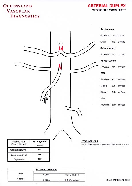
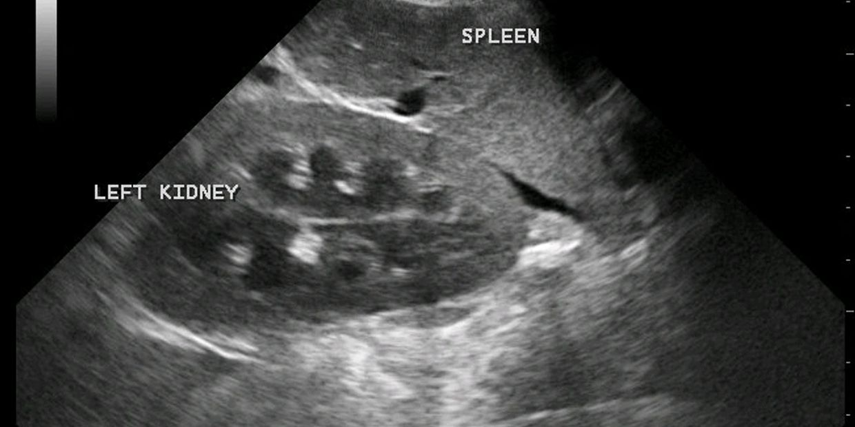
Ultrasound is a useful way of examining many of the body's internal organs, including but not limited to the:heart and blood vessels, including the abdominal aorta and its major branches.liver.gallbladder.spleen.pancreas.kidneys.bladder.uterus, ovaries, and unborn child (fetus) in pregnant patients.More items...
Why are pulses used in ultrasound?
The time between a pulse of sound being transmitted and detected and the speed of sound in water can be used to calculate the distance of the reflecting surface or object. The process is very similar to ultrasound imaging.
What is a cycle in ultrasound?
1:584:23Ultrasound 6 Duty Cycle - YouTubeYouTubeStart of suggested clipEnd of suggested clipOkay so it's essentially this idea of how long is the pulse on versus how long is the entire pulse.MoreOkay so it's essentially this idea of how long is the pulse on versus how long is the entire pulse. Period okay so again usually primarily non-thermal um we can target both superficial.
How does the ultrasound transducer work?
An ultrasound transducer is the handheld device that the technician or doctor moves on or over the body of the patient. A cord connects it to a computer. The device sends sound waves and receives the echoes as they bounce off the body tissue and organs of the patient.Mar 29, 2018
What are 4 uses of ultrasound?
Diagnostics: Doctors can use ultrasounds to diagnose conditions, including those in the heart, blood vessels, liver, gallbladder, spleen, pancreas, kidneys, bladder, uterus, ovaries, eyes, thyroid, and testicles.
What does not show up on an ultrasound?
Ultrasound images are not as detailed as those from CT or MRI scans. Ultrasound cannot tell whether a tumor is cancer. Its use is also limited in some parts of the body because the sound waves can't go through air (such as in the lungs) or through bone.Nov 30, 2015
How do I prepare for a pregnancy ultrasound?
There is no special preparation for the ultrasound test. Some doctors require you to drink 4 to 6 glasses of water before the test, so your bladder is full. This will help the doctor view the baby better on the ultrasound. You will be asked to refrain from urinating until after the test.Jan 1, 2018
How is ultrasound produced?
The ultrasound image is produced based on the reflection of the waves off of the body structures. The strength (amplitude) of the sound signal and the time it takes for the wave to travel through the body provide the information necessary to produce an image.
What is ultrasound imaging?
Ultrasound imaging (sonography) uses high-frequency sound waves to view inside the body. Because ultrasound images are captured in real-time, they can also show movement of the body's internal organs as well as blood flowing through the blood vessels. Unlike X-ray imaging, there is no ionizing radiation exposure associated with ultrasound imaging.
Why is ultrasound important for pregnant women?
Because ultrasound is not based on ionizing radiation, it is particularly useful for women of child-bearing age when CT or other imaging methods would otherwise result in exposure to radiation. Expectant Mothers. Ultrasound is the most widely used medical imaging method for viewing the fetus during pregnancy.
What is ALARA in ultrasound?
As with all other imaging modalities, the principles of As Low As Reasonably Achievable (ALARA) should be practiced by health care providers.
Is ultrasound safe for health care?
Although ultrasound imaging is generally considered safe when used prudently by appropriately trained health care providers, ultrasound energy has the potential to produce biological effects on the body. Ultrasound waves can heat the tissues slightly.
What is a 1004?
1004 - Repurchase, repairs, or replacement of electronic products. 1005 - Importation of electronic products. There are no federal radiation safety performance standards for diagnostic ultrasound. Because they are medical devices, ultrasound imaging equipment must also comply with the medical device regulations.
Does ultrasound have energy?
Ultrasound imaging does introduce energy into the body, and laboratory studies have shown that diagnostic levels of ultrasound can produce physical effects in tissue, such as pressure oscillations with subsequent mechanical effects and rise in temperature.
How to do an ultrasound?
Before your ultrasound begins, you may be asked to do the following: 1 Remove any jewelry from the area being examined. 2 Remove some or all of your clothing. 3 Change into a gown.
How long does it take to get an ultrasound?
A typical ultrasound exam takes from 30 minutes to an hour.
Why do we need ultrasound?
Ultrasound is used for many reasons, including to: View the uterus and ovaries during pregnancy and monitor the developing baby's health. Diagnose gallbladder disease. Evaluate blood flow. Guide a needle for biopsy or tumor treatment. Examine a breast lump.
What is ultrasound used for?
The images can provide valuable information for diagnosing and treating a variety of diseases and conditions. Most ultrasound examinations are done using an ultrasound device outside your body, though some involve placing a device inside your body.
How does ultrasound help with tumors?
These images show how ultrasound can help guide a needle into a tumor (left), where material is injected (right) to destroy tumor cells. During a transvaginal ultrasound, your doctor or a medical technician inserts a wandlike device (transducer) into your vagina while you are positioned on an exam table.
What is gel used for?
During the procedure. Gel is applied to your skin over the area being examined. It helps prevent air pockets, which can block the sound waves that create the images. This water-based gel is easy to remove from skin and, if needed, clothing.
How does a transducer work?
The transducer sends sound waves into your body, collects the ones that bounce back and sends them to a computer, which creates the images. Sometimes, ultrasounds are done inside your body. In this case, the transducer is attached to a probe that's inserted into a natural opening in your body. Examples include:
What is the effect of ultrasound on quartz?
When ultrasound is purposed for cleaning things, a high-frequency alternating electricity supply sends power to piezoelectric transducers. If mechanical pressure is applied to one pair of opposite faces of certain crystals like quartz, equal and opposite electrical charges appear across its other faces. This effect is called the piezoelectric ...
What is ultrasonic cleaning?
Not least among these is ultrasonic cleaning, which uses ultrasonic sound waves traveling through liquid to produce cavitation bubbles that clean more thoroughly than solvents and scrubbing alone.
What is the effect of an electric field on a crystal?
This effect is called the piezoelectric effect. The converse of piezoelectric effect is also true, in that if an electric field is applied to one pair of faces, the corresponding changes in the dimensions of the other pair of faces of the crystal are produced. This effect is known as inverse piezoelectric effect.
What is ultrasound ultrasound?
Fetal ultrasound. Ultrasound is sound waves with frequencies higher than the upper audible limit of human hearing. Ultrasound is not different from "normal" (audible) sound in its physical properties, ...
When was ultrasound first used?
The first technological application of ultrasound was an attempt to detect submarines by Paul Langevin in 1917. The piezoelectric effect, discovered by Jacques and Pierre Curie in 1880, was useful in transducers to generate and detect ultrasonic waves in air and water.
Why is the upper frequency limit 20 kHz?
The upper frequency limit in humans (approximately 20 kHz) is due to limitations of the middle ear. Auditory sensation can occur if high‐intensity ultrasound is fed directly into the human skull and reaches the cochlea through bone conduction, without passing through the middle ear.
How many kHz can fish hear?
Several types of fish can detect ultrasound. In the order Clupeiformes, members of the subfamily Alosinae ( shad) have been shown to be able to detect sounds up to 180 kHz, while the other subfamilies (e.g. herrings) can hear only up to 4 kHz.
Why is ultrasound used in cattle?
Ultrasound is used to evaluate fat thickness, rib eye area, and intramuscular fat in living animals. It is also used to evaluate the health and characteristics of unborn calves.
What is the frequency of ultrasound?
Ultrasound is defined by the American National Standards Institute as " sound at frequencies greater than 20 kHz". In air at atmospheric pressure, ultrasonic waves have wavelengths of 1.9 cm or less.
How to measure fluid flow?
In rheology, an acoustic rheometer relies on the principle of ultrasound. In fluid mechanics, fluid flow can be measured using an ultrasonic flow meter .
What is an ultrasound?
Photo: An ultrasound (echocardiograph) image of a beating human heart. NASA (the US space agency) uses this equipment to study the long-term effects of space travel on astronauts. Photo courtesy of NASA Marshall Space Flight Center (NASA-MSFC) .
How do ultrasonics work?
Another popular use for ultrasonics is in ships, both for navigation and for locating objects underwater. Sound travels faster through water than through air, which is very helpful because light hardly travels through water at all. Most people know that whales can use low-frequency sound to communicate across entire oceans. Submarines use a similar trick with a type of navigation called sonar (sound navigation and ranging), which is a bit like an underwater equivalent of radar.
How does a piezoelectric crystal work?
Piezoelectric crystals also work in the reverse way: if ultrasound waves traveling through the air happen to collide with a piezoelectric crystal, they squeeze its surface very slightly, causing a brief burst of electricity to flow through it.
What is high power ultrasound?
High-power ultrasound. Relatively low-strength ultrasound waves are used for medical scans and non-destructive testing. Much stronger ultrasound waves have very different uses. If you have a painful kidney stone, firing powerful ultrasound waves from outside your body can make the stone vibrate and break apart.
How to detect illness?
To save having to open up your body to detect an illness, doctors can simply run an ultrasound scanner over your skin to see inside. The scanner probe often looks a bit like a computer mouse . It has a built in transducer that beams harmless, ultrasound waves down into your body.
Can you see cracks in metal?
If there's a crack deep inside a metal, inspecting it from the inside won't reveal the problem. But if you run an ultrasound scanner over the outside of the metal, the crack inside will disturb and reflect back some of the ultrasound waves— so the defect will show up on your testing monitor.
What frequency does a towfish use?
Photo: A typical side-scan sonar towfish. This one uses ultrasound at a frequency of 600 kHz, which is well above the limit of human hearing. Here, it's being hooked up to equipment onboard a scientific research ship before being lowered into the water to be dragged alongside.
What are the parts of an ultrasound machine?
A basic ultrasound machine has the following parts: 1 Transducer probe - probe that sends and receives the sound waves 2 Central processing unit (CPU) - computer that does all of the calculations and contains the electrical power supplies for itself and the transducer probe 3 Transducer pulse controls - changes the amplitude, frequency and duration of the pulses emitted from the transducer probe 4 Display - displays the image from the ultrasound data processed by the CPU 5 Keyboard/cursor - inputs data and takes measurements from the display 6 Disk storage device (hard, floppy, CD) - stores the acquired images 7 Printer - prints the image from the displayed data
What is the principle of ultrasound?
The transducer probe generates and receives sound waves using a principle called the piezoelectric ( pressure electricity) effect , which was discovered by Pierre and Jacques Curie in 1880. In the probe, there are one or more quartz crystals called piezoelectric crystals.
What is a transducer probe?
Transducer probe - probe that sends and receives the sound waves. Central processing unit (CPU) - computer that does all of the calculations and contains the electrical power supplies for itself and the transducer probe.
How does a CPU work?
The CPU sends electrical currents to the transducer probe to emit sound waves, and also receives the electrical pulses from the probes that were created from the returning echoes. The CPU does all of the calculations involved in processing the data. Once the raw data are processed, the CPU forms the image on the monitor.

Overview
- Diagnostic ultrasound, also called sonography or diagnostic medical sonography, is an imaging method that uses high-frequency sound waves to produce images of structures within your body. The images can provide valuable information for diagnosing and treating a variety of diseases and conditions. Most ultrasound examinations are done using an ultrasound device outside you…
Why It's Done
- Ultrasound is used for many reasons, including to: 1. View the uterus and ovaries during pregnancy and monitor the developing baby's health 2. Diagnose gallbladder disease 3. Evaluate blood flow 4. Guide a needle for biopsy or tumor treatment 5. Examine a breast lump 6. Check your thyroid gland 7. Detect genital and prostate problems 8. Assess joint inflammation (synoviti…
Risks
- Diagnostic ultrasound is a safe procedure that uses low-power sound waves. There are no known risks. Ultrasound is a valuable tool, but it has limitations. Sound doesn't travel well through air or bone, so ultrasound isn't effective at imaging body parts that have gas in them or are hidden by bone, such as the lungs or head. To view these areas, your doctor may order other imaging tests…
How You Prepare
- Most ultrasound exams require no preparation. However, there are a few exceptions: 1. For some scans, such as a gallbladder ultrasound, your doctor may ask that you not eat or drink for certain period of time before the exam. 2. Others, such as a pelvic ultrasound, may require a full bladder. Your doctor will let you know how much water you need to drink before the exam. Do not urinat…
What You Can Expect
- Before the procedure
Before your ultrasound begins, you may be asked to do the following: 1. Remove any jewelry from the area being examined. 2. Remove some or all of your clothing. 3. Change into a gown. You'll be asked to lie on an examination table. - During the procedure
Gel is applied to your skin over the area being examined. It helps prevent air pockets, which can block the sound waves that create the images. This water-based gel is easy to remove from skin and, if needed, clothing. A trained technician (sonographer) presses a small, hand-held device (tr…
Results
- When your exam is complete, a doctor trained to interpret imaging studies (radiologist) analyzes the images and sends a report to your doctor. Your doctor will share the results with you. You should be able to return to normal activities immediately after an ultrasound.
Clinical Trials
- Explore Mayo Clinic studiesof tests and procedures to help prevent, detect, treat or manage conditions.