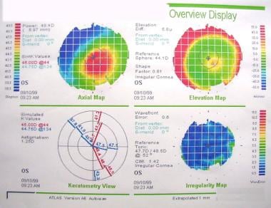
A corneal topographer projects a series of illuminated rings, referred to as a Placido disc, onto the surface of the cornea. The rings are reflected back into the instrument. After analyzing the reflected rings of light, the computer generates a topographical map of the cornea.
What is a corneal topography test?
The cornea is the outer layer of the eye, responsible for about 70 percent of the eye’s focusing power. A corneal topography test provides detailed 3D maps of the cornea’s shape and curvature and enables detection of corneal diseases, and irregular corneal conditions, such as swelling, scarring, abrasions, deformities, and irregular astigmatisms.
What is a corneal mapping?
Corneal topography, computer-assisted videokeratography (CAVK), or corneal mapping is a computer-assisted diagnostic imaging technique that helps in creating visuals of the corneal surface so that the doctor can view the visuals and analyze the entire shape of the cornea for diagnosis and treatment.
What are the clinical uses of topography?
We will also review 5 clinical uses for topography that will prepare you well for cornea clinic. This is the technical distinction between topography and tomography: 1) Corneal to p ography is a non-invasive imaging technique for mapping the surface curvature and shape of the anterior corneal surface.
What to expect during a corneal topography scan?
What to Expect During a Corneal Topography Scan You will be seated facing a large bowl with lighted circles inside it. The chin and forehead rests keep your head secure to get the clearest images. You will be asked to stare at a fixed target in the bowl while the pictures are taken.

What is corneal topography testing?
Corneal topography is a special photography technique that maps the surface of the clear, front window of the eye (the cornea). It works much like a 3D (three-dimensional) map of the world, that helps identify features like mountains and valleys.
How much is a corneal topography?
How Much Does a Computerized Corneal Topography Cost? On MDsave, the cost of a Computerized Corneal Topography ranges from $38 to $69. Those on high deductible health plans or without insurance can save when they buy their procedure upfront through MDsave. Read more about how MDsave works.
Is corneal topography harmful?
Corneal topography is a painless, non-contact technique, meaning that the corneal topography device will not touch your eye during the measurement. If your eye is dry you may have a few moistening drops instilled into your eye to improve the quality of the topography image.
Is corneal topography painful?
A corneal topography test is quick and painless. During the test, you will sit in front of a lighted bowl that contains a pattern of rings, and rest your head against a bar.
Is corneal topography necessary?
Since the cornea is normally responsible for approximately 70% of the eye's refractive power, its topography is of critical importance in determining the quality of vision. Therefore, corneal topography is an important diagnostic test.
How long does a corneal topography take?
Now that I have my total vision back I can show off my “beautiful eyes” thanks to Dr. James Pasternack. He was so gentle; the procedure took less than 20 minutes and pain free. I would recommend everyone to have this procedure done and be set free from glasses forever!
Is corneal topography covered by insurance?
Corneal topography is a covered service for the above indications when medically reasonable and necessary only if the results will assist in defining further treatment. It is not covered for routine follow-up testing.
Does map dystrophy cause blindness?
This condition is common, treatable, and rarely leads to significant vision loss.
Is corneal topography covered by insurance?
Corneal topography is a covered service for the above indications when medically reasonable and necessary only if the results will assist in defining further treatment. It is not covered for routine follow-up testing.
How much does a pentacam cost?
Some slit-scanning devices: Orbscan (Bausch + Lomb); Pentacam (Oculus); and Galilei G2 (Ziemer). The average cost of these instruments is $50,000. Some manufacturers also offer instruments that combine corneal topography with full optical wavefront aberrometry.
How is keratoconus diagnosed on topography?
Topographic diagnosis of keratoconus is suggested by:High central corneal power.Large difference between the power of the corneal apex and periphery.Differences in steepness between the two corneas of a given patient.
What is the difference between topography and tomography?
Results: Topography is the study of the shape of the corneal surface, while tomography allows a three-dimensional section of the cornea to be presented.
What is the corneal topography?
Corneal topography, also known as corneal mapping, is a diagnostic tool that provides 3-D images of the cornea. The cornea is the outer layer of the eye, responsible for about 70 percent of the eye’s focusing power.
Why is the corneal map important?
This map is used to determine the true shape of the cornea and is crucial for selecting the best contact lens design for an irregular cornea. This map display is especially important when deciding between a scleral gas permeable (GP) lens or a corneal contact lens.
Is topography more accurate than other maps?
However, since it collects the averages of the data to produce a smooth map, it is considered less accurate than the other maps.
Which error is associated with steeper central corneal curvature?
4) Refractive error. Myopia is associated with steeper central corneal curvature. Hyperopia is associated with flatter central corneal curvature. However, these are not hard and fast rules, as axial length plays a big role in the overall myopia/hyperopia of the eye.
What are the colors of topographic maps?
Colored Maps: You will see a rainbow of colors on every topographic map. These range from warm colors ( red, orange, yellow ), to neutrals ( green) to cool colors ( blue, purple ). On our representative Pentacam images below, you will see four different types of maps.
Can refractive surgery cause corneal ectasia?
Refractive surgery itself can induce corneal ectasia. In preoperative planning, percent tissue altered (PTA) is used to estimate the risk of inducing a cornea ectasia. Generally, a PTA < 40% is accepted as a lower risk in a normal eye. 5.
What is corneal topography used for?
Corneal topography is most commonly used for the following purposes. Refractive surgery: To screen candidates for normal corneal shape, patterns and ruling out suspicious or keratoconic patterns .
What is the history of corneal topography?
History of Corneal topography. In early 17th century, Schiener used reflection of marbles from the cornea as perhaps the earliest corneal topography. Placido’s disc was a major advancement in the late 19th century. Placido disc has stood the test of time and the current placido based topographers work on the same principle ...
What is the non-planar shape of cornea?
The non planar shape of cornea can potentially lead to spurious results and therefore the use of schiempflug principle in corneal imaging is a welcome new change. Theodre Scheimpflug , an Austrian army man worked extensively on a method for correcting arial skew distortion in perspective photographs. Even though the technique was described before him, his development of the principle let his name to be associated with the principle. In an ideal scenario , the lens plane and the image plane are parallel. Therefore a linear object will form a plane of focus parallel to the lens plane and thus can be focused totally on the image plane (Figure 3a). Consider a situation , when the object is not parallel to the prospective image plane .It will not be possible to focus all the image on a plane parallel to image plane (Figure 3b). Thus this may lead to image distortion. However, according to the Scheimpflug principle, when a planar subject is not parallel to the image plane , an oblique tangent can be drawn from the image, object and lens planes, and the point of intersection is called schiempflug intersection (Figure 3c) .Using this orientation, A careful manipulation of the image plane and the lens plane can however lead to a focused and sharp image on the non parallel object.
What is the primary objective of the cornea?
The primary optical aim of cornea is refraction and focusing of the light rays as it acts as a covering lens overall. However , all non-ideal refracting surfaces reflect some light off them. This is the principle used for Purkinje imaging as well in the Placido discs.
Why is topography important for keratoconus?
In cases with established keratoconus, the role of topography is paramount for monitoring progression and doing a timely collagen cross linking , and in contact lens fitting.
Why is topography printout so daunting?
A modern topography printout can be quite daunting for the beginner because of the volumes of data it contains . This is a major shift from the rather simplistic videokeratographs and keratometers. A stepwise approach is very handy in interpretation. Step 1.
Can corneal topography be affected by corneal artifacts?
It should always be kept in mind that corneal topography can be effected by corneal artifacts and therefore interpretative value is decreased in cases such as nebulomacular corneal opacities, dry eye , corneal neovascularisations and corneal scars.
Why do we do corneal topography?
A corneal topography scan provides insight into any curvature or surface abnormalities that may be present. Such abnormalities can indicate diseases, scarring and other conditions that may affect the health of your cornea. Corneal topography is used for several reasons, including the following: Diagnosing, monitoring and treating eye conditions ...
What is the name of the tool that creates a color-coded, three-dimensional map of the cornea
Corneal topography (also known as computerized corneal mapping or computer-assisted videokeratoscopy) is a diagnostic tool that creates a color-coded, three-dimensional map of the surface of the cornea — the eye’s outermost layer.
What is corneal crosslinking?
Corneal crosslinking is a procedure performed to strengthen the corneal tissue of people who have keratoconus. In these cases, corneal topography scans are also used postoperatively to monitor the eye. In refractive surgeries such as LASIK, the cornea is reshaped to correct refractive errors such as myopia (nearsightedness).
What is astigmatism after corneal transplant?
Astigmatism, especially after corneal transplant surgery (keratoplasty). Keratoconus (detecting early signs of the disease and monitoring progression). If you have an eye disease that requires steady monitoring, your eye doctor will let you know how frequently you’ll need a corneal topography exam.
What is the best way to fit contact lenses?
Contact lens fitting via corneal topography is recommended for people who need contact lenses after a refractive surgery, as well as those who have astigmatism or keratoconus — a progressive eye disease that causes the cornea to thin and bulge into a cone-like shape.
What are the conditions that ophthalmologists monitor?
Ophthalmologists may recommend an exam to detect and/or monitor the following: Corneal growths such as pterygium (surfer’s eye). Corneal abrasions and ulcers. Corneal trauma, or scarring near the cornea that can result in a change in shape. Astigmatism, especially after corneal transplant surgery (keratoplasty).
Do you have to remove contacts before a corneal topography?
Contact lenses should be removed before a scan. During a corneal topography scan, you will be seated in front of a large, bowl-like instrument (called a corneal topographer) with lighted circles inside. This structure has a forehead and chin rest, which are used to keep your head and eyes aligned during the scan.
What is corneal topography?
Emiliano Pane/Getty Images. Corneal topography is a procedure used to monitor and measure changes that may occur to the shape and integrity of the cornea of your eye.
What is the cornea?
The cornea is characterized as the transparent, dome-shaped tissue covering the iris and the pupil. The cornea provides 2/3 of the refracting power to the eye. The cornea is a remarkable piece of tissue made up of specialized cells. There are no blood vessels in the cornea to provide it nourishment.
What is the purpose of a keratometer?
Keratometry. Before computerized corneal topographers were invented, a keratometer was used to measure a small area in the central cornea. It gives the doctor two measurements about the steepness of the cornea. A keratometer is older technology but you will still find at least one in every doctor's office still today.
Where does the cornea get its nourishment?
The cornea receives most of its nourishment directly from the tears on the surface and through the aqueous humor (a fluid that fills the anterior chamber of the eye) from inside the eye. Because the cornea is like a lens, it must be completely transparent, as blood vessels would interfere with the focusing process.
Do contact lenses fit your eyes?
Contact Lens Fitting. Your eye doctor wants your contact lenses to fit your eyes as well as possible, and knowing the exact shape of your cornea is extremely important. Contact lenses that are too tight may constrict normal tear flow, creating an unhealthy environment for normal cell function.
How to do corneal topography?
How corneal topography is performed? 1 The corneal topography usually has a computer linked to a lighted bowl, which has a pattern of concentric rings. 2 The patient is usually asked to sit in front of the bowl with their head pressed against a bar. 3 Multiple light concentric rings are then projected on the cornea and the reflected image is captured on a charge-coupled device (CCD) camera. 4 The computer software then digitizes these data points and prints out the corneal shape using different color representations to identify different elevations. 5 The cool shades of blue and green are usually used to indicate flatter areas of the cornea. 6 Warmer shades of orange and red may indicate steeper areas.
What is the advantage of corneal topography?
The greatest advantage of the corneal topography is its ability to detect conditions. Corneal topography provides doctors or ophthalmologists with the most detailed possible information about the curvature of the cornea (the transparent part in the front of the eye), potential eyesight issues, and eye diseases.
What is the name of the sore on the cornea?
Corneal Ulcer. A corneal ulcer is an open sore on the cornea. Infection is a common cause of corneal ulcer. Symptoms and signs of corneal ulcer include redness, eye pain and discharge, blurred vision, photophobia, and a gray or white spot on the cornea. Treatment depends upon the cause of the corneal ulcer.
How can you tell if your cornea is damaged?
If it is damaged by disease, infection, or injury, vision problems may occur. Corneal problems can be detected by having an eye exam. Corneal problems can be prevented by protecting the eyes from injury and avoiding contact with people who have eye infections.
What is the cornea?
What is a cornea? The cornea is the transparent dome-shaped covering the front of the eye and protecting the pupil, iris, and eye chamber. The cornea plays a key role in a patient’s vision quality and optical health. The cornea works with the lens and anterior chamber to focus light to help you see clearly.
What are the symptoms of eye strain?
Eye strain is a symptom caused by looking at something for a long time. Symptoms and signs include redness, light sensitivity, headaches, and blurred vision. Symptoms may be treated by closing the eyes and taking a break from the visual task.
Is corneal topography useful for keratoconus?
However, both are very useful in the diagnosis of keratoconus. Corneal topography usually provides a map of the front surface of the cornea that is used to evaluate its curvature. Corneal tomography also provides a map of the front surface, as well as additional information about the curvature of the back surface of the cornea (inside of the eye).

Types of Corneal Topography
Types of Topographic Maps
- Axial display map This is the most traditional way of viewing a topography image, as it is known for its overview of the corneal power. However, since it collects the averages of the data to produce a smooth map, it is considered less accurate than the other maps. Axial maps are a helpful tool for selecting the base curve of a soft contact lens because the average of the centra…
What Should I Expect During A Corneal Topography Test?
- A corneal topography test is quick and painless. During the test, you will sit in front of a lighted bowl that contains a pattern of rings, and rest your head against a bar. A series of data points will be collected, and a color coded image of your corneal shape will be generated on a computer screen. The images will contain different colors to dif...
When Is Corneal Topography used?
- Corneal topography can be used for a variety of reasons: 1. Keratoconus 2. Planning refractive surgery 3. Monitoring ocular health post refractive surgery 4. Determining appropriate intraocular lens for cataract surgery 5. Evaluating and treating astigmatismpost-keratoplasty 6. Detecting corneal conditions such as pterygia, corneal scars, and Salzmann nodules 7. Monitoring ocular …
History of Corneal Imaging
Uses
- Corneal topography and tomography is most commonly used for the following purposes 1. Refractive surgery: To screen candidates for normal corneal shape, patterns and ruling out suspicious or keratoconic patterns . Post operatively , imaging can help to assess the dioptric change created at corneal level ( thus the effective change in the cornea) , ruling out de-centere…
General Principles
- Corneal imaging uses three of the following principles 1. Placido disc reflection 2. Scanning slit 3. Scheimpflug photography
Interpretation of A Topography Printout
- A modern topography printout can be quite daunting for the beginner because of the volumes of data it contains. This is a major shift from the rather simplistic videokeratographs and keratometers. A stepwise approach is useful in interpretation. Step 1. Correctly identify the patient, age and eye. Step 2 . Start by looking at the quad or multi map option as it gives the bes…