What is CSF and its function?
Cerebrospinal fluid (CSF) is a clear, colorless liquid that surrounds the brain and spinal cord. While the primary function of CSF is to cushion the brain within the skull and serve as a shock absorber for the central nervous system, CSF also circulates nutrients and chemicals filtered from the blood and removes waste products from the brain.
What does CSF stand for in project management?
Critical success factor (CSF) is a management term for an element that is necessary for an organization or project to achieve its mission. To achieve their goals they need to be aware about each key success factor (KSF) and the variations between the keys and the different roles key result area (KRA).
How much CSF is produced in an hour?
The normal rate of CSF production is approximately 20 mL per hour. In respect to this, how is CSF produced and circulated? According to the traditional understanding of cerebrospinal fluid (CSF) physiology, the majority of CSF is produced by the choroid plexus, circulates through the ventricles, the cisterns, and the subarachnoid space to be absorbed into the blood by the arachnoid villi.
What does CSF consist of?
what does CSF consist of? Cerebrospinal fluid ( CSF) is a clear, colorless body fluid found in the brain and spinal cord. It is produced by specialised ependymal cells in the choroid plexuses of the ventricles of the brain, and absorbed in the arachnoid granulations. You may ask, Which diagnostic test evaluates cerebrospinal fluid?

Where and how is CSF made?
According to the traditional understanding of cerebrospinal fluid (CSF) physiology, the majority of CSF is produced by the choroid plexus, circulates through the ventricles, the cisterns, and the subarachnoid space to be absorbed into the blood by the arachnoid villi.
How is CSF formed?
CSF is produced mainly by the choroid plexus epithelium and ependymal cells of the ventricles and flows into interconnecting chambers; namely, the cisterns and the subarachnoid spaces.
How is CSF examination done?
A spinal needle will be inserted. An opening pressure is sometimes taken. An abnormal pressure can suggest an infection or other problem. Once the needle is in position, the CSF pressure is measured and a sample of 1 to 10 milliliters (mL) of CSF is collected in 4 vials.
What is CSF made up of?
Cerebrospinal fluid (CSF) is a clear, colourless ultrafiltrate of plasma with low protein content and few cells. The CSF is mainly produced by the choroid plexus, but also by the ependymal lining cells of the brain's ventricular system.
What Colour is brain fluid?
Cerebrospinal fluid (CSF) is a clear liquid that surrounds the brain and spinal cord. It provides a cushion for delicate brain and spinal tissue.
Where does CSF drain?
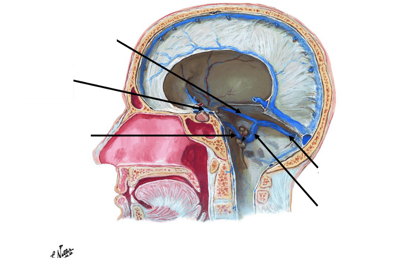
CSF drains into the lymphatic circulation, through lymph ducts contiguous to the olfactory duct as it passes through the cribriform plate.
Is CSF test painful?
You will lie on your side or sit on an exam table. A provider will clean your back and inject an anesthetic into your skin, so you won't feel pain during the procedure.
How painful is a lumbar puncture?
A lumbar puncture is where a thin needle is inserted between the bones in your lower spine. It should not be painful, but you may have a headache and some back pain for a few days.
Why CSF test is done?
CSF Analysis in Acute Demyelinating/Autoimmune Diseases CSF analysis can be very useful in diagnosis of certain diseases and can aid in progress of disease and prognostication following therapy. The demyelinating diseases include multiple sclerosis, acute disseminated encephalomyelitis, and neuromyelitis optica (NMO).
What are the 5 functions of CSF?
CSF performs vital functions including: Support; Shock absorber; Homeostasis; Nutrition; Immune function. Adult CSF volume is estimated to be 150 ml with a distribution of 125 ml within the subarachnoid spaces and 25 ml within the ventricles.
What cells are found in CSF?
The nucleated cells seen in normal adult CSF are predominantly lymphocytes and monocyte/macrophages. A rare neutrophil may be seen. An increased number of lymphocytes, monocytes, or neutrophils in CSF is termed pleocytosis. Morphologically normal cells can be seen in abnormal numbers in meningitis and inflammation.
Where is CSF found?
Summary. The cerebrospinal fluid (CSF) is contained in the brain ventricles and the cranial and spinal subarachnoid spaces. The mean CSF volume is 150 ml, with 25 ml in the ventricles and 125 ml in subarachnoid spaces.
How quickly does CSF regenerate?
The CSF is continually produced, and all of it is replaced every six to eight hours. The fluid is eventually absorbed into the veins; it leaves the cerebrospinal spaces in a variety of locations, including spaces around the spinal roots and the cranial nerves.
Where is CSF produced ventricles?
CSF is primarily produced by the choroid plexus of the ventricles (≤70% of the volume); most of it is formed by the choroid plexus of the lateral ventricles. The rest of the CSF production is the result of transependymal flow from the brain to the ventricles.
Which ventricle is CSF produced?
CSF is produced mainly by a structure called the choroid plexus in the lateral, third and fourth ventricles.
What increases CSF production?
The increased CSF production is the result of an increased activity of Na+-K+ ATPase at the choroid plexus level, which establishes a sodium gradient across the choroid epithelial cells, as well as of an elevated CBF (66).
When to use CSF?
CSF analysis may be ordered if you’ve had CNS trauma. It may also be used if you have cancer and your doctor wants to see if the cancer has spread to the CNS.
What is CSF analysis?
Cerebrospinal fluid (CSF) analysis is a way of looking for conditions that affect your brain and spine. It’s a series of laboratory tests performed on a sample of CSF. CSF is the clear fluid that cushions and delivers nutrients to your central nervous system (CNS). The CNS consists of the brain and spinal cord.
What does CSF show in MS?
CSF analysis may be done to rule out other medical conditions that have symptoms similar to MS. The fluid may also show signs that your immune system isn’t functioning normally. This can include high levels of IgG (a type of antibody) and the presence of certain proteins that form when myelin breaks down. About 85 to 90 percent of people with MS have these abnormalities in their cerebral spinal fluid.
How often is CSF replaced?
The fluid is completely replaced every few hours. In addition to delivering nutrients, CSF flows around your brain and spinal column, providing protection and carrying away waste. A CSF sample is commonly collected by performing a lumbar puncture, which is also known as a spinal tap.
What is the purpose of ventricular shunt?
A ventricular shunt or drain can collect CSF from a tube that your doctor places in your brain. This is done to release high fluid pressure.
How to measure CSF pressure?
First, the pressure inside the skull is measured using a manometer. Both high and low CSF pressure can be signs of certain conditions. Fluid samples are then taken through the needle. When fluid collection is complete, the needle is removed.
What does normal mean in spinal fluid?
Normal results mean that nothing abnormal was found in the spinal fluid. All measured levels of CSF components were found to be within normal range.
What is the CSF test for?
CSF tests for infections look at white blood cells, bacteria, and other substances in the cerebrospinal fluid. Autoimmune disorders, such as Guillain-Barré Syndrome and multiple sclerosis (MS). CSF tests for these disorders look for high levels of certain proteins in the cerebrospinal fluid. These tests are called albumin protein and igG/albumin.
What happens during a CSF analysis?
Your cerebrospinal fluid will be collected through a procedure called a spinal tap, also known as a lumbar puncture. A spinal tap is usually done in a hospital. During the procedure:
What is a cerebrospinal fluid (CSF) analysis?
Cerebrospinal fluid (CSF) is a clear, colorless liquid found in your brain and spinal cord. The brain and spinal cord make up your central nervous system. Your central nervous system controls and coordinates everything you do including, muscle movement, organ function, and even complex thinking and planning. CSF helps protect this system by acting like a cushion against sudden impact or injury to the brain or spinal cord. CSF also removes waste products from the brain and helps your central nervous system work properly.
Why do I need a CSF analysis?
You may need a CSF analysis if you have symptoms of an infection of the brain or spinal cord, or of an autoimmune disorder, such as multiple sclerosis (MS).
How long does it take for a cerebrospinal fluid to be withdrawn?
Your provider will withdraw a small amount of cerebrospinal fluid for testing. This will take about five minutes. You'll need to stay very still while the fluid is being withdrawn. Your provider may ask you to lie on your back for an hour or two after the procedure.
How is CSF produced?
Firstly, a filtered form of plasma moves from fenestrated capillaries in the choroid plexus into an interstitial space , with movement guided by a difference in pressure between the blood in the capillaries and the interstitial fluid. This fluid then needs to pass through the epithelium cells lining the choroid plexus into the ventricles, an active process requiring the transport of sodium, potassium and chloride that draws water into CSF by creating osmotic pressure. Unlike blood passing from the capillaries into the choroid plexus, the epithelial cells lining the choroid plexus contain tight junctions between cells, which act to prevent most substances flowing freely into CSF. Cilia on the apical surfaces of the ependymal cells beat to help transport the CSF.
Where is CSF found?
Cerebrospinal fluid ( CSF) is a clear, colorless body fluid found within the tissue that surrounds the brain and spinal cord of all vertebrates. It replaces the body fluid found outside the cells of all bilateral animals. The CSF is produced by specialised ependymal cells in the choroid plexuses of the ventricles of the brain, ...
What is the purpose of a CSF test?
Testing often includes observing the colour of the fluid, measuring CSF pressure , and counting and identifying white and red blood cells within the fluid; measuring protein and glucose levels; and culturing the fluid. The presence of red blood cells and xanthochromia may indicate subarachnoid hemorrhage; whereas central nervous system infections such as meningitis, may be indicated by elevated white blood cell levels. A CSF culture may yield the microorganism that has caused the infection, or PCR may be used to identify a viral cause. Investigations to the total type and nature of proteins reveal point to specific diseases, including multiple sclerosis, paraneoplastic syndromes, systemic lupus erythematosus, neurosarcoidosis, cerebral angiitis; and specific antibodies such as Aquaporin 4 may be tested for to assist in the diagnosis of autoimmune conditions. A lumbar puncture that drains CSF may also be used as part of treatment for some conditions, including idiopathic intracranial hypertension and normal pressure hydrocephalus.
What causes CSF to leak?
CSF can leak from the dura as a result of different causes such as physical trauma or a lumbar puncture, or from no known cause when it is termed a spontaneous cerebrospinal fluid leak. It is usually associated with intracranial hypotension: low CSF pressure. It can cause headaches, made worse by standing, moving and coughing, as the low CSF pressure causes the brain to "sag" downwards and put pressure on its lower structures. If a leak is identified, a beta-2 transferrin test of the leaking fluid, when positive, is highly specific and sensitive for the detection for CSF leakage. Medical imaging such as CT scans and MRI scans can be used to investigate for a presumed CSF leak when no obvious leak is found but low CSF pressure is identified. Caffeine, given either orally or intravenously, often offers symptomatic relief. Treatment of an identified leak may include injection of a person's blood into the epidural space (an epidural blood patch ), spinal surgery, or fibrin glue.
How does CSF protect the brain?
Protection: CSF protects the brain tissue from injury when jolted or hit, by providing a fluid buffer that acts as a shock absorber from some forms of mechanical injury. Prevention of brain ischemia: The prevention of brain ischemia is aided by decreasing the amount of CSF in the limited space inside the skull.
How much cerebrospinal fluid is produced in the brain?
The brain produces roughly 500 mL of cerebrospinal fluid per day , at a rate of about 25 mL an hour. This transcellular fluid is constantly reabsorbed, so that only 125–150 mL is present at any one time. CSF volume is higher on a mL/kg basis in children compared to adults.
What test is used to detect a CSF leak?
If a leak is identified, a beta-2 transferrin test of the leaking fluid, when positive, is highly specific and sensitive for the detection for CSF leakage. Medical imaging such as CT scans and MRI scans can be used to investigate for a presumed CSF leak when no obvious leak is found but low CSF pressure is identified.
How is CSF propelled?
CSF is propelled along the neuroaxis from the site of secretion to the site of absorption, mainly by the rhythmic systolic pulse wave within the choroidal arteries.
Where is CSF secreted?
CSF is secreted by the CPs located within the ventricles of the brain, with the two lateral ventricles being the primary producers. CSF flows throughout the ventricular system unidirectionally in a rostral to caudal manner.
What is CSF 2021?
Cerebrospinal fluid (CSF) is an ultrafiltrate of plasma contained within the ventricles of the brain and the subarachnoid spaces of the cranium and spine .[1] . It performs vital functions, including providing nourishment, waste removal, and protection to the brain.[2] .
What is the CSF turnover?
The reduction of CSF turnover may contribute to the accumulation of metabolites seen in aging and neurodegenerative diseases. The composition of CSF is strictly regulated, and any variation can be useful for diagnostic purposes.[1] Cerebrospinal fluid (CSF) is an ultrafiltrate of plasma contained within the ventricles of the brain and ...
What causes CSF to accumulate in the brain?
Hydrocephalus is a pathological condition in which CSF abnormally accumulates due to increased CSF production, blockage of flow, or decreased absorption. The ventricles distend to accommodate elevated CSF volumes, potentially causing damage to the brain by pressing its tissue against the boney skull. Hydrocephalus may be congenital or acquired. Blocked CSF flow throughout the ventricles is classified as non-communicating, or obstructive, hydrocephalus. The blockage is often a mass such as a tumor or an abscess located within a foramen. Because CSF secretion is constant, obstruction of flow will lead to CSF build up in front of the blockage. For example, stenosis of the cerebral aqueduct, one of the most common causes of obstructive hydrocephalus , leads to enlargement of both lateral ventricles as well as the third ventricle. If the flow of CSF becomes obstructed outside the ventricles, in either the subarachnoid space or site of absorption, it classifies as communicating, or non-obstructive, hydrocephalus.
How does CSF help the brain?
CSF assists the brain by providing protection, nourishment, and waste removal. CSF provides hydromechanical protection of the neuroaxis through two mechanisms. First, CSF acts as a shock absorber, cushioning the brain against the skull. Second, CSF allows the brain and spinal cord to become buoyant, reducing the effective weight of the brain from its normal 1,500 grams to a much lesser 50 grams. The reduction in weight lessens the force applied to the brain parenchyma and cerebral vessels during mechanical injury. Another function of CSF is to maintain homeostasis of the interstitial fluid of the brain. A stable environment for brain parenchyma is imperative for maintaining normal neuronal function.
What percent of CSF is produced by a network of modified ependymal cells?
The composition of CSF is strictly regulated, and any variation can be useful for diagnostic purposes. [1] Cellular. Seventy to eighty percent of CSF production is via a network of modified ependymal cells known as the choroid plexus (CP).[1] .
Why is CSF analysis important?
Hence analysis of CSF by various methods will help in diagnosis as well as prognostication and response to therapy . CSF analysis is particularly useful in various acute neurological conditions and helps in rapid diagnosis of the conditions and initiate therapeutic measures .
How much CSF is produced in a day?
CSF is produced at a rate of 0.2–0.7 mL per minute or 500–700 mL per day.1The main function of the CSF is to reduce buoyancy of the brain. It also supplies nutrients as well as helps in removal of various substances like amino acids, neurotransmitters, metabolic byproducts and cells.
What is cerebral fluid?
Cerebrospinal fluid (CSF) is a clear fluid circulating in the intracranial and spinal compartments. Under normal conditions, the composition of CSF remains constant. However, in various neurological disease especially in acute conditions, the composition, quantity and its pressure can be altered. By measuring the levels ...
What are the indications for LP and CSF?
Patients with suspected meningitis is one of the major indication for LP and CSF study. Meningitis can be community acquired or hospital acquired and caused by various micro organisms ranging from bacteria, virus, fungus, protozoa, etc.9,10Aseptic meningitis is a condition that needs to be distinguished from other forms of meningitis that need a CSF analysis.11Presentation of meningitis varies from acute debilitating illness or chronic symptoms as in tuberculosis. Patients suspected to have acute meningitis usually present with altered consciousness, fever and neck stiffness. The classic triad is seen only in 46% of patients. In others one or two of the signs of triad may be present. In addition patients can present with nausea, vomiting, headache and photophobia. In patients with meningoencephalitis additional clinical signs at presentation include altered sensorium, confusion, behavioural changes, seizures, focal neurological deficits.
Where is cerebral fluid secreted?
CSF is present in both the intracranial and spinal compartments. It is continuously being secreted by the choroid plexus at a constant rate inside the ventricles of the brain and circulates in the subarachnoid space of the brain ...
Can a manometer be used to measure CSF?
A manometer can be connected if CSF opening pressure measurement is planned. Color of CSF is noted and if blood stained due to traumatic puncture, it may be needed to wait for the blood to be cleared before samples are collected. Samples are usually collected in three to four test tubes each of 3–5 mL CSF for analysis.
What is the function of CSF?
The function of CSF has been one focus of mechanistic study, and the study of disease states which influence production, absorption, or CSF composition. Other than its mechanical role, CSF plays a significant role in biochemical homeostasis throughout the CNS. It has playfully been called a “nourishing liquor,” among, others for its filtering functions.[12] New techniques to analyze proteins, lipids, hormones, and microRNAs suggest the robust diversity of CSF constituents, their diffusion, and their active transport across patient cohorts, within a patient over development or time, or dependent on a disease state.[18] Some CSF biomolecules, such as secreted growth factors, neurotransmitters, morphogens, cytokines, extracellular matrix proteins, proteins involved in permeability, binding proteins, and adhesion molecules can influence production, absorption, and periventricular tissue and CSF homeostasis. Similarly, the microenvironment composition surrounding periventricular cells, and their activity, are manipulated by changes in solute transporters and CSF pathologies.[18]
What is CSF composed of?
CSF is mainly composed of water (99%), with the remaining 1% accounted for by proteins, ions, neurotransmitters, and glucose.[13] The concentration of each of these proteins, the total viscosity, and the CSF surface tension varies with pathology.[14,15] On the apical side, epithelial cells are anchored together by tight junctions which restrict the movement of these molecules; this and intercellular gap junctions give rise to the blood–CSF barrier. The composition of CSF varies from that of serum due to the differential expression of membrane-associated channels and transport proteins, ultimately resulting in the unidirectional nature of the choroidal epithelium.[1] The apical side of the epithelium is covered in microvilli that beat with the motion of the CSF, while the basolateral side is packed with folds and creases which increase the cells’ surface area, making it more suited for absorption. Compared to plasma, CSF generally contains a higher concentration of sodium, chloride, and magnesium and lower concentrations of potassium and calcium.[16] This difference is conferred by active transport from the interstitial compartment that is propagated by cytoplasmic carbonic anhydrases which produce the H+and HCO3−ions that are exchanged for Na+and Cl−by basolateral transport proteins.[1] On the apical side, active transport pumps release the ions into the ventricular spaces. Movement of water across the apical membrane has been shown to be due to the presence of aquaporin-1 (AQ-1); in fact, a study conducted by Mobasheri and Marples revealed that choroid plexus was among the tissues with the highest expression of AQ-1 in the body.[17] The method for water transport across the basolateral membrane remains to be inconclusive; many studies have seen diverging results pertaining to AQ-4, which was believed to be the prime candidate for the mechanism.
What happens if CSF is disrupted?
Obstruction anywhere in the ventricular system may result in increased intracranial pressure, which can create cascades of brain abnormalities, including cell death, inflammatory cell response, and cell shedding from the ventricular wall, or manipulation of the biochemical response of the cells.[28] Common the result of obstructive hydrocephalus include tumors, intraventricular hemorrhages, and congenital webs.[29] Blockage of the ventricular system proximally at the third ventricle, aqueduct of Sylvius, or fourth ventricle prevents absorption of CSF through the classical pathway and alternative pathways such as extracranial lymphatics. Alternatively, hydrocephalus caused by decreased absorption of CSF is commonly the result of infection, meningitis, subarachnoid hemorrhage, and trauma.[29] Infection, meningitis, and subarachnoid hemorrhage lead to an inflammatory response that causes scarring and obstruction of arachnoid granulations with a resultant decrease in CSF absorption and dysregulation of CSF homeostasis. Posttraumatic hydrocephalus is a little more complex and may be multifactorial. In the event of a patient with traumatic brain injury (TBI), ventriculomegaly may result from neuronal loss, ischemic events, or increased brain compliance after a craniectomy.[30,31] In the case of craniectomy, the dura is not closed and the bone flap may be left off for weeks to months before cranioplasty which leads to decreased resistance to CSF flow and a resultant increase in brain compliance.[20,32] Cranioplasty may result in the resolution of these changes or the alterations in brain compliance may not be readily reversed leading to the need for ventricular shunting of the excess CSF.[31,33] It is the increased intracranial pressure from the hydrocephalus not the ventriculomegaly that is the problem. Ex vacuo hydrocephalus, or enlarged ventricles due to loss of brain matter, is commonly seen secondary to brain atrophy. Common causes in addition to the neuronal loss seen from traumatic brain injury are any pathologies resulting in a significant neuronal loss such as dementia, alcoholism, and advanced age. In ex vacuo hydrocephalus, the ventricles are enlarged, but the brain compliance and CSF outflow resistance are not increased.
How is CSF absorbed?
Studies in rabbit and ovine models have revealed that CSF may also be significantly absorbed by way of cervical lymphatics. [6] CSF not reabsorbed through arachnoid granulations may reach the cervical lymphatics through two potential pathways. The first is along the subarachnoid space of exiting cranial nerves.[6,21] This provides a direct route in which CSF may be transferred from the cisterns to the to the extracranial lymphatics. The second pathway by which CSF may reach lymphatics is along the Virchow–Robin space of arteries and veins penetrating brain parenchyma.[22] The Virchow–Robin space is the potential space surrounding penetrating arteries and veins of the brain parenchyma that may vary in size depending on pathology. When CSF is not absorbed through the classical pathway, it may enter the Virchow–Robin space or be shunted to the ISF. The ISF is a compartment with the bidirectional flow to the Virchow–Robin and subarachnoid space that is believed to be mediated by AQs; but this is the topic of ongoing research. If CSF enters the ISF, it will ultimately be reabsorbed into the bloodstream, enter the Virchow–Robin space, or reenter the subarachnoid space. From the Virchow–Robin space, CSF can reenter the subarachnoid space or be reabsorbed by cervical lymphatics dependent on the forces exerted by cardiac pulsations and pulmonary respiration.
What is the effect of a choroid plexectomy on CSF?
According to the classical theory, a choroid plexectomy should significantly reduce the overall secretion of CSF, therefore providing some pressure relief in patients who have hydrocephalus. However, this is not always the outcome of the procedure; in fact, research shows that two-thirds of patients who receive the treatment should be shunted due to the recurrence of hydrocephalus.[8] In addition, Orešković and Klarica cite a study conducted by Hammock and Millhorat on rhesus monkeys in which a choroid plexectomy was performed, yet the biochemical composition of the fluid remained normal; this suggests a lesser role for the choroid plexus in molecular transport.[9] Bearing in mind nearly a century of a century of CSF research, a critical, new theory emerged in an attempt to reconcile the apparent inconsistencies of the classical theory. The new theory takes a more systematic approach, it shifts attention to the Virchow–Robin spaces (also known as perivascular spaces), which exist between where the cerebral vasculature descends from the subarachnoid space into the CNS, perforating the pia mater.[10] It is at this junction that the formation and absorption of both interstitial and CSFs occur, driven by both hydrostatic and osmotic pressure differences between the CSF circulation system and surrounding tissue. This would indicate that CSF is continually produced throughout the circulatory route and not in localized secretory organs, and any changes in the volume of CSF are influenced by the CSF osmolarity.[9] Interestingly, however, osmolarity changes can be particularly acute, where CSF volume flow can return to normal despite hypotonic serum (sink action).[11]
What is the CSF of the nervous system?
Cerebrospinal fluid (CSF) is a clear, proteinaceous fluid that exists in the surrounding spaces of mammalian central nervous systems (CNS). It is a multifaceted marvel, able to continuously support the nervous system through the lifespan of the organism. In the average adult human, there is roughly 150 mL of CSF circulating at any given moment. The ventricular portion amounts to roughly 17% of the total fluid volume, the rest of which lies in the cisterns and subarachnoid space. CSF forms at a rate of about 0.3–0.4 mL/min; translating to 18-25 mL/hour and 430–530 mL/day.[1] The classic thought is that CSF flows due to the forces generated by cardiac pulsations and pulmonary respiration. In this review, we will outline the physiology of CSF in the typical adult, as well as the pathologies associated with CSF circulation, malabsorption, and production. The existence of CSF has been known for centuries. Hippocrates was among the first to describe the fluid as water that surrounded the brain.[2] The constant production of fluid was hypothesized, but anatomists could not describe, nor pinpoint, the means of production. It was not until Cushing published his paper “Studies on the Cerebro-Spinal Fluid” in 1914 that a source for CSF, the choroid plexus, had come to be acknowledged.[3] Dandy, soon after, conducted an experiment in which he ablated the choroid plexus of one lateral ventricle in a dog, then obstructed the foramen leading into the third ventricle; he discovered that the ventricle that was ablated and evacuated of CSF would collapse, while the ventricle that was not manipulated would expand.[4] This led to the belief that the choroid plexus is the main generator of CSF. Since then, this theory has been taken as fact, and many studies conducted on the choroid plexus and CSF secretion have revolved around this concept. The original theory of CSF production views 75% of all CSF being produced by the choroid plexus epithelium, while the remaining quarter being produced by other CNS structures such as the ependymal wall, cerebral parenchyma, and interstitial fluid (ISF).[5] Recently, however, there has been criticism regarding the design of experiments conducted by Cushing and Dandy on the choroid plexuses– calling into question the veracity of what we know about CSF.
Where does CSF flow?
Next, it flows through the aqueduct of Sylvius into the fourth ventricle. From the fourth ventricle, the CSF may exit through the foramen of Lushka laterally, or the foramen of Magendie medially to the subarachnoid space. Passing through the foramen of Magendie results in filling of the spinal subarachnoid space. CSF egressing through the foramen of Lushka travels into the subarachnoid space of the cisterns and subarachnoid space overlying the cerebral cortex. The CSF from the subarachnoid space is eventually reabsorbed through outpouchings into the superior sagittal sinus (SSS) known as the arachnoid granulations. Arachnoid granulations act as an avenue for CSF reabsorption into the blood circulation through a pressure-dependent gradient.[6,20] The arachnoid granulations appear as outpouchings into the SSS due to the pressure in the subarachnoid space being greater than the venous sinus pressure (NB: direct visualization of arachnoid granulations intraoperatively would reveal the inverse).
What is the CSF?
Cerebrospinal fluid (CSF) is a clear, colorless plasma-like fluid that bathes the central nervous system (CNS). Cerebrospinal fluid circulates through a system of cavities found within the brain and spinal cord; ventricles, subarachnoid space of the brain and spinal cord and the central canal of the spinal cord. Most CSF is secreted by the specialized tissue called the choroid plexus, which is located within the lateral, third and fourth ventricles. The secretion of CSF equals its removal, so there is around 150-270 milliliters of cerebrospinal fluid within the CNS at all times.
Where is CSF obtained?
CSF is also very useful for clinical diagnosis, and its samples are usually obtained from the subarachnoid space (SAS) by lumbar puncture. This article will discuss the anatomy and functions of the cerebrospinal fluid flow. Key facts about cerebrospinal fluid flow. Secretion.
How does CSF exit the subarachnoid space?
The CSF exits the subarachnoid space by diffusing through the walls of arachnoid granulations. The arachnoid granulations provide a valvular mechanism for the flow of CSF, which allows the inflow of CSF into the bloodstream without permitting the backflow of blood into the CSF. Normally the pressure of the CSF is higher than that of the venous system, so CSF flows through the villi and granulations into the venous blood.
What is the CSF fluid?
Thus, the CSF fluid is not simply an ultrafiltrate of blood but differs from it in terms of its electrolyte, glucose and protein content.
How much CSF is produced in a day?
Cerebrospinal fluid is constantly produced at a secretion rate of 0.2-0.7 ml/min, meaning that there is 600–700 ml of newly produced CSF per day. Since the total volume of CSF averages around 150-270 mL, this means that the entire volume of CSF is replaced around 4 times per day.
Which system does CSF exit?
There are three recognized routes through which CSF exits the subarachnoid space (SAS) to enter the cerebral venous system; arachnoid granulations, minute channels that pass through the cribriform plate of ethmoid bone, and the glymphatic system.
Where is the most CSF secreted?
Most CSF is secreted by the specialized tissue called the choroid plexus, which is located within the lateral, third and fourth ventricles. The secretion of CSF equals its removal, so there is around 150-270 milliliters of cerebrospinal fluid within the CNS at all times.
What is CSF in the brain?
CSF washes out impurities from the brain, transfers nutrients and provides protective cushioning to the brain and spinal cord.
How to check for CSF leak?
If your doctor suspects a CSF leak, he or she may recommend the following tests: 1 Analysis of the nasal fluid: This test is used to detect beta-2 transferrin,a protein found almost exclusively in CSF. 2 Pledget study: This simple test involves inserting pledgets (small cotton pads) into your nose. A pledget study can confirm the presence of CSF draining into the nose from the skull. 3 CT scan: This noninvasive diagnostic imaging procedure uses a combination of X-rays and computer technology to produce detailed imaging of bones and different planes of the brain. 4 MRI scan: This method combines a large magnet, radiofrequencies and a computer to produce detailed images of organs and structures within the body. MRI scans can help determine the location and severity of a CSF leak. 5 A myelogram is a scan that involves injecting a contrasting substance into the spinal cord and using MRI or CT scans to look for tears or ruptures in the dura. 6 A cisternogram involves injecting a radioactive medium into the spinal fluid through a spinal tap and then performing CT scans. This test may help identify the origin of the CSF leak in the tissues adjacent to the spine or into the nasal cavities.
What is cerebrospinal fluid?
Cerebrospinal fluid (CSF) is a clear liquid that surrounds the brain and spinal cord. It provides a cushion for delicate brain and spinal tissue. Reduced cerebrospinal fluid, as in the case of a leak, requires immediate care by a trained expert.
What is CT scan for CSF leak?
A CT cisternogram may help identify the site of a CSF leak into the nasal cavities or mastoid bone. During this test, a contrast medium is injected into the spinal fluid through a spinal tap, and then a CT scan is performed.
How to fix a CSF leak in the ear?
Ear CSF leak closure requires cutting the skin behind the ear and removing portions of mastoid (honeycomb-like, bony tissue) to access the source of the CSF leak around the ear. Using your own tissue or a biomaterial graft, the surgeon repairs the leak and seals the surgical opening.
Why do CSF tears need to be repaired?
A CSF leak is a very serious condition, and patients who have tears in their dura with persistent CSF leaks need repair as soon as possible to reduce headache pain and the chance of meningitis.
What test is used to detect CSF leak?
If your doctor suspects a CSF leak, he or she may recommend the following tests: Analysis of the nasal fluid: This test is used to detect beta-2 transferrin,a protein found almost exclusively in CSF. Pledget study: This simple test involves inserting pledgets (small cotton pads) into your nose.
What is CSF test?
Cerebrospinal fluid (CSF) collection is a test to look at the fluid that surrounds the brain and spinal cord. CSF acts as a cushion, protecting the brain and spine from injury. The fluid is normally clear. It has the same consistency as water.
Why does CSF increase?
Increased CSF pressure may be due to increased intracranial pressure (pressure within the skull).
Why is it important to stay in position for a CSF test?
It may be uncomfortable to stay in position for the test. Staying still is important because movement may lead to injury of the spinal cord. You may be told to straighten your position slightly after the needle is in place. This is to help measure the CSF pressure. The anesthetic will sting or burn when first injected.
How to get a CSF sample?
There are different ways to get a sample of CSF. Lumbar puncture (spinal tap) is the most common method. To have the test: You will lie on your side with your knees pulled up toward the chest, and chin tucked downward. Sometimes the test is done sitting up, but bent forward.
What does it mean when you have red blood cells in your CSF?
Red blood cells in the CSF sample may be a sign of bleeding into the spinal fluid or the result of a traumatic lumbar puncture.
What is the purpose of a CSF pressure test?
This test is done to measure pressures within the CSF and to collect a sample of the fluid for further testing.
Why is my CSF glucose low?
Decreased CSF glucose may be due to hypoglycemia (low blood sugar), bacterial or fungal infection (such as meningitis ), tuberculosis, or certain other types of meningitis.
Overview
- What is a CSF analysis?
Cerebrospinal fluid (CSF) analysis is a way of looking for conditions that affect your brain and spine. It’s a series of laboratory tests performed on a sample of CSF. CSF is the clear fluid that cushions and delivers nutrients to your central nervous system (CNS). The CNS consists of the b… - CSF is produced by the choroid plexus in the brain and then reabsorbed into your bloodstream. …
A CSF sample is commonly collected by performing a lumbar puncture, which is also known as a spinal tap. An analysis of the sample involves the measurement of and examination for:
Related procedures
- Sometimes a person can’t have a lumbar puncture because of a back deformity, infection, or pos…
During a ventricular puncture, your doctor drills a hole into your skull and inserts a needle directly into one of the ventricles of your brain. - During a cisternal puncture, your doctor inserts a needle into the back of your skull.
A ventricular shunt or drain can collect CSF from a tube that your doctor places in your brain. This is done to release high fluid pressure.
Infectious diseases
- Viruses, bacteria, fungi, and parasites can all infect the CNS. Certain infections can be found by …
eastern equine encephalitis virus (EEEV)
Hemorrhaging
- Intracranial bleeding can be detected by CSF analysis. However, isolating the exact cause of bleeding may require additional scans or tests. Common causes include high blood pressure, stroke, or an aneurysm.
Immune response disorders
- CSF analysis can detect immune response disorders. The immune system can cause damage t…
Common diseases of this type include:
Tumors
- CSF analysis can detect primary tumors in the brain or spine. It can also detect metastatic canc…
CSF analysis and multiple sclerosis - CSF analysis may also be used to help diagnose multiple sclerosis (MS). MS is a chronic conditi…
CSF analysis may be done to rule out other medical conditions that have symptoms similar to MS. The fluid may also show signs that your immune system isn’t functioning normally. This can include high levels of IgG (a type of antibody) and the presence of certain proteins that form whe…
Overview
Cerebrospinal fluid (CSF) is a clear, colorless body fluid found within the tissue that surrounds the brain and spinal cord of all vertebrates.
CSF is produced by specialised ependymal cells in the choroid plexus of the ventricles of the brain, and absorbed in the arachnoid granulations. There is about 125 mL of CSF at any one time, and about 500 mL is generated every day. CSF acts as a shock absorber, cushion or buffer, providin…
Structure
There is about 125–150 mL of CSF at any one time. This CSF circulates within the ventricular system of the brain. The ventricles are a series of cavities filled with CSF. The majority of CSF is produced from within the two lateral ventricles. From here, CSF passes through the interventricular foramina to the third ventricle, then the cerebral aqueduct to the fourth ventricle. From the fourth ventricle, the fluid passes into the subarachnoid space through four openings – the central canal o…
Development
At around the third week of development, the embryo is a three-layered disc, covered with ectoderm, mesoderm and endoderm. A tube-like formation develops in the midline, called the notochord. The notochord releases extracellular molecules that affect the transformation of the overlying ectoderm into nervous tissue. The neural tube, forming from the ectoderm, contains CSF prior to the development of the choroid plexuses. The open neuropores of the neural tube close after the …
Physiology
CSF serves several purposes:
1. Buoyancy: The actual mass of the human brain is about 1400–1500 grams; however, the net weight of the brain suspended in CSF is equivalent to a mass of 25-50 grams. The brain therefore exists in neutral buoyancy, which allows the brain to maintain its density without being impaired by its own weight, which would cut off blood supply and kill neurons in the lower sections without C…
Clinical significance
CSF pressure, as measured by lumbar puncture, is 10–18 cmH2O (8–15 mmHg or 1.1–2 kPa) with the patient lying on the side and 20–30 cmH2O (16–24 mmHg or 2.1–3.2 kPa) with the patient sitting up. In newborns, CSF pressure ranges from 8 to 10 cmH2O (4.4–7.3 mmHg or 0.78–0.98 kPa). Most variations are due to coughing or internal compression of jugular veins in the neck. When lying down, the CSF pressure as estimated by lumbar puncture is similar to the intracrania…
History
Various comments by ancient physicians have been read as referring to CSF. Hippocrates discussed "water" surrounding the brain when describing congenital hydrocephalus, and Galen referred to "excremental liquid" in the ventricles of the brain, which he believed was purged into the nose. But for some 16 intervening centuries of ongoing anatomical study, CSF remained unmentioned in the literature. This is perhaps because of the prevailing autopsy technique, whic…
Other animals
During phylogenesis, CSF is present within the neuraxis before it circulates. The CSF of Teleostei fish is contained within the ventricles of the brains, but not in a nonexistent subarachnoid space. In mammals, where a subarachnoid space is present, CSF is present in it. Absorption of CSF is seen in amniotes and more complex species, and as species become progressively more complex, the system of absorption becomes progressively more enhanced, and the role of spinal epidural …
See also
• Neuroglobin
• Pandy's test
• Reissner's fiber
• Syrinx (medicine)