Where and how is CSF produced?
CSF is produced mainly by a structure called the choroid plexus in the lateral, third and fourth ventricles. CSF flows from the lateral ventricle to the third ventricle through the interventricular foramen (also called the foramen of Monro).
How is CSF produced and circulated?
Abstract. According to the traditional understanding of cerebrospinal fluid (CSF) physiology, the majority of CSF is produced by the choroid plexus, circulates through the ventricles, the cisterns, and the subarachnoid space to be absorbed into the blood by the arachnoid villi.
How much CSF does the brain produce per day?
between 400 to 600 ml per dayCerebrospinal fluid secretion in adults CSF secretion in adults varies between 400 to 600 ml per day, depending on the subject and the method used to study CSF secretion.
What drains CSF from the brain?
The choroid plexus mostly synthesizes CSF. Arachnoid granulations are responsible for CSF resorption; they drain CSF into the dural venous sinuses. CSF drains into the lymphatic circulation, through lymph ducts contiguous to the olfactory duct as it passes through the cribriform plate.
What is CSF explain about its function and circulation?
According to the traditional understanding of cerebrospinal fluid (CSF) physiology, the majority of CSF is produced by the choroid plexus, circulates through the ventricles, the cisterns, and the subarachnoid space to be absorbed into the blood by the arachnoid villi.
Where does the cerebrospinal fluid CSF circulate quizlet?
CSF flows from the subarachnoid space at the base of the brain rostrally over the cerebral hemispheres or down the spinal cord.
How long does it take CSF to circulate?
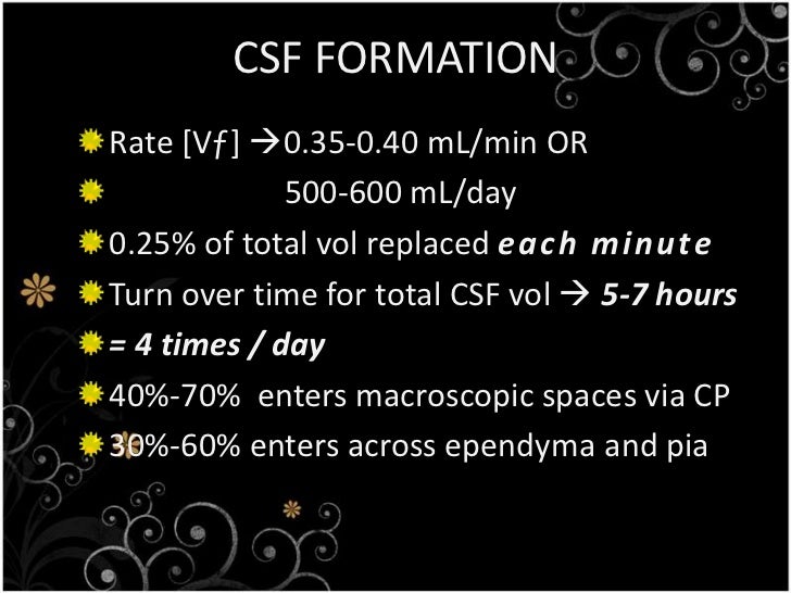
CSF forms at a rate of about 0.3–0.4 mL/min; translating to 18-25 mL/hour and 430–530 mL/day. [1] The classic thought is that CSF flows due to the forces generated by cardiac pulsations and pulmonary respiration.
Where is CSF produced quizlet?
CSF is produced by the choroid plexus in the ventricles. CSF flows from the 3rd ventricle through the cerebral aqueduct into the 4th ventricle. CSF then flows into the subarachnoid space by passing through the paired lateral apertures or the single median aperture and into the central canal of the spinal cord.
Where is CSF obtained?
CSF is also very useful for clinical diagnosis, and its samples are usually obtained from the subarachnoid space (SAS) by lumbar puncture. This article will discuss the anatomy and functions of the cerebrospinal fluid flow. Key facts about cerebrospinal fluid flow. Secretion.
What is the CSF?
Cerebrospinal fluid (CSF) is a clear, colorless plasma-like fluid that bathes the central nervous system (CNS). Cerebrospinal fluid circulates through a system of cavities found within the brain and spinal cord; ventricles, subarachnoid space of the brain and spinal cord and the central canal of the spinal cord. Most CSF is secreted by the specialized tissue called the choroid plexus, which is located within the lateral, third and fourth ventricles. The secretion of CSF equals its removal, so there is around 150-270 milliliters of cerebrospinal fluid within the CNS at all times.
How does CSF exit the subarachnoid space?
The CSF exits the subarachnoid space by diffusing through the walls of arachnoid granulations. The arachnoid granulations provide a valvular mechanism for the flow of CSF, which allows the inflow of CSF into the bloodstream without permitting the backflow of blood into the CSF. Normally the pressure of the CSF is higher than that of the venous system, so CSF flows through the villi and granulations into the venous blood.
What is the CSF fluid?
Thus, the CSF fluid is not simply an ultrafiltrate of blood but differs from it in terms of its electrolyte, glucose and protein content.
How much CSF is produced in a day?
Cerebrospinal fluid is constantly produced at a secretion rate of 0.2-0.7 ml/min, meaning that there is 600–700 ml of newly produced CSF per day. Since the total volume of CSF averages around 150-270 mL, this means that the entire volume of CSF is replaced around 4 times per day.
Which system does CSF exit?
There are three recognized routes through which CSF exits the subarachnoid space (SAS) to enter the cerebral venous system; arachnoid granulations, minute channels that pass through the cribriform plate of ethmoid bone, and the glymphatic system.
Where is the most CSF secreted?
Most CSF is secreted by the specialized tissue called the choroid plexus, which is located within the lateral, third and fourth ventricles. The secretion of CSF equals its removal, so there is around 150-270 milliliters of cerebrospinal fluid within the CNS at all times.
What is the CSF produced by?
The cerebrospinal fluid (CSF) is produced from arterial blood by the choroid plexuses of the lateral and fourth ventricles by a combined process of diffusion, pinocytosis and active transfer. A small amount is also produced by ependymal cells.The CSF cushions the brain and serves also as a “sink” that receives products generated by brain catabolism and synaptic function. The choroid plexus consists of tufts of capillaries with thin fenestrated endothelial cells. These are covered by modified ependymal cells with bulbous microvilli. The epithelial cells of the choroid plexus have tight junctions and form the blood-CSF barrier (BCSFB), which controls the movement of water and solutes into the CSF. The apical surface of choroid plexus epithelial cells contains Aquaporin1 (AQP1), a membrane protein (water channel) that facilitates movement of water across cell membranes. The choroid plexus epithelial cells contain also carbonic anhydrase, a hydrolytic metalloenzyme that is involved in CSF secretion.The total volume of CSF in the adult ranges from140 to 270 ml. The volume of the ventricles is about 25 ml. CSF is produced at a rate of 0.2 - 0.7 ml per minute or 600-700 ml per day. The circulation of CSF is aided by the pulsations of the choroid plexus and by the motion of the cilia of ependymal cells. CSF is absorbed across the arachnoid villi into the venous circulation and a significant amount probably also drains through the cribriform plate and spinal canal into cervical and spinal lymph nodes. The arachnoid villi act as one-way valves between the subarachnoid space and the dural sinuses. The rate of absorption correlates with the CSF pressure. CSF acts as a cushion that protects the brain from shocks and supports the venous sinuses (primarily the superior sagittal sinus, opening when CSF pressure exceeds venous pressure). It also plays an important role in the homeostasis and metabolism of the central nervous system.
Where does CSF go in the body?
CSF is absorbed across the arachnoid villi into the venous circulation and a significant amount probably also drains through the cribriform plate and spinal canal into cervical and spinal lymph nodes. The arachnoid villi act as one-way valves between the subarachnoid space and the dural sinuses.
How does blood stain CSF?
Blood: Blood may be spilled into the CSF by accidental puncture of a leptomeningeal vein during entry of the LP needle. Such blood stains the fluid that is drawn initially and clears gradually. If it does not clear, blood indicates subarachnoid hemorrhage. Erythrocytes from subarachnoid hemorrhage are cleared in 3 to 7 days. A few neutrophils and mononuclear cells may also be present as a result of meningeal irritation. Xanthochromia (blonde color) of the CSF following subarachnoid hemorrhage is due to oxyhemoglobin which appears in 4 to 6 hours and bilirubin which appears in two days. Xanthochromia may also be seen with hemorrhagic infarcts, brain tumors, and jaundice.
What is the CSF secretion rate of choroid plexus epithelial cells?
The volume of the ventricles is about 25 ml. CSF is produced at a rate of 0.2 - 0.7 ml per minute or 600-700 ml per day.
What is the role of CSF in the brain?
CSF acts as a cushion that protects the brain from shocks and supports the venous sinuses (primarily the superior sagittal sinus, opening when CSF pressure exceeds venous pressure). It also plays an important role in the homeostasis and metabolism of the central nervous system.
What is the protein concentration in lumbar CSF?
CSF from the lumbar region contains 15 to 45 mg/dl protein (lower in childen) and 50-80 mg/dl glucose (two-thirds of blood glucose). Protein concentration in cisternal and ventricular CSF is lower. Normal CSF contains 0-5 mononuclear cells. The CSF pressure, measured at lumbar puncture (LP), is 100-180 mm of H2O (8-15 mm Hg) with the patient lying on the side and 200-300 mm with the patient sitting up.
Which cell controls the movement of water and solutes into the CSF?
The epithelial cells of the choroid plexus have tight junctions and form the blood-CSF barrier (BCSFB), which controls the movement of water and solutes into the CSF. The apical surface of choroid plexus epithelial cells contains Aquaporin1 (AQP1), a membrane protein (water channel) that facilitates movement of water across cell membranes.
How is CSF propelled?
CSF is propelled along the neuroaxis from the site of secretion to the site of absorption, mainly by the rhythmic systolic pulse wave within the choroidal arteries.
Where is CSF secreted?
CSF is secreted by the CPs located within the ventricles of the brain, with the two lateral ventricles being the primary producers. CSF flows throughout the ventricular system unidirectionally in a rostral to caudal manner.
What is CSF 2021?
Cerebrospinal fluid (CSF) is an ultrafiltrate of plasma contained within the ventricles of the brain and the subarachnoid spaces of the cranium and spine .[1] . It performs vital functions, including providing nourishment, waste removal, and protection to the brain.[2] .
What is the CSF turnover?
The reduction of CSF turnover may contribute to the accumulation of metabolites seen in aging and neurodegenerative diseases. The composition of CSF is strictly regulated, and any variation can be useful for diagnostic purposes.[1] Cerebrospinal fluid (CSF) is an ultrafiltrate of plasma contained within the ventricles of the brain and ...
What causes CSF to accumulate in the brain?
Hydrocephalus is a pathological condition in which CSF abnormally accumulates due to increased CSF production, blockage of flow, or decreased absorption. The ventricles distend to accommodate elevated CSF volumes, potentially causing damage to the brain by pressing its tissue against the boney skull. Hydrocephalus may be congenital or acquired. Blocked CSF flow throughout the ventricles is classified as non-communicating, or obstructive, hydrocephalus. The blockage is often a mass such as a tumor or an abscess located within a foramen. Because CSF secretion is constant, obstruction of flow will lead to CSF build up in front of the blockage. For example, stenosis of the cerebral aqueduct, one of the most common causes of obstructive hydrocephalus , leads to enlargement of both lateral ventricles as well as the third ventricle. If the flow of CSF becomes obstructed outside the ventricles, in either the subarachnoid space or site of absorption, it classifies as communicating, or non-obstructive, hydrocephalus.
How does CSF help the brain?
CSF assists the brain by providing protection, nourishment, and waste removal. CSF provides hydromechanical protection of the neuroaxis through two mechanisms. First, CSF acts as a shock absorber, cushioning the brain against the skull. Second, CSF allows the brain and spinal cord to become buoyant, reducing the effective weight of the brain from its normal 1,500 grams to a much lesser 50 grams. The reduction in weight lessens the force applied to the brain parenchyma and cerebral vessels during mechanical injury. Another function of CSF is to maintain homeostasis of the interstitial fluid of the brain. A stable environment for brain parenchyma is imperative for maintaining normal neuronal function.
What percent of CSF is produced by a network of modified ependymal cells?
The composition of CSF is strictly regulated, and any variation can be useful for diagnostic purposes. [1] Cellular. Seventy to eighty percent of CSF production is via a network of modified ependymal cells known as the choroid plexus (CP).[1] .
Where is CSF formed?
Multisource evidence indicates that 70% to 80% of CSF is formed by the plexuses in the lateral, third, and fourth ventricles. Human CSF elaboration is mechanistically similar to that in many mammalian species. Extrachoroidal formation of a CSF-like fluid occurs at the cerebral capillary wall,5but it is less efficient compared with choroid plexus generation. A lavish turnover of fluid at the blood-CSF interface (choroidal epithelium) is engendered by a high blood flow to the plexus, substantial activities of Na+,K+-ATPase6,7and carbonic anhydrase,8,9and a plethora of ion transporters. Stable composition is the hallmark of CSF. Extracellular ion stability is essential to neurotransmission.
How does CSF affect the brain?
Because CSF dynamics have an impact on brain metabolism , it is important to assess optimal CSF formation rate in health. Adult humans normally form CSF at about 0.35 mL/min. 1,2 Nocturnally elevated CSF production 13 may relate to altered cerebral metabolism during sleep. CSF formation also fluctuates in disease. 14 Neurosurgeons face pathophysiologic situations in which the choroid plexus forms too much or not enough fluid. With hypersecreting choroid plexus papillomas, 15,16 surgical excision often reduces ICP. With hyposecreting choroid plexus, as in normal-pressure hydrocephalus (NPH) and Alzheimer’s disease (AD), the stagnated CSF turnover rate 17,18 may contribute to cerebral dysfunction. Disease-modified fluid formation by the choroid plexus can thus harm CSF dynamics and the extracellular environment of neurons. 11 Accordingly, imbalanced CSF formation necessitates surgical or pharmacologic remediation.
What is the watery medium of CSF?
CSF is 99% water. The watery medium of CSF enables multiple buffering, distributive, and excretory functions. 1Therefore, it is important to thoroughly characterize water movement across the blood-CSF interface. After Na+, Cl−, and HCO3−transport into CSF, water chases these osmotically active ions into the ventricles by diffusing down its chemical potential gradient through aquaporin 1 (AQP1) channels in the apical membrane.37AQP1 channel involvement in CSF formation is deduced from AQP1 knockout mice displaying substantially reduced fluid movement into the ventricles.38As a result, ICP is lowered.39Transcellular water diffusion across the choroid plexus is potentially a drug target in modulating CSF dynamics.
How do hydrophilic diuretics reduce CSF?
The strategy is to suppress fluid formation without altering CSF composition by interfering with choroid plexus ion transporters at apical or basolateral membranes. One desirable clinical outcome is to lower ICP pressure by decreasing fluid input to the ventricles. To interpret hydrophilic drug effects, a significant factor is whether the inhibiting agent is administered on the blood side (intravenously or intraperitoneally) or the CSF side (intracerebroventricularly). Tight junctions between choroid epithelial cells limit penetration of water-soluble agents by diffusion across the blood-CSF barrier. Hydrophilic agents such as ouabain, a potent inhibitor of apical Na+,K+-ATPase, therefore do not inhibit CSF formation when they are presented on the blood side of the barrier.7Drug access to the transporter target is critical.
What is cerebrospinal fluid?
Continually generated and flowing cerebrospinal fluid (CSF) is vital for brain homeostasis. 1 Day and night, the choroid plexus churns out fluid into the ventricles. CSF formation is about 0.4 mL/min per gram of choroid plexus. Nascent CSF is more than a passive ultrafiltrate of plasma. 2 Active secretion by choroidal epithelium is exquisitely modulated so that intracranial pressure (ICP) is stable if CSF absorption is normal. A tenth of the choroidal blood flow of about 4 mL/min per gram 3,4 becomes new CSF in the ventricular spaces.
What is the main anion in CSF?
As the main anion in CSF secretion, Cl−is actively transported across the basolateral membrane in exchange for cellular HCO3−.31This pulls plasma Cl−into the epithelium for accumulation above electrochemical equilibrium.32Under some conditions, intraepithelial Cl−diffuses into CSF through the efflux arm of the Na+-K+-Cl−cotransporter.33However, the downhill diffusion of Cl−into CSF by way of apical Cl−channels is likely to be the main pathway by which Cl−accesses the ventricles to sustain fluid formation.26
Which plexus forms cerebral fluid?
Mechanisms of Cerebrospinal Fluid Formation by Choroid Plexus
What is CSF in the brain?
CSF washes out impurities from the brain, transfers nutrients and provides protective cushioning to the brain and spinal cord.
How to check for CSF leak?
If your doctor suspects a CSF leak, he or she may recommend the following tests: 1 Analysis of the nasal fluid: This test is used to detect beta-2 transferrin,a protein found almost exclusively in CSF. 2 Pledget study: This simple test involves inserting pledgets (small cotton pads) into your nose. A pledget study can confirm the presence of CSF draining into the nose from the skull. 3 CT scan: This noninvasive diagnostic imaging procedure uses a combination of X-rays and computer technology to produce detailed imaging of bones and different planes of the brain. 4 MRI scan: This method combines a large magnet, radiofrequencies and a computer to produce detailed images of organs and structures within the body. MRI scans can help determine the location and severity of a CSF leak. 5 A myelogram is a scan that involves injecting a contrasting substance into the spinal cord and using MRI or CT scans to look for tears or ruptures in the dura. 6 A cisternogram involves injecting a radioactive medium into the spinal fluid through a spinal tap and then performing CT scans. This test may help identify the origin of the CSF leak in the tissues adjacent to the spine or into the nasal cavities.
What is cerebrospinal fluid?
Cerebrospinal fluid (CSF) is a clear liquid that surrounds the brain and spinal cord. It provides a cushion for delicate brain and spinal tissue. Reduced cerebrospinal fluid, as in the case of a leak, requires immediate care by a trained expert.
What is CT scan for CSF leak?
A CT cisternogram may help identify the site of a CSF leak into the nasal cavities or mastoid bone. During this test, a contrast medium is injected into the spinal fluid through a spinal tap, and then a CT scan is performed.
How to fix a CSF leak in the ear?
Ear CSF leak closure requires cutting the skin behind the ear and removing portions of mastoid (honeycomb-like, bony tissue) to access the source of the CSF leak around the ear. Using your own tissue or a biomaterial graft, the surgeon repairs the leak and seals the surgical opening.
Why do CSF tears need to be repaired?
A CSF leak is a very serious condition, and patients who have tears in their dura with persistent CSF leaks need repair as soon as possible to reduce headache pain and the chance of meningitis.
What test is used to detect CSF leak?
If your doctor suspects a CSF leak, he or she may recommend the following tests: Analysis of the nasal fluid: This test is used to detect beta-2 transferrin,a protein found almost exclusively in CSF. Pledget study: This simple test involves inserting pledgets (small cotton pads) into your nose.
Where is CSF formed?
CSF is formed in the choroid plexus by both filtration and active transport. In normal adults, the CSF volume is 90 to 200 mL [ 1 ]; approximately 20 percent of the CSF is contained in the ventricles; the rest is contained in the subarachnoid space in the cranium and spinal cord. The normal rate of CSF production is approximately 20 mL per hour.
Why is cerebrospinal fluid important?
Examination of the cerebrospinal fluid (CSF) may provide critically important diagnostic information in a number of infectious and noninfectious medical conditions. Knowledge of the normal physiology and pathophysiology of CSF production and flow is useful in interpreting such test results.
