What tests are used to diagnose diffuse axonal injury?
CT scans may results in false negatives, so can’t be relied on to give definitive results when it comes to diffuse axonal injury. Evoked Potentials – Commonly called the SSEP, BAER, and VEP, these tests look at the visual, auditory, and sensory pathways in the brain.
What are the symptoms of diffuse axonal injury?
The shearing can also release chemicals which can contribute to additional brain injury. The main symptom of diffuse axonal injury is lack of consciousness, which can last up to six hours or more.
Is open surgery an option for patients with diffuse axonal injury?
Surgery is not an option for those who have sustained a diffuse axonal injury. If the patient has sustained a mild or moderate diffuse axonal injury, the rehabilitation phase will follow once the patient is stabilized and awake.
Does CT head show diffuse axonal injury?
Radiographically, computed tomography (CT) head findings of small punctate hemorrhages to white matter tracts can indicate diffuse axonal injury in the setting of an appropriate clinical presentation. Overall, CT head has a low yield in detecting diffuse axonal injury-related injuries.

Does diffuse axonal injury show up on MRI?
Magnetic resonance imaging (MRI) is the preferred examination for DAI (particularly with gradient-echo sequences), although CT scanning may demonstrate findings suggestive of DAI and is more practical and available. Studies have indicated that MRI can play a role in predicting the length of coma in DAI patients.
What are the symptoms of diffuse axonal injury?
Diffuse Axonal Injury SymptomsCognitive deficits such as difficulty with memory, problem solving, and decision making. ... Confusion may occur as a result of cognitive deficits.Severe headaches may occur as a result of inflammation of the brain.More items...•
How do you get diffuse axonal injury?
A DAI is caused by shaking or strong rotation of the head by physical forces, such as with a car crash. Injury occurs because the unmoving brain lags behind the movement of the skull, causing nerve structures to tear. The tearing of the nerve tissue disrupts the brain's regular communication and chemical processes.
Is diffuse axonal injury curable?
In select cases, a course of steroids will be given to reduce the swelling. There is no surgery available to people who have sustained a DAI. If the injury is severe, there is a likelihood of a vegetative state or even death. But if the DAI is mild to moderate, rehabilitation is possible.
Does anoxic brain injury show on MRI?
Magnetic resonance imaging (MRI) is more sensitive than computed tomography at detecting stroke in the early phase, subtle abnormalities related to anoxic/hypoxic encephalopathy, and diffuse axonal injury (DAI) in patients with TBI.
How is brain damage diagnosed?
The extent and effect of brain damage is determined by a neurological exam, neuroimaging testing such as MRI or CT scans, and neuropsychological assessments.
Can MRI show old brain injury?
MRI might show brain atrophy long after the injury, which results when injured or dead brain tissue is reabsorbed after TBI. Since injured brain tissue might not fully recover after TBI, doctors might detect TBI-related changes many years following an injury.
Can you fully recover from a DAI?
Mild DAI was observed in 44.9% of the patients and severe DAI in 35.9%. Six months after trauma, 30.8% of the patients had died, and 45.1% had shown full recovery according to the GOS-E.
Can a fall cause diffuse axonal injury?
Abstract. 82 cases of diffuse axonal injury were found at necropsy in 635 patients with fatal nonmissile head injuries. 13 of these injuries were attributable to falls, and in all the patients fell from a considerable height.
What is a grade 3 diffuse axonal injury?
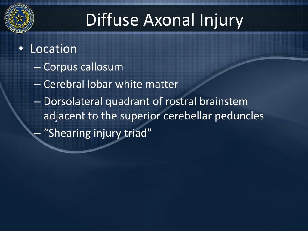
grade 3: focal lesions in both the corpus callosum and dorsolateral quadrant of the rostral brainstem, in addition to diffuse axonal damage.
What is DAI diagnosis?
Briefly, a diagnosis of DAI is made when a patient has experienced a LOC of more than 6 hours after head trauma, has shown related clinical manifestations, and has DAI lesions on a conventional brain MRI [4,8,10].
Can the brain repair itself after injury?
And the answer is yes. The brain is incredibly resilient and possesses the ability to repair itself through the process of neuroplasticity. This phenomenon is the reason why many brain injury survivors can make astounding recoveries.
What Are The Causes of Diffuse Axonal Injury
Diffuse axonal injury isn’t the result of a blow to the head. Instead, it results from the brain moving back and forth in the skull as a result of...
Symptoms of Diffuse Axonal Injury
The main symptom of diffuse axonal injury is lack of consciousness, which can last up to six hours or more. A person with a mild or moderate diffus...
Diagnosing Diffuse Axonal Injury
If the patient has sustained a mild diffuse axonal injury and is conscious, he or she will be asked a variety of questions including how the injury...
Treatment of Diffuse Axonal Injury
Immediate measures will be taken to reduce swelling inside the brain, which can cause additional damage. In most cases, a course of steroids or oth...
Prognosis of Diffuse Axonal Injury
It is thought that diffuse axonal injury can occur in just about every level of severity, with concussion thought to be one of the milder forms. In...
What are the symptoms of a brain injury?
They can include: disorientation or confusion. headache. nausea or vomiting. drowsiness or fatigue. trouble sleeping. sleeping longer than normal. loss of balance or dizziness.
Can you have surgery for a DAI?
There is no surgery available to people who have sustained a DAI. If the injury is severe, there is a likelihood of a vegetative state or even death. But if the DAI is mild to moderate, rehabilitation is possible. A recovery program will depend on the individual, but may include: speech therapy. physical therapy.
Can a CT scan detect traumatic brain injury?
The changes in the brain are often very tiny and can be difficult to detect using CT or MRI scans. It is one of the most common types of traumatic brain injury and also one of the most devastating.
Is DAI a serious injury?
Outlook. DAI is a serious but common type of traumatic brain injury. It can be fatal, but it is also possible to regain consciousness after a DAI. For those who recover, intensive rehabilitation will be needed. Last medically reviewed on October 19, 2017.
Where is DAI in MRI?
DAI can be inferred when MRI, especially SWI or GRE sequences that are exquisitely sensitive to paramagnetic blood products, shows several small 1-15 mm ovoid or linear regions of susceptibility artifact (i.e., hemorrhage) at the grey-white matter junction (most commonly the frontal and temporal lobes), in the corpus callosum, and in more severe cases in the brainstem and pons, surrounded by FLAIR hyperintensity
How long does it take for edema to resolve on a SWI?
Surrounding edema increases in the first few days and usually resolves in 3 months on FLAIR images, whereas hemorrhagic changes on SWI may take 12 months to resolve; this is to be expected as edema is faster to resolve than hemorrhage. Pearls.
Why is diffuse axonal injury difficult to detect?
Because most diffuse axonal injuries result in microscopic tears, damage can be difficult to detect with imaging.
What is diffuse axonal injury?
Generally, diffuse axonal injuries are a severe type of traumatic brain injury that affects multiple areas of the brain. As a result, various connections within the brain may become disrupted, leading to a wide range of secondary effects. Fortunately, many individuals with diffuse axonal injuries have the potential to recover affected functions and improve their quality of life.
What is the term for the damage caused by the brain's axons?
Diffuse axonal injury occurs when the brain quickly moves inside the skull as a result of a traumatic injury, like a car accident. As the brain repeatedly hits against the inside of the skull from the force of the injury, the long connecting fibers in the brain, known as axons, can tear and potentially cause severe damage.
What causes diffuse axonal shearing?
Some of the most common causes of diffuse axonal injury include: Car accidents. Sports injuries. Falls. Domestic abuse. The severity of the traumatic event is related to a higher potential for axonal shearing.
What can a peech therapist do for a diffuse axonal injury?
In addition, s peech therapists can teach you various social communication skills that you might lack after DAI, such as learning how to begin and end a conversation.
What is the presence of neurological reflexes?
The presence of neurological reflexes is typically viewed as a positive sign of diffuse axonal injury recovery . Following a DAI, physicians may look for:
How does cognitive behavioral therapy help with axonal brain injury?
Cognitive-behavioral therapy helps individuals understand what is causing their negative thoughts or behaviors and teaches them more effective ways to cope.
What is diffuse axonal injury?
A diffuse axonal injury falls under the category of a diffuse brain injury. This means that instead of occurring in a specific area, like a focal brain injury, it occurs over a more widespread area. In addition to being one of the most common types of brain injuries, it’s also one of the most devastating. As a matter of fact, severe diffuse axonal ...
How long does a diffuse axonal injury last?
Symptoms of Diffuse Axonal Injury. The main symptom of diffuse axonal injury is lack of consciousness, which can last up to six hours or more. A person with a mild or moderate diffuse axonal injury who is conscious may also show other signs of brain damage, depending upon which area of the brain is most affected.
What causes swelling in the brain after a head injury?
This causes the lesions that are responsible for unconsciousness, as well as the vegetative state that occurs after a severe head injury. A diffuse axonal injury also causes brain cells to die, which cause swelling in the brain. This increased pressure in the brain can cause decreased blood flow to the brain, as well as additional injury.
What is a traumatic brain injury?
A traumatic brain injury (TBI) occurs when there is a “bump, blow, or jolt to the head” that causes issues with the functions of the ...
What is a CT scan?
CT Scan – This test uses an x-ray machine and a computer monitor to show detailed images of the interior of the brain. CT scans may results in false negatives, so can’t be relied on to give definitive results when it comes to diffuse axonal injury.
What is the test called for evoked potentials?
Evoked Potentials – Commonly called the SSEP, BAER, and VEP, these tests look at the visual, auditory, and sensory pathways in the brain.
Can a concussion be a diffuse axonal injury?
It is thought that diffuse axonal injury can occur in just about every level of severity, with concussion thought to be one of the milder forms. In mild to moderate forms of diffuse axonal injury, recovery is possible, with the mildest forms of diffuse axonal injury often resulting in few if any long-term issues.
What causes axonal separation?
While it was once thought that the main cause of axonal separation was tearing due to mechanical forces during the trauma , it is now understood that axons are not typically torn upon impact; rather, secondary biochemical cascades, which occur in response to the primary injury (which occurs as the result of mechanical forces at the moment of trauma) and take place hours to days after the initial injury, are largely responsible for the damage to axons.
What happens when you stretch your axons?
Though the processes involved in secondary brain injury are still poorly understood, it is now accepted that stretching of axons during injury causes physical disruption to and proteolytic degradation of the cytoskeleton. It also opens sodium channels in the axolemma, which causes voltage-gated calcium channels to open and Ca 2+ to flow into the cell. The intracellular presence of Ca 2+ triggers several different pathways, including activating phospholipases and proteolytic enzymes, damaging mitochondria and the cytoskeleton, and activating secondary messengers, which can lead to separation of the axon and death of the cell.
What is the term for axons that are white?
Immediate disconnection of axons could be observed in severe brain injury, but the major damage of DAI is delayed secondary axon disconnections slowly developed over an extended time course. Tracts of axons, which appear white due to myelination, are referred to as white matter. Lesions in both grey and white matters are found in postmortem brains in CT and MRI exams.
How long does it take for a wallerian degeneration to occur?
When the axon is transected, Wallerian degeneration, in which the part of the axon distal to the break degrades, takes place within one to two days after injury. The axolemma disintegrates, myelin breaks down and begins to detach from cells in an anterograde direction (from the body of the cell toward the end of the axon), and nearby cells begin phagocytic activity, engulfing debris.
How long does it take for a brain injury to heal?
Mitochondria, dendrites, and parts of the cytoskeleton damaged in the injury have a limited ability to heal and regenerate, a process which occurs over 2 or more weeks. After the injury, astrocytes can shrink, causing parts of the brain to atrophy.
Is a concussion a diffuse axonal injury?
DAI can occur across the spectrum of traumatic brain injury (TBI) severity, wherein the burden of injury increases from mild to severe. Concussion may be a milder type of diffuse axonal injury.
Can a DAI be detected on a CT scan?
However, there are characteristics typical of DAI that may or may not show up on a CT scan. Diffuse injury has more microscopic injury than macroscopic injury and is difficult to detect with CT and MRI, but its presence can be inferred when small bleeds are visible in the corpus callosum or the cerebral cortex. MRI is more useful than CT for detecting characteristics of diffuse axonal injury in the subacute and chronic time frames. Newer studies such as Diffusion Tensor Imaging are able to demonstrate the degree of white matter fiber tract injury even when the standard MRI is negative. Since axonal damage in DAI is largely a result of secondary biochemical cascades, it has a delayed onset, so a person with DAI who initially appears well may deteriorate later. Thus injury is frequently more severe than is realized, and medical professionals should suspect DAI in any patients whose CT scans appear normal but who have symptoms like unconsciousness.