
Each myelin sheath is formed by the concentric wrapping of an oligodendrocyte (CNS) or Schwann cell (PNS) process (a limb-like extension from the cell body) around the axon. Myelin Myelin is a lipid-rich substance formed in the central nervous system by glial cells called oligodendrocytes, and in the peripheral nervous system by Schwann cells. Myelin insulates nerve cell axons to increase the speed at which information travels from one nerve cell body to another or, for example, from a nerve cell body to a muscle. The myelinated axon can be likened to an electrical wire with insulati…Myelin
What does myelin actually do?
Myelin sheath is the covering of nerves and the axons play an important role in conveying nervous stimuli. Regaining their working capacity Nerve Renew makes the nervous system active like before. The root cause of neuropathy is nerve damage and it shows up the symptoms like pain, tingling, and numbness.
How can I naturally repair myelin?
Top 55 Ways to Increase Myelin Naturally (and surprising facts)
- Gotu Kola Gotu kola helps rats make a more rapid functional recovery and a greater numbers of myelinated axons following nerve damage ( R ).
- Uridine Uridine can help treat myelin sheath lesions ( R ).
- Ashwagandha Ashwagandha has an active component called withanoside IV. ...
- SAMe and Methylation S-adenosylmethionine (SAMe) helps regulate DNA methylation. ...
How long does it take for myelin to regenerate?
We find restoration of the normal number of oligodendrocytes and robust remyelination approximately two weeks after induction of cell ablation, whereby myelinated axon number is restored to control levels. Remarkably, we find that myelin sheaths of normal length and thickness are regenerated during this time.
What is myelination and why is it so important?
Myelin enables nerve cells to transmit information faster and allows for more complex brain processes. The myelination process is vitally important to healthy central nervous system functioning. What does myelination mean? Myelination: The formation of the myelin sheath around a nerve fiber. Also known as myelinization.
What is myelin and how is it formed?
Myelin is an insulating layer, or sheath that forms around nerves, including those in the brain and spinal cord. It is made up of protein and fatty substances. This myelin sheath allows electrical impulses to transmit quickly and efficiently along the nerve cells. If myelin is damaged, these impulses slow down.
How is myelin formed in the central nervous system?
Each myelin sheath is formed by the concentric wrapping of an oligodendrocyte (CNS) or Schwann cell (PNS) process (a limb-like extension from the cell body) around the axon. Myelin reduces the capacitance of the axonal membrane.
What is the process of myelination?
Myelination is the formation of a myelin sheath. Myelin sheaths are made of myelin, and myelin is produced by different types of neuroglia: oligodendrocytes and Schwann cells, where oligodendrocytes myelinate axons in the central nervous system, and Schwann cells myelinate axons in the peripheral nervous system.
What cells create myelin sheath?
The myelin sheath is a greatly extended and modified plasma membrane wrapped around the nerve axon in a spiral fashion [1]. The myelin membranes originate from and are a part of the Schwann cells in the peripheral nervous system (PNS) and the oligodendroglial cells in the central nervous system (CNS) (see Chap.
Does myelin regenerate?
Our brains have a natural ability to regenerate myelin. This repair involves special myelin-making cells in the brain called oligodendrocytes. These cells are made from a type of stem cell found in our brains, called oligodendrocyte progenitor cells (OPCs). But as we age, this regeneration happens less.
Why does myelination occur?
Myelination allows more rapid transmission of neural information along neural fibers and is particularly critical in a cerebral nervous system dependent on several long axon connections between hemispheres, lobes, and cortical and subcortical structures.
How do neurons in the brain become more myelinated?
The myelination of axons throughout the nervous system is one such crucial maturation process. In the central nervous system (CNS), glial cells called oligodendrocytes extend many processes into their surrounding environment, which concentrically wrap membrane around axons to form myelin sheaths.
What increases myelination?
High-fat diet in combination with exercise training increases myelin protein expression. PLP and MBP levels were highest in the group that exercised and consumed a high-fat diet. Exercise training or high fat consumption alone also increased PLP.
What forms a fatty substance called myelin?
Myelin is made by oligodendrocytes in the central nervous system (CNS) and Schwann cells in the peripheral nervous system (PNS).
How do oligodendrocytes make myelin?
This particular composition confers to the oligodendrocyte's capacity to isolate axons from each other and mostly to allow fast nerve signal conduction. In order to do so, the oligodendrocyte extends parts of its membrane to the axon and twists around it thereby forming a wrap of myelin sheaths around each axon.
Which chemical stimulate formation of myelin sheath?
Myelin sheath is a fatty acid containing a white creamy layer that forms cover the axons of myelinated fibers. It is mainly composed of phospholipids and copper is essential for phospholipid biosynthesis. Thus, the correct answer is option C.
Which tissues of the central nervous system contain myelin?
White matter is found in the deeper tissues of the brain (subcortical). It contains nerve fibers (axons), which are extensions of nerve cells (neurons). Many of these nerve fibers are surrounded by a type of sheath or covering called myelin.
Q.1. What is Myelin?
Ans: Myelin is a concentric layer around the axon, which is made up of mostly lipids. It provides insulation to the axon.
Q.2. What are Unmyelinated Neurons?
Ans: Unmyelinated neurons are the neurons that do not have myelin sheath. These neurons perform slow conductance of the action potential, they are...
Q.3. What is Myelin Sheath?
Ans: Myelin sheath is the bundle of multiple concentric layers of myelin secreted by the glial cell.
What is the bundle of multiple concentric layers of myelin secreted by the glial cell?
Ans: Myelin sheath is the bundle of multiple concentric layers of myelin secreted by the glial cell.
What is the role of myelin in neuron?
The addition of the myelin layer increases the surface area of the neuron , this increased surface area leads to higher resistance and low capacitance. That is, it increases the separation of cation and anion. Because of this increased resistance, there is a reduction in leak current across the transmembrane. On the other hand, the ion channels can only be found on nodes of Ranvier, which allows the action potential to travel in the saltatory manner (hopping), this combined results in faster conduction of the potential.
What is the mechanism of action of the neuron myelin sheath?
Mechanism of Action. The neuron myelin sheath contains sphingolipids that contribute to the dielectric behavior of the cell. The addition of the myelin layer increases the surface area of the neuron, this increased surface area leads to higher resistance and low capacitance.
What is the function of a neuroglial cell?
They are also known as the neuroglial cell, they act as a supporting cell that helps maintain the functioning of the neuron. It is the glial cell that attributes the regenerative property of the neurons of the PNS. Neuroglial cells are not excitatory but proliferative in nature.
Which cell is the most important in the human body?
2. Schwann Cell- These are one of the most important cells of the human body, they are similar to the oligodendrocytes of the CNS. They perform myelination of the neuron, the outermost layer of the myelin sheath is known as neurolemma. The neurilemma of the neuron formed by these cells contains the nucleus and cytoplasm of the cell, which attributes to the regenerative property of the neurons of PNS. It is important to note that one Schwann cell myelinates one internodal area between nodes of Ranvier.
What is the layer around the axon?
Ans: Myelin is a concentric layer around the axon, which is made up of mostly lipids. It provides insulation to the axon.
What is the least abundant type of cell?
1. Microglial- They are specialized macrophages. They are of non-nervous origin, that is they originated from the mesodermal layer rather than ectoderm. They are the least abundant type of cells.
What is the cell that wraps around the axon called?
Myelin is considered a defining characteristic of the jawed vertebrates ( gnathostomes ), though axons are ensheathed by a type of cell, called glial cells, in invertebrates. These glial wraps are quite different from vertebrate compact myelin, formed, as indicated above, by concentric wrapping of the myelinating cell process multiple times around the axon. Myelin was first described in 1854 by Rudolf Virchow, although it was over a century later, following the development of electron microscopy, that its glial cell origin and its ultrastructure became apparent.
What is the process of myelination?
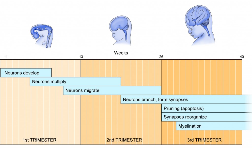
The process of generating myelin is called myelination or myelinogenesis. In the CNS, cells called oligodendrocyte precursor cells (OPCs; the precursors of oligodendrocytes) differentiate into mature oligodendrocytes, which form myelin. In humans, myelination begins early in the 3rd trimester, although only little myelin is present in either the CNS or the PNS at the time of birth. During infancy, myelination progresses rapidly, with increasing numbers of axons acquiring myelin sheaths. This corresponds with the development of cognitive and motor skills, including language comprehension, speech acquisition, crawling and walking. Myelination continues through adolescence and early adulthood and although largely complete at this time, myelin sheaths can be added in grey matter regions such as the cerebral cortex, throughout life.
What is the discontinuous structure of the myelin sheath?
The discontinuous structure of the myelin sheath results in saltatory conduction, where by the action potential "jumps" from one node of Ranvier, over a long myelinated stretch of the axon called the internode, before "recharging" at the next node of Ranvier, and so on, until it reaches the axon terminal.
How to repair myelin sheaths?
Research to repair damaged myelin sheaths is ongoing. Techniques include surgically implanting oligodendrocyte precursor cells in the central nervous system and inducing myelin repair with certain antibodies. While results in mice have been encouraging (via stem cell transplantation), whether this technique can be effective in replacing myelin loss in humans is still unknown. Cholinergic treatments, such as acetylcholinesterase inhibitors (AChEIs), may have beneficial effects on myelination, myelin repair, and myelin integrity. Increasing cholinergic stimulation also may act through subtle trophic effects on brain developmental processes and particularly on oligodendrocytes and the lifelong myelination process they support. Increasing oligodendrocyte cholinergic stimulation, AChEIs, and other cholinergic treatments, such as nicotine, possibly could promote myelination during development and myelin repair in older age. Glycogen synthase kinase 3β inhibitors such as lithium chloride have been found to promote myelination in mice with damaged facial nerves. Cholesterol is a necessary nutrient for the myelin sheath, along with vitamin B12.
How is myelin formed?
Myelin is formed in the central nervous system (CNS; brain, spinal cord and optic nerve) by glial cells called oligodendrocytes and in the peripheral nervous system (PNS) by glial cells called Schwann cells . In the CNS, axons carry electrical signals from one nerve cell body to another. In the PNS, axons carry signals to muscles and glands or from sensory organs such as the skin. Each myelin sheath is formed by the concentric wrapping of an oligodendrocyte (CNS) or Schwann cell (PNS) process (a limb-like extension from the cell body) around the axon. Myelin reduces the capacitance of the axonal membrane. On a molecular level, in the internodes it increases the distance between extracellular and intracellular ions, reducing the accumulation of charges. The discontinuous structure of the myelin sheath results in saltatory conduction, whereby the action potential "jumps" from one node of Ranvier, over a long myelinated stretch of the axon called the internode, before "recharging" at the next node of Ranvier, and so on, until it reaches the axon terminal. Nodes of Ranvier are the short (c. 1 micron) unmyelinated regions of the axon between adjacent long (c. 0.2 mm – >1 mm) myelinated internodes. Once it reaches the axon terminal, this electrical signal provokes the release of a chemical message or neurotransmitter that binds to receptors on the adjacent post-synaptic cell (e.g., nerve cell in the CNS or muscle cell in the PNS) at specialised regions called synapses .
Why is action potential faster in myelinated neurons than in unmyelinated neurons?
Action potential propagation in myelinated neurons is faster than in unmyelinated neurons because of Saltatory conduction. The main purpose of myelin is to increase the speed at which electrical impulses propagate along the myelinated fiber. In unmyelinated fibers, electrical impulses ( action potentials) travel as continuous waves, but, ...
What is the name of the substance that surrounds nerve cells?
Nervous system. Identifiers. FMA. 62977. Anatomical terminology. Myelin is a lipid -rich (fatty) substance that surrounds nerve cell axons (the nervous system's "wires") to insulate them and increase the rate at which electrical impulses (called action potentials) are passed along the axon. The myelinated axon can be likened to an electrical wire ...
What is the discontinuous sheath of the nerve fiber?
In the 1870s, French physician Louis-Antoine Ranvier noted that the myelin sheath is discontinuous, covering most of the nerve fiber but with gaps at regular intervals along the axon. Scientists later learned that charged particles called ions can cross the axon only at these myelin gaps, which became known as the “ nodes of Ranvier .”
What is myelin made of?
Made of lipids and proteins, myelin was later found to wrap around the axons of neurons. Myelin is made by two different types of support cells. In the central nervous system (CNS) — the brain and spinal cord — cells called oligodendrocytes wrap their branch-like extensions around axons to create a myelin sheath.
What is the name of the mouse that has a defective myelin protein?
Researchers developed mouse models that had defective myelin proteins, resulting in a myelin deficiency. One such mouse is the “shiverer” mouse, named after its body tremors. Mice like the shiverer mouse have provided researchers with a model system for studying myelin’s function in the healthy nervous system and its dysfunction in demyelinating diseases.
What is myelin in neurology?
Myelin: An Overview. Myelin is a fatty material that wraps around nerve cell projections. In this image, myelin can be seen on either end of the nerve fibers. The gaps in the middle of the fibers are called nodes, which help transmit electrical signals in neurons. Desmazieres, et al. Journal of Neuroscience, 2014.
What are the gaps in the middle of the fibers called?
The gaps in the middle of the fibers are called nodes , which help transmit electrical signals in neurons. Desmazieres, et al. Journal of Neuroscience, 2014. In this illustration of a neuron, myelin is shown in yellow. In the nerves outside of the brain and spinal cord, myelin is produced by support cells called Schwann cells.
How many people have MS?
MS is a chronic, disabling disease of the CNS that affects more than 2.3 million people worldwide. MS results from the accumulation of damage to myelin and the underlying nerve fibers it insulates and protects.
What is the purpose of myelin?
Myelin is a fatty substance that wraps around nerve fibers and serves to increase the speed of electrical communication between neurons. While the function of myelin remained elusive for many years, today scientists are putting their knowledge about this insulating substance to use ...
How does PI3K signaling affect myelination?
Inhibiting PI3K signaling is known to stimulate the formation of new layers of myelin by acting on AKT, mammalian target of rapamycin (mTOR), and other substrates to promote cell polarization, glial process outgrowth, and myelination. PIP3 is antagonized by the phosphatase and tenesin homolog (PTEN), which dephosphorylates PIP3 to PIP2. Previously members of this research team found that myelinating cells lacking PTEN have elevated PIP3 levels and hypermyelination, even when induced in mature oligodendrocytes ( Goebbels et al., 2010 ). Here Snaidero and colleagues report that when myelin synthesis is stimulated in this way (by conditional inactivation of Pten, which elevates PI (3,4,5)P3 levels) the number of cytoplasmic channels increased with the increase in myelination. Moreover, a large number of cytoplasmic rich inclusions were seen advancing along the length of the myelin sheath when viewed in long-section, explaining how new layers of myelin can be laid down underneath the existing layers of compact myelin.
What is the purpose of a dense layer of myelin?
The formation of dense layers of highly compacted cell membrane creates an impediment in delivering proteins and lipids to replace those lost from the compacted myelin she ath and to supply the inner tongue of uncompacted membrane where new layers of myelin are formed. The lateral cytoplasmic domains at the edge of each myelin layer remain uncompacted and in contact with the axonal membrane. These tubes of cytoplasm at the edge of each sheet move in a continuous helix around the axon toward the future node of Ranvier, where they stack up and form the paranodal loops as seen in cross section flanking the node. This long spiraling cytoplasmic channel provides a long distance pathway for transporting material from the cell body. Transport is also facilitated by fenestrated pockets of cytoplasm intruding between the layers of otherwise compacted myelin. In addition to providing a conduit for transmitting cellular constituents across the compacted myelin, these cytoplasmic channels are thought to allow dynamic regulation of the myelin sheath to participate “in a dynamic process whereby the myelin lamellae are continually parting and coming together during life in response to physiological stresses and strains” (Robertson, 1958, as quoted in Velumian et al., 2011 ). Filling the cytoplasmic channels with the fluorescent dye Lucifer yellow shows that they can be in open or closed states, presumably associated with myelin stability and dynamics ( Velumian et al., 2011 ). Snaidero et al., provide an important advance by showing that these channels can be regulated by stimulating myelin synthesis.
How is myelin formed?
In the central nervous system, myelin is formed by multipolar glia, oligodendrocytes, that can extend dozens of slender cell processes to ensheath multiple axons simultaneously. Wrapping multiple layers of membrane around an axon as one would wind electrical tape on a wire is a topological impossibility for a multipolar cell. Myelin is formed in the PNS (peripheral nervous system) and CNS by the innermost sheet-like glial process in contact with the axon spiraling around it and spinning out multiple layers of overlapping membrane. Cytoplasm becomes expelled from all but the innermost and outermost layers of the myelin sheath. In the intervening layers, the cell membranes come together to form compact myelin by the action of myelin basic protein (MBP), found preferentially in the compacted layers of myelin. The process of myelination begins when an oligodendrocyte cell process contacts an axon and forms a specialized membrane junction “spot weld,” as described by Luse in 1959. This junction is now understood to be a specialized membrane domain for intercellular communication between the glial cell process and axon ( Wake et al., 2011 ). The glial process then expands laterally along the axon and begins to encircle it in a nonuniform manner ( Luse, 1959 ). Because the segment of myelin between each node of Ranvier is several times larger than an oligodendrocyte, as it wraps, the glial cell process expands laterally into a ribbon that broadens in width to wrap the entire internodal length. This can be seen in live imaging studies, where the process has been likened to making a croissant from a triangular piece of dough ( Sobottka et al., 2011 ). Using similar methods and serial block face imaging of myelination in zebrafish, Snaidero et al., provide data consistent with this mechanism of myelin formation ( Figure 1 ).
What is the role of oligodendrocytes in the cell body?
Oligodendrocytes are highly polarized cells that synthesize vast quantities of specialized membrane to ensheath axons. Consequently, trafficking of vesicles, specific mRNAs, and proteins is highly polarized and precisely sorted in oligodendrocytes to generate and maintain the unique composition of the myelin sheath and cell body membrane domains. Vesicular stomatitis virus glycoprotein (VSC-G), a marker of trafficking to the basolateral region of cells, is trafficked away from the cell body and accumulates selectively in the myelin sheath subcellular domain of oligodendrocytes in cell culture ( Baron et al., 1999 ). Delivery of VSC to the membrane depends on submembrane F-actin at the leading edge, as shown by disrupting the cytoskeleton or altering actin polymerization with protein kinases. Snaidero et al., replicate these cell culture results and show that this also occurs in vivo by injecting the virus into the brain during myelination of the corpus callosum and observing VSC accumulating at the inner tongue of myelin adjacent to the axon membrane.
How did the insulating sheath of the axon affect the transmission of neural impulses?
The myelin sheath transformed the way neural impulses are transmitted, by forcing action potentials to “jump” rapidly between periodic breaks in myelin (nodes of Ranvier), thus dramatically increasing transmission speed and elevating nervous function well beyond that of invertebrates. Not until the development of electron microscopy was the surprising submicroscopic structure of myelin revealed. Rather than being a secretion of the axon, myelin was found to be a thick wrapping of highly compacted layers of cell membrane spun around the axon by nonneuronal cells (glia). Myelin and the nodes of Ranvier are the most complex cell-cell junctions known, requiring precise cell-cell recognition, synthesis of vast quantities of specialized cell membrane, and intricate cell motility to wrap up to 100 layers of membrane around axons. Damage to myelin is the source of much disease and disability, and recently, myelin has attracted attention as a possible new cellular mechanism participating in learning ( Fields, 2010 ). The studies by Snaidero et al. (2014), provide new information on the cellular dynamics and molecular signaling controlling myelin formation and remodeling. The work advances understanding of how myelin membrane is added to the existing sheath, which has significance for nervous system development, disease, and understanding of how myelin may be remodeled to optimize function.
What is myelin insulation?
Myelin is a multilayer wrapping of insulation formed by glial cells around axons that is essential for rapid impulse transmission, but how glial cells accomplish this cellular choreography has long intrigued researchers. In this issue of Cell, Snaidero et al., provide new insights into how myelin forms and is remodeled.
Does myelin remodeling affect learning?
There is current interest in the possibility that myelin remodeling could participate in learning, cognitive function, and psychiatric illness by adjusting con duction velocity for optimal function in an activity-dependent manner ( Field s, 2010 ). Changes in anisotropy of water diffusion seen by diffusion tensor imaging in white matter regions of individuals after learning ( Zatorre et al., 2012) could reflect changes in myelination or occur more rapidly from altered water diffusion through these cytoplasmic channels opened after learning.
What causes myelin to be damaged?
Other than multiple sclerosis, damage to myelin can be caused by any number of common and uncommon conditions. These include: 3 1 Stroke 2 Infections 3 Inflammation 4 Metabolic disorders 5 Certain medications 6 Immune disorders 7 Excessive alcohol use 8 Carbon monoxide poisoning 9 Vitamin B12 deficiency
What is the term for the destruction of the myelin sheath?
Demyelination is the term used to describe the destruction of the myelin sheath, the protective covering surrounding nerve fibers. This damage causes nerve signals to slow down or stop, resulting in neurological impairment.
What happens when myelin is scarred?
Repeated attacks eventually lead to scarring. When myelin is scarred, nerve impulses cannot be properly transmitted; they either travel too slowly or not at all. Eventually, axons degenerate as a result of the chronic myelin loss, leading to nerve cell death. 2 .
What is the role of myelin in the nervous system?
Myelin is vital to a healthy nervous system, affecting everything from movement to cognition. In multiple sclerosis (MS), the most common disease 1 associated with myelin damage, immune cells attack myelin—and eventually, the axons—in the brain and spinal cord. Repeated attacks eventually lead to scarring.
What is the function of myelin sheath?
These thin projections are called axons and most of them are protected by the myelin sheath, which allows nerve impulses to travel rapidly and effectively. Myelin is vital to a healthy nervous system, ...
What is myelin made of?
Myelin is made of fat and protein and it's wrapped in numerous layers around many of the nerves in the central nervous system (CNS), which includes your brain, spinal cord, and the optic (eye) nerves, as well as in the peripheral nervous system (PNS), which contains all the nerves outside of the CNS. Myelin is created by specific types of glial ...
What is the protective layer of nerve fibers?
The myelin sheath is the protective, fatty coating surrounding your nerve fibers, similar to the protective insulation around electrical wires. This coating enables the electrical impulses between nerve cells to travel back and forth rapidly. When myelin becomes damaged, these electrical signals are interrupted and may even stop altogether.
Does Myelination Occur Throughout Life?
Myelination first occurs during embryonic development and is then a continuous process from birth, maturing at about 2 years of age. Once at this stage, motor and sensory systems are matured and cerebral myelination is mostly complete.
How many arms does an oligodendrocyte have?
Oligodendrocytes are star-shaped cells which have about 15 arms coming out of their cell body, meaning it is able to myelinate multiple axons at one time. In a similar fashion to Schwann cells, oligodendrocytes spiral around the axons of neurons to form a myelin sheath.
Why is myelin sheath important?
Since myelin sheath provides insulation to axons, this allows these axons to conduct electrical signals at a higher speed than if they were not insulated by myelin. Thus, the more thoroughly myelinated an axon is, the higher the speed of electrical transmission.
How fast can an axon conduct impulses?
One of the most myelinated axons, for instance, can conduct impulses at a speed of approximately 70 to 120 m/s, the speed of a race car. Similarly, myelin sheath around an axon is able to prevent electrical impulses from traveling through the sheath and out of the axon.
What is the protective sleeve that wraps around the axon of neurons?
Myelin sheath consists of lipids and proteins which make up a fatty substance and is white in appearance. This forms the protective sleeve that wraps around the axon of neurons. The sheath is made up of many concentric layers of plasma membrane, wrapped tightly around the axon.
Why is the myelin sheath wrapped around the axons of neurons?
Myelin sheath is the protective layer that wraps around the axons of neurons to aid in insulating the neurons, and to increase the number of electrical signals being transferred. An axon is usually wrapped by the myelin sheath around its whole length in order to increase the speed of these electrical signals, allowing all actions ...
What are the breaks in the myelin sheath called?
There are breaks of between 0.2 and 2 mm. in the myelin sheath, these are called nodes of Ranvier. Action potentials (nerve impulses) traveling down the axon "jump" from node to node. This speeds up the transmission.
What are the two main categories of demyelinating disease?
There are two main categories of demyelinating disease: demyelinating disease of the central nervous system (CNS) and demyelinating disease of the peripheral nervous system (PNS).
What is the term for a severe bout of inflammation in which swelling damages the myelin on cells in the?
Acute disseminated encephalomyelitis (ADEM): ADEM is a severe bout of inflammation in which swelling damages the myelin on cells in the brain, spinal cord, and sometimes the optic nerves.
How to treat ALD in children?
One effective treatment for childhood ALD is hematopoietic stem cell transplant, which is a bone marrow transplant. People with Addison’s disease may experience some benefit from taking steroids. Some people may also take seizure medications or go to physical therapy to help with muscle spasms and weakness.
What is the best treatment for ADEM?
Intravenous steroids like methylprednisolone or oral steroids can help reduce the inflammation caused by ADEM. Plasmapheresis may also be an option with severe cases of this condition.
What is myelin insulation?
Myelin is essentially an insulating layer of lipids and proteins that covers many of your nerves. If that coating becomes damaged or wears away, it causes problems with your nerves’ ability to send and receive electrical messages normally.
What is CIDP in a patient?
Chronic inflammatory demyelinating polyn europathy: Also known as chronic relapsing polyneuropathy, CIDP causes progressive muscle weakness and affects roughly 5 to 7 out of every 100,000 people.
How many people have MS?
Multiple sclerosis (MS): MS is the most common type of demyelinating disease of the central nervous system, and affects about 1 million people in the United States.

Overview
Myelin is a lipid-rich material that surrounds nerve cell axons (the nervous system's "wires") to insulate them and increase the rate at which electrical impulses (called action potentials) are passed along the axon. The myelinated axon can be likened to an electrical wire (the axon) with insulating material (myelin) around it. However, unlike the plastic covering on an electrical wire, m…
Development
The process of generating myelin is called myelination or myelinogenesis. In the CNS, oligodendrocyte progenitor cells (OPCs) differentiate into mature oligodendrocytes, which form myelin. In humans, myelination begins early in the 3rd trimester, although only little myelin is present in either the CNS or the PNS at the time of birth. During infancy, myelination progresses rapidly, with increasing numbers of axons acquiring myelin sheaths. This corresponds with the d…
Species distribution
Myelin is considered a defining characteristic of the jawed vertebrates (gnathostomes), though axons are ensheathed by a type of cell, called glial cells, in invertebrates. These glial wraps are quite different from vertebrate compact myelin, formed, as indicated above, by concentric wrapping of the myelinating cell process multiple times around the axon. Myelin was first described in 1854 by Rudolf Virchow, although it was over a century later, following the develop…
Composition
CNS myelin differs slightly in composition and configuration from PNS myelin, but both perform the same "insulating" function (see above). Being rich in lipid, myelin appears white, hence the name given to the "white matter" of the CNS. Both CNS white matter tracts (e.g. the optic nerve, corticospinal tract and corpus callosum) and PNS nerves (e.g. the sciatic nerve and the auditory nerve, which …
Function
The main purpose of myelin is to increase the speed at which electrical impulses (known as action potentials) propagate along the myelinated fiber. In unmyelinated fibers, action potentials travel as continuous waves, but, in myelinated fibers, they "hop" or propagate by saltatory conduction. The latter is markedly faster than the former, at least for axons over a certain diameter. Myelin decreases capacitance and increases electrical resistance across the axonal membrane (the axole…
Clinical significance
Demyelination is the loss of the myelin sheath insulating the nerves, and is the hallmark of some neurodegenerative autoimmune diseases, including multiple sclerosis, acute disseminated encephalomyelitis, neuromyelitis optica, transverse myelitis, chronic inflammatory demyelinating polyneuropathy, Guillain–Barré syndrome, central pontine myelinosis, inherited demyelinating diseases such as leukodystrophy, and Charcot–Marie–Tooth disease. Sufferers of pernicious an…
Invertebrate myelin
Functionally equivalent myelin-like sheaths are found in several invertebrate taxa including oligochaetes, penaeids, palaemonids, and calanoids. These myelin-like sheaths share several structural features with the sheaths found in vertebrates including multiplicity of membranes, condensation of membrane, and nodes. However, the nodes in vertebrates are annular; i.e. they encircle the axon. In contrast, nodes found in the sheaths of invertebrates are either annular or f…
See also
• Lesional demyelinations of the central nervous system
• Myelin-associated glycoprotein
• Myelin incisure
• The Myelin Project, project to regenerate myelin
The Nervous System’S Insulation

Insight Into Myelin’S Role in Health and Disease
- Removing Myelin Disrupts Neural Communication
With knowledge of myelin’s role in neural communication, researchers aimed to find out what happens when myelin is disrupted. In the 1980s, researchers used animal models to assess how electrical nerve signals are altered after axons were stripped of the myelin (demyelinated). Whe… - Myelin Loss in Disease
Loss of myelin is a problem for many CNS disorders, including stroke, spinal cord injury, and, most notably, multiple sclerosis (MS). MS is a chronic, disabling disease of the CNS that affects more than 2.3 million people worldwide. MS results from the accumulation of damage to myelin and t…
New Treatment Possibilities For Demyelinating Diseases
- Research understanding the components of myelin, how it is produced, and how it functions has paved the way for new therapeutic possibilities in myelin-degenerative diseases like MS.