Common Causes
In the patients with proptosis of less than 21 mm, there was a significant difference in the measurements between the three methods; the highest value was obtained by CT followed by the 3D reconstruction software and Hertel exophthalmometer ( p < 0.001).
Related Conditions
We have developed a computed tomography (CT)-based three-dimensional (3D) reconstruction software to measure the degree of proptosis. To verify clinical usefulness and reliability, the degree of proptosis was measured in … The evaluation of proptosis is essential for the diagnosis of orbital disease.
What is the best way to measure proptosis?
Testing. Proptosis can be confirmed with exophthalmometry, which measures the distance between the lateral angle of the bony orbit and the cornea; normal values are < 20 mm in whites and < 22 mm in blacks. CT or MRI is often useful to confirm the diagnosis and to identify structural causes of unilateral proptosis.
How can we measure the degree of proptosis in orbital disease?
To compensate for these limitations, computed tomography (CT) has been used to measure proptosis and is reported to produce more accurate data 10, 11, 12, 13, 14; however, proptosis measurement using CT is also associated with some limitations.
How do you test for proptosis in dogs?
How accurate is CT for measuring proptosis?

How is radiology proptosis measured?
The reference line for measurement of proptosis is the interzygomatic line (a line is drawn at the anterior portions of the zygomatic bones): the upper limit of normal distance from this line to the anterior surface of the globe is 23 mm, above which indicates proptosis.
What is normal proptosis?
It is suggested that the following "upper limits of normal" be used when clinically estimating proptosis: 19 and 21 mm for white female and male patients, respectively; and 23 and 24 mm for black female and male patients, respectively.
How do you measure Hertel?
0:000:37Hertel measurement and Hertel exophthalmometer - YouTubeYouTubeStart of suggested clipEnd of suggested clipRight eye should be determined with the examiner's. Left eye and vice versa. The device measures theMoreRight eye should be determined with the examiner's. Left eye and vice versa. The device measures the distance between the lateral orbital rim and the most anterior portion of the cornea.
What kind of equipment is used for exophthalmos degree measuring?
An exophthalmometer is an instrument used for measuring the degree of forward displacement of the eye in exophthalmos. The device allows measurement of the forward distance of the lateral orbital rim to the front of the cornea.
What is the most common cause of proptosis in adults?
What are the most common causes of proptosis? The most common cause of bulging eyes is an autoimmune disorder in which the body's immune system attacks cells in the thyroid gland and the tissue behind the eye. Proptosis in people with thyroid issues is also called thyroid eye disease.
What does proptosis look like?
Evaluation of Proptosis Ocular examination findings typical of hyperthyroidism but unrelated to infiltrative eye disease include eyelid retraction, eyelid lag, temporal flare of the upper eyelid, and staring. Other signs include eyelid erythema and conjunctival hyperemia.
What is the difference between Exophthalmos and Proptosis?
Proptosis can describe any organ that is displaced forward, while exophthalmos refers to only the eyes. Proptosis can include any directional forward displacement.
What instrument measures the forward protrusion of the eye?
Measurements will be recorded using the Hertel exophthalmometer. This noninvasive tool is an instrument designed to measure the forward protrusion of the eye and may be used normally during a general eye exam.
How is Enophthalmos measured?
A horizontal line (baseline) is drawn to connect the outermost of the bilateral anterior tips of the lateral walls. A vertical line is then drawn from the most prominent corneal part to the baseline. The length of this line is measured.
What is non axial Proptosis?
Pneumosinus Dilatans (PD) is a rare condition characterized by abnormal enlargement of one or more paranasal sinuses that can lead to different functional and cosmetic presentations.
What is Hypoglobus?
Hypoglobus is an abnormal lowering of the globe usually due to a deficient anterior orbital floor. Proptosis is an abnormally anteriorly positioned globe with respect to the bony orbit.
What is Pseudoproptosis?
Definition. Pseudoproptosis is the simulation of abnormal prominence or a true asymmetry that is not associated with displacement of the globe.
What is the difference between exophthalmos and proptosis?
Proptosis can describe any organ that is displaced forward, while exophthalmos refers to only the eyes. Proptosis can include any directional forward displacement.
What is proptosis in Ophthalmopathy?
Proptosis is spontaneous decompression resulting from enlargement of the extraocular muscles and adipose tissue, as well as orbital fat deposits and the infiltration of orbital tissues by GAGs and leukocytes (Figure 2). TAO is the most common cause of unilateral and bilateral proptosis in adults.
How to measure proptosis?
The severity of proptosis can be measured by various methods. The Hertel exophthalmometer uses a system of prisms to project a lateral view of the eye forward. A millimetre-scale is superimposed on this view and is read by the opposite eye of the examiner. The patient is made to sit at the eye level of the examiner. The two ends of the instrument are placed at the lateral orbital margins of each eye. Subsequently, the observer uses his opposite eye to look at the distance of the apex of the cornea on the scale by excluding parallax. A millimetre-scale is provided on the top of the instrument to measure the distance between the lateral orbital margins of the two eyes; this reading can be noted for future reference. Errors in measurement are caused by thickness of subcutaneous tissue over the bone, facial asymmetry, parallax and examiner skills. 6,31 Apart from Hertel’s exophthalmometer, a simple plastic scale at the lateral orbital margin, or a Luedde exophthalmometer, can be used. The latter is a thick plastic ruler with a notch to fit in the lateral orbital margin and markings in millimetres on both sides of the scale to avoid parallax. In exophthalmometry, normal readings are taken as less than 21 mm between the lateral orbital rim and apex of the cornea, or a difference of less than 2 mm between the two eyes. 1,6
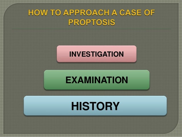
What are the methods of investigating proptosis?
The methods for investigating proptosis have been classified into primary, secondary, pathological and laboratory techniques. The article extensively discusses thyroid eye disease, including the current hypothesis of pathophysiology, mechanisms, stages, clinical features, classification and management of this disease.
What is P roptosis?
Overview. P roptosis is an abnormal prominence of the eyeball beyond the confines of the bony orbit. It can appear in various systemic, as well as orbital or peri-orbital, disorders. Not only is proptosis potentially vision-threatening, but also it could be a manifestation of life-threatening conditions, such as metastatic carcinomas.
What is the term for an abnormal protrusion of the eyeball beyond the boundaries of the bony orbit?
Article: An abnormal protrusion of the eyeball, beyond the boundaries of the bony orbit, is termed ‘proptosis ’. A similar appearance, seen in endocrine dysfunction, especially thyroid disorders, is called ‘exophthalmos’.
Which direction does proptosis occur?
Direction of proptosis. Proptosis can be axial, when it occurs in the anteroposterior direction or can be in the direction opposite to the causative lesion . Thus, a lesion in the upper-lateral aspect of the orbit, such as a lacrimal gland tumour, will push the eyeball downwards and medially.
Why is time important in proptosis?
In certain situations, time is of vital importance to prevent optic nerve compression and permanent loss of vision. This review provides a simplified pathway to approach proptosis and to improve patient care and outcomes. 16
When should visual acuity be assessed?
An assessment of visual acuity should be conducted at the first visit and on follow-up visits. If the patient is admitted to the ward, frequent monitoring of visual acuity and colour vision may be required. The mechanisms of visual loss in such scenarios include central retinal artery occlusion, direct compressive optic neuropathy, compression of small nutrient vessels of the optic nerve leading to ischemic optic neuropathy. Stretching of the nerve and posterior globe tenting aggravate the situation. 25 Vision may also be affected by exposure keratopathy as explained above.
Overview
Proptosis is the bulging of one or both or your eyes from their natural position. The condition can affect your appearance, leaving you with a startled expression that doesn’t go away.
Symptoms and Causes
The most common cause of bulging eyes is an autoimmune disorder in which the body’s immune system attacks cells in the thyroid gland and the tissue behind the eye. Proptosis in people with thyroid issues is also called thyroid eye disease.
Diagnosis and Tests
Your healthcare provider will ask about your symptoms and medical history to determine potential causes for your symptoms.
Management and Treatment
Artificial tears, including drops or gel to relieve dry eyes and protect the cornea.
Living With
Protruding eyes can affect your appearance and cause issues with your confidence and self-esteem. If it affects your vision, you may experience unexpected changes in your daily life. These changes can leave you feeling upset, anxious or depressed.
How to measure proptosis?
Measurements were taken with the patient’s head in the primary position and the examiner’s eye at the same level as the patient’s eyes in a well-lit room. The measurement was the distance between the point on the temporal orbital rim at the deepest palpable point of the angle and the apex of the cornea. Right and left eye readings were performed sequentially without removing the instrument from the orbital rims. The measurements were recorded to the nearest 0.5 mm 18.
What is the best way to measure proptosis?
Measurement of ocular proptosis is essential for the diagnosis of orbital diseases such as Graves’ orbitopathy (GO), orbital tumor, and orbital fracture. There are various types of devices available for measuring the degree of proptosis. The Hertel exophthalmometer, invented by Hertel in 1905, is the most widely used device to date 1, 2. It estimates the degree of proptosis from the lateral orbital rim to the corneal surface, perpendicular to the frontal plane 3; however, the Hertel exophthalmometer has been criticized for its low reliability and accuracy 4. Musch et al. 5 showed a statistically significant inter-observer difference with the Hertel exophthalmometer, with a 61–80% agreement. Furthermore, differences in proptosis measurements have been reported between Hertel exophthalmometers made by different companies 1, 6, 7. The differences in readings may result from misplacement of the foot plates, strabismus, asymmetry of the lateral orbital rims, compression of soft tissues, parallax errors, or the lack of a uniform measurement technique 8, 9.
What is the intra-observer correlation coefficient of Hertel exophthalmometer?
The intra-observer correlation coefficients of the 3D reconstruction software, CT, and Hertel exophthalmometer measurements were 0.997, 0.942, and 0.953, respectively. In strabismus patients, the intra-observer correlation coefficients for the 3D reconstruction software, CT, and Hertel exophthalmometer were 0.996, 0.895, and 0.920, respectively. The ICCs for CT and 3D reconstruction software between three different ophthalmologists were 0.742 and 0.846, respectively. This ICC for 3D reconstruction software is interpreted as excellent agreement. Our study was limited by the fact that we did not evaluate the inter-observer variability of the Hertel exophthalmometer; however, a high variability in the Hertel exophthalmometer measurements between different observers has been widely reported, from 30 to 80% 3, 5. Considering these results, 3D reconstruction software is the most reliable of the tested methods and would be useful when there is a large difference between the Hertel exophthalmometer and CT measurements, such as strabismus, or if other test are less reliable due to inconsistent measurements.
Can a CT scan be used to measure proptosis?
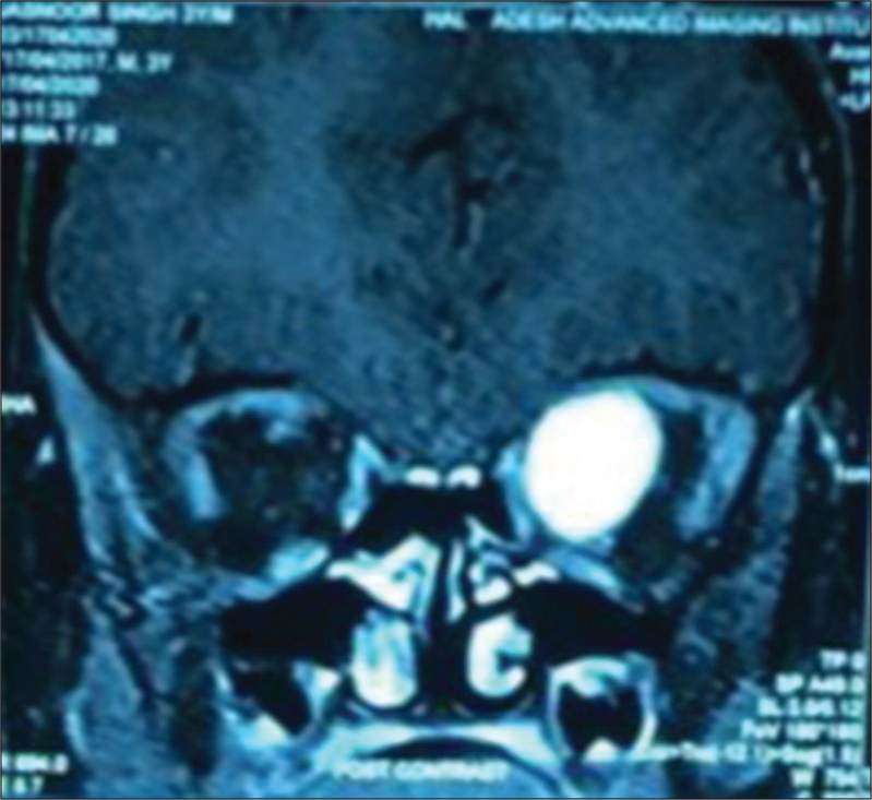
Since the apex of the cornea and interzygomatic line cannot be included in the same plane on a two-dimensional (2D) CT scan, the level of the measured CT slice may not correspond to the area of maximal proptosis . In addition, it can cause errors in the process of manually specifying the point of interest and measuring the distance. Lastly, the measured distance is limited to only a 2D space. Errors in measurements can result from an eyeball position that is not centered by aligning the mid-sagittal line perpendicular to the straight line, which is typical in patients with vertical strabismus.
What test is used to check for bulging in the eye?
Tests may include an MRI, bloodwork, an ultrasound, or even a biopsy.
Can proptosis cause permanent vision loss?
Because the eyelids may not be able to close completely during normal blinking or sleeping, the cornea may dry out significantly. This dryness is not only uncomfortable but can also cause scarring which could lead to permanent vision loss.
What is the term for the protrusion of the eyeball?
DR P. MARAZZI/SCIENCE PHOTO LIBRARY. Proptosis is protrusion of the eyeball. Exophthalmos means the same thing, and this term is usually used when describing proptosis due to Graves disease. Disorders that may cause changes in the appearance of the face and eyes that resemble proptosis but are not include hyperthyroidism without infiltrative eye ...
How to treat Graves exophthalmos?
Lubrication to protect the cornea is required in severe cases. When lubrication is not sufficient, surgery to provide better coverage of the eye surface or to reduce proptosis may be required. Systemic corticosteroids (eg, prednisone 1 mg/kg orally once a day for 1 week , tapered over ≥ 1 month) are often helpful in controlling edema and orbital congestion due to thyroid eye disease or inflammatory orbital pseudotumor. Other interventions vary by etiology. Graves exophthalmos is not affected by treatment of the thyroid condition but may lessen over time. Tumors must be surgically removed. Selective embolization or, rarely, trapping procedures may be effective in cases of arteriovenous fistulas involving the cavernous sinus.