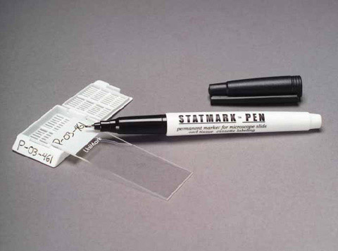
Do microscope slides expire?
It is no longer suitable for use. Following the storage instructions and instructions for use will ensure the quality of the microscope slides until the Use- by date is reached. According to EN ISO 15223-1 standard, the Use-by date is the date after which the microscope slides are not be used anymore.
How do you preserve a microscope slide?
To keep your prepared microscope slides in good condition, always store them in a container made for the purpose and away from heat and bright light. The ideal storage area is a cool, dark location, such as a closed cabinet in a temperature-controlled room. Stained slides naturally fade over time.
Are microscope slides reusable?
Do not re-use or recycle glass slides. Discard chipped or scratched slides. When using cleaned slides, use those that were cleaned the earliest and not those that were cleaned most recently.Jan 1, 2016
How long do if slides last?
Thus a reliable storage period for keeping renal IF slides, should be well established. Storage periods for DIF stains related to skin diagnosis has been found adequate for only 2 to 9 months from 1975 to 1985 [11-13], and is found to last longer, up to 11 months, in a 2003 study [14].
How do you make a microscope slide permanent?
8:3910:20Making dry-mounted permanent microscopy slides - YouTubeYouTubeStart of suggested clipEnd of suggested clipThen one possibility could be is that you place it into concentrated alcohol for several days andMoreThen one possibility could be is that you place it into concentrated alcohol for several days and then you allow the alcohol to evaporate.
How do you make temporary slides permanent?
I have made temporary slides more permanent by painting round the cover-slip with clear nail varnish. This has been useful for water-based slides to stop them drying out and enable them to be preserved for another day.
Can you reuse microscope slide covers?
If you're using cover slips, you should also wash those between every use. Johnny L. The common type of microscope slides are the simple glass ones used for compound light microscopes, and yes, they can be used repeatedly. Just make sure you wash and dry the slide very well between each use.Dec 8, 2014
How do you clean a microscope slide?
0:040:54Clean a microscope slide - YouTubeYouTubeStart of suggested clipEnd of suggested clipSolution a few drops should be placed on the upper surface of the slide rubbing downward with aMoreSolution a few drops should be placed on the upper surface of the slide rubbing downward with a clean piece of lens. Paper rogue dust the front and the back sides of this line with a wet.
How do you clean a slide?
1:062:13Cleaning Slide Film: A Museum How-to - YouTubeYouTubeStart of suggested clipEnd of suggested clipGet our swab. Get our swab wet now you don't want to dip the uh. The slide directly into the alcoholMoreGet our swab. Get our swab wet now you don't want to dip the uh. The slide directly into the alcohol that will uh that that will harm the slide. And also might harm the case.
How long can immunofluorescence slides last?
Loss of immunofluorescence was limited to reactants initially read as 1+ intensity on a scale of 1 to 4+. Prior studies of preservation of DIF slides at room temperature are limited. Grimwood and Proffer2 examined 22 cases stored at room temperature and reported that they can be preserved up to 30 months.
How long can you store immunofluorescence slides?
I found that ~ 6 months with vectashield in -20C might be ok.
How long are IHC slides good for?
Slides can be safely stored for 6-12 months at -80°C until ready for staining.
Most recent answer
For the correct preservation of the slides, several criteria are important. The fluorochrome coupled to the antibody, the mounting medium, the storage temperature.
Popular Answers (1)
Generally it is going to come down to how well your sample is sealed. Oxidation is the biggest killer of fluorescence in fluorescent samples.
All Answers (13)
Generally it is going to come down to how well your sample is sealed. Oxidation is the biggest killer of fluorescence in fluorescent samples.
What is a wet mount slide?
Wet mount slides are most commonly used where you are observing live specimens that either live in water or their movement is most easily observed in a liquid medium. For example, you could technically observe bacteria under dry mount slide however this will greatly reduce the ability of the bacteria to move.
Why does my wet mount slide have bubbles?
Air bubbles can form if your slide coverslip closes too rapidly causing air to be trapped under the coverslip or a displacement in the water that is rapidly filled by air.
Why does my coverslip float?
Floating Coverslip. In some cases, you may find that if you add too much water your coverslip will float after you place it over the water droplet. This can cause you slide coverslip to move to one side or the other of the slide and can also disrupt your sample in some cases.
Why do you add a quieting solution to a specimen?
If you are observing a specimen that has very rapid movements or is difficult to keep in the field of view because it is constantly moving, you can add a quieting solution that will actually slow the movement of some microorganisms.
What is a saline wet mount?
For example, saline wet mounts are the preferred method to view movements of certain protozoa during the feeding cycle stage. Saline wet mounts are prepared the same way a water wet mount is prepared except instead of water, of course, you use a saline solution.
Who is Brandon from Microscopy?
Brandon is an enthusiast, hobbyist, and amateur in the world of microscopy. His love for science and all things microscopic moves him to share everything he knows about microscopy and microbiology.
Can you add contaminated water to a slide?
Contamination can occur if you add contaminated water to your slide that contains other microorganisms or even chemicals that may affect you intended specimen. To avoid contamination, you need to understand your specimen and what things in your water source may cause contamination or unintended consequences.
How long does it take to freeze a tissue?
It usually takes 10 to 20 minutes. The fresh tissue is grossly examined by the pathologist to decide which part of it should be looked at under the microscope. Instead of processing the tissue in wax blocks, the tissue is quickly frozen in a special solution that forms what looks like an ice cube around the tissue.
How are biopsies processed?
After its removal, the biopsy specimen is put in a container with a mixture of water and formaldehyde (formalin) or some other fluid to preserve it . The container is labeled with the patient’s name and other identifying information (hospital number and birth date, for example) and the site of biopsy ...
Why is a gross examination important?
The gross examination is important since the pathologist may see features that suggest cancer . It also helps the pathologist decide which parts ...
What is a cassette in a biopsy?
For small biopsies, such as a punch biopsy or a core needle biopsy, the entire specimen is usually looked at under a microscope. The tissue is put into small containers called cassettes. The cassettes hold the tissue securely while it’s processed.
What is frozen section exam?
Sometimes information about a tissue sample is needed during surgery to make immediate decisions. If the surgeon can’t wait the day or more that it takes for routine processing and histology, he or she will request an intra-operative (during surgery) pathology consult. This is often called a frozen section exam.
What is Mohs surgery?
Process for Mohs surgery (microscopically controlled surgery) This procedure is used to treat certain kinds of skin cancer. In Mohs surgery, the surgeon removes a thin layer of the skin that the tumor might have invaded and then checks the sample under a microscope. If cancer cells are seen, more layers are removed and checked ...
How does paraffin wax work?
The wax cools to form a solid block that protects the tissue. This paraffin wax block with the embedded tissue is cut into very thin slices using an instrument called a microtome. These thin slices of the specimen are placed on glass slides, and dipped into a series of stains or dyes to change the color of the tissue.
Most recent answer
It depends on the fixation and mounting protocol. I usually fix animal cells or sections with 4% PFA in PBS or methanol. For embedding I normally use an embedding medium containing glycerol and N-propyl-gallate (anti-fading reagent). With this protocol the fluorescence will stay relatively constant for several months.
Popular Answers (1)
Hi Hosam, Tony is right, depends on fixation and mounting. I use FFPE tissue and mount with Fluoromount G (also ProLong medium is very good), which is a aqueous-based medium that keeps fluorescence for a really long time. If you stain sections with quality Alexa Fluor or cyanine-conjugated antibodies you can re-check your slides for months.
All Answers (36)
I believe it depends on your fixation method, and the mounting medium that you use. I think it also depends on the sensitivity of what you're doing.
Similar questions and discussions
How long can we comfortably store a slide with immunofluorescent material (cells/tissues) to avoid signal decaying and developing other issues? I found that ~ 6 months with vectashield in -20C might be ok. Funny things could start occurring later.
