What is a myelogram?
Why is a myelogram important?
How does a myelogram show nerve roots?
What kind of scan is needed for spine?
How long after spinal surgery can you stop taking medications?
How long does it take to get a CT scan of your spine?
How long do you have to stop taking clot medications?
See 4 more
About this website

How long do you have to lay flat after a myelogram?
Most patients are asked to lie down for two hours after the procedure. If you need to urinate, you may need to do so in a bedpan or urinal during the time that you need to stay flat. You will be asked to drink extra fluids to rehydrate after the procedure.
Are you sedated during a CT myelogram?
A myelogram is an outpatient procedure that takes about an hour. You will be asked to remove clothing and jewelry that may interfere with the test. You will lie down on a padded table and will receive sedation (medication to make you drowsy and relaxed).
Can you drive home after a myelogram?
After the Myelogram Average recovery time is two hours. Once you are stable, you'll be allowed to leave. You will NOT be allowed to drive home, so please arrange for someone to take you home. To minimize the risk of having a headache, bed rest is recommended for the remainder of the day.
Are you put to sleep during a myelogram?
What is a myelogram like? You will be awake during the procedure. You will lie on your stomach. You will be given a numbing injection that may sting for a few seconds.
How painful is a myelogram?
How does it feel? You will feel a quick sting from a small needle that has medicine to numb the skin on your back. You will also feel some pressure as the long, thin spinal needle is put into your spinal canal. You may feel a quick, sharp pain down your buttock or leg when the needle is moved in your spine.
What is the prep for a myelogram?
Before Your Myelogram You should not eat any solid food after midnight the day before your exam. You can drink clear liquids as instructed. You should let your healthcare provider know if you are or may be pregnant, have any bleeding issues, take any medications, have allergies or have had back surgery or pain.
How do you sleep after a myelogram?
It is important to keep your head slightly elevated for 24 hours following the myelogram. Use 1-2 pillows on the bed. Do not lie flat or allow your head to be lower than the rest of your body until the next morning. Drink extra fluids for the remainder of the day.
How will I feel after myelogram?
A myelogram may increase your risk for a headache, neck or back pain, nausea, or vomiting. You may have bleeding or spinal fluid may leak from the injection site. The procedure may cause injury to a disc, nerves, or your spinal cord. The dye used during the procedure may cause and allergy, seizure, or brain problems.
Where is the needle placed for a lumbar myelogram?
A lumbar puncture is performed by inserting a hollow needle into the subarachnoid space in the lumbar area (lower back) of the spinal column. The subarachnoid space is the canal in the spinal column that carries cerebrospinal fluid (CSF) between the brain and the spinal cord.
How long do you hurt after a myelogram?
It's normal to have a mild headache for up to 24 hours after the test. Take a pain reliever such as acetaminophen (Tylenol). Follow the instructions on the bottle. Do not lie flat or let your head be lower than the rest of your body for the first 24 hours after the test.
Why was my myelogram so painful?
Myelography is a very safe procedure with minimal possible risks. Nerve injury. Nerve injury occurring at the time of the lumbar puncture can result in shooting pain into the buttocks or down the leg and may persist as a dull ache or resolve over days to weeks.
Does a myelogram show the entire spine?
What can you see from a myelogram? A myelogram is able to show your spinal cord, spinal nerves, nerve roots, and bones in the spine by injecting contrast into your spinal fluid. As a result, it will also reveal whether anything is pressing against your spinal cord or nerves.
What can I expect from a CT myelogram?
During the test Cleaning your lower back with an antiseptic and giving you a local anesthetic. Once the area becomes numb, the radiologist will introduce a needle into your spinal canal and then inject a contrast material into the fluid-filled sac containing your spinal cord and nerve roots.
Where is the needle placed for a lumbar myelogram?
A lumbar puncture is performed by inserting a hollow needle into the subarachnoid space in the lumbar area (lower back) of the spinal column. The subarachnoid space is the canal in the spinal column that carries cerebrospinal fluid (CSF) between the brain and the spinal cord.
What does a myelogram feel like?
The compression on a mammogram should feel like pressure and may be uncomfortable for a short period of time, but shouldn't be painful. Every patient's tolerance of the compression varies and the technologists will work with the patient to keep them as comfortable as possible while achieving quality diagnostic images.
Is a CT myelogram the same as a spinal tap?
A myelogram is performed first in a separate procedure. This is similar to a lumbar puncture, or spinal tap, where the fluid space around the spinal cord (within the spinal canal) is accessed with local anesthesia and contrast (usually 12cc non-ionic iodinated contrast) is administered.
Why can't I have something to sedate me for the myelogram. I…
Why can't I have something to sedate me for the myelogram. I have been in pain so long (and more recently increased pain to the point that I was unable to walk) that I am on edge, can't lie flat (either back or stomach) from left leg pain.
Myelogram Updated Drug List - Patient Care at UVA Health
CT Myelogram Drugs to Avoid Hold for 48 Hours Before and 12 Hours After Your Myelogram UVA Neuroradiology . 2 . Isocarboxazid (Marplan) Lacosamide
What are the side effects of a myelogram? - HealthTap
Risks: Any procedure has risk of infection, reaction to injectate, bleeding, nerve injury or paralysis.However, those risk are rare for this procedure. The most common side effect would be a spinal headache which is treatable and reversible if it occurs at all.
When does CT myelography add value beyond MRI for lumbar ... - PubMed
Background context: In patients with lumbar spinal stenosis, it is crucial for clinicians to identify all symptomatic levels. Prior studies have demonstrated that CT myelography has a greater sensitivity in revealing stenosis (94.4%) compared to MRI (75.9%).
Myelogram (Aftercare Instructions) - Drugs.com
Drugs.com provides accurate and independent information on more than 24,000 prescription drugs, over-the-counter medicines and natural products. This material is provided for educational purposes only and is not intended for medical advice, diagnosis or treatment. Data sources include IBM Watson Micromedex (updated 1 Nov 2022), Cerner Multum™ (updated 25 Oct 2022), ASHP (updated 12 Oct 2022 ...
CT Myelogram: Reasons, Preparation and Procedure - Medical Health Tests
The ct myelogram procedure begins with a spinal tap performed on the lumbar region of the spine. Read more on how to prepare and conduct a ct myelogram.
What happens during a myelogram?
A myelogram may be done on an outpatient basis or as part of your stay in a hospital. For outpatients, please allow four hours for the preparation, procedure, and recovery time.
What is a myelogram used for?
Myelograms may be used to evaluate many diseases, including: Herniated discs (discs that bulge and press on nerves and/ or the spinal cord) Spinal cord or brain tumors. Infection and/or inflammation of tissues around the spinal cord and brain.
Why might I need a myelogram?
A myelogram may be done to assess the spinal cord, subarachnoid space, or other structures for changes or abnormalities. It may be used when another type of exam, such as a standard X-ray, does not give clear answers about the cause of back or spine problems. Myelograms may be used to evaluate many diseases, including:
What are the risks of a myelogram?
There is a risk of an allergic reaction to the contrast dye. Be sure to let your healthcare provider know if you have ever had a reaction to any contrast dye.
How is a spinal fluid needle inserted?
A needle will be inserted through the numbed skin and into the space where the spinal fluid is located . You will feel some pressure while the needle goes in, but you must remain still.
How long after a neuroradiology procedure can you have headaches?
If you go home, usually your healthcare provider will advise you to rest for the remainder of the day. If the headaches persist for more than 24 hours after the procedure, or are worse when you change positions, contact the neuroradiology team with the phone number provided on your discharge instructions.
How long to drink water before a syringe?
EAT/DRINK: Try to increase your fluid intake (such as water and juice) for the two days leading up to your procedure unless a medical condition does not allow you to safely do so. If you are not sure if it is safe for you, contact your primary care provider or referring provider.
How long should you keep your head elevated after a myelogram?
After the procedure, you will be brought into the Radiology holding area for observation. It is important to keep your head slightly elevated for 24 hours following the myelogram. Use 1-2 pillows on the bed.
What is the term for the examination of the spinal cord?
Myelography is an imaging examination that shows the passage of contrast material in the space around the spinal cord (the subarachnoid space) using a real-time form of x-ray called fluoroscopy, in which organs can be seen over time.
How long before a blood thinning procedure can you stop taking aspirin?
Please bring a list of your current medications with you (out-patient). Stop taking Aspirin or aspirin-containing products at least 5 days prior to the procedure. If you are taking other blood thinning medications, contact your doctor to discuss.
Who must accompany you home after a medical exam?
For outpatients, a responsible adult must accompany you home after the exam.
Why do doctors do myelograms?
Myelograms are now carried out much less often than they were 20 years ago. They are used to examine the spinal cord and nerves that come out of the spinal cord to look at spinal and disc problems. Advances in technology allow radiologists (specialist doctors) to see the nerves and cord directly, using magnetic resonance imaging (MRI) instead of X-rays. In some special circumstances, a myelogram is still used to investigate problems involving the spinal cord and spinal nerves. These include:
What medications are used for myelogram?
When you make your appointment for the myelogram, you need to let the radiology clinic or department know if you are taking any blood thinning medication, such as warfarin, clopidogrel, dabigatran, prasugrel, dipyridamole or asasantin (for more information about these medications, go to NPS: www.nps.org.au/medicines ).
Why is myelography important?
Myelography can be very helpful for diagnosis and management of acute or chronic spinal problems, and diagnosis of CSF leakage.
How to inject contrast in the spinal cord?
The X-ray machine will be used to guide the radiologist locating the place for the injection and passing the needle into this spot. Iodine containing contrast medium, usually approximately 10 mL, is then injected into the fluid around the spinal cord . The table may be tipped a little so that your feet are a bit lower than your head when the contrast is injected to make the contrast run downwards into the lower back if you have lower back problems. The needle used to inject the contrast is then removed and a number of X-rays are taken. You will be asked to roll slightly onto each side, onto your back, and to stand while these pictures are taken. The table may also be tipped so that your head is lower than your feet in order to make the contrast that has been injected run upwards into your neck or into your head. This is commonly done if you have neck problems or a question of leakage of CSF into your nose.
What is the room called when you are taken into a hospital for a fluoroscopy?
You will be taken into the room where the procedure is carried out, called a fluoroscopy suite.
How long does blood thinner medication need to be stopped?
Blood thinning medications may need to be stopped for a period of days, or your normal dose reduced, before this procedure is carried out. It is very important that you do not stop any of these medications or change the dose without consulting both the radiology clinic or department and your own doctor.
Where is local anaesthetic injected?
Local anaesthetic is injected through a small needle in the middle of your lower back. This will numb the skin and deeper tissues. This is uncomfortable for a few seconds, producing a pin prick and a stinging sensation. You will be awake and only the area where the myelogram is being carried out will be numb.
How long does it take to get a myelogram done?
About 2 hours: Most myelograms are performed in a radiology facility by a radiologist trained in interventional procedures. The procedure may involve a tilting radiology table, in order to investigate the flow of the injected dye. This procedure often includes a post- myelogram ct scan, allowing the radiologist to obtain information similar to that of a MRI scan.
How long does it take to get a doctor's answer on HealthTap?
Doctors typically provide answers within 24 hours. Educational text answers on HealthTap are not intended for individual diagnosis, treatment or prescription. For these, please consult a doctor (virtually or in person). For potential or actual medical emergencies, immediately call 911 or your local emergency service.
What is a myelogram?
WHAT YOU SHOULD KNOW: Myelogram, also called myelography (mie-LOG-rah-fe), is an x-ray procedure to examine the spinal canal. This is done using a contrast material (dye). The spinal canal contains the spinal cord, which carries messages between your brain and your body, and cerebrospinal fluid (CSF). CSF is a clear fluid that also flows ...
Why do you need a myelogram?
Myelogram is usually done to diagnose a slipped disc, tumor, or problems causing a block in CSF flow. It may be used to check the spine after surgery or in patients with a slow wearing away of the bones. This procedure may not be done if you bleed easily.
Where is dye injected in a myelogram?
During a myelogram, dye is injected into the spinal canal and x-ray pictures of the spine are taken. The dye may be made of oil or water. Dye made of water can be absorbed by the body. This helps caregivers see the structures in your spine, such as nerves, spinal cord, discs, and bones better. The dye is usually put in the lower back area.
How to know if you have a syringe?
SEEK CARE IMMEDIATELY IF: 1 You have a headache that is very bad and does not get better. 2 You have a fever. 3 You have a stiff neck or have trouble thinking clearly. 4 Your legs, feet, or other parts below the waist feel numb, tingly, or weak.
What is a myelogram?
A myelogram is an imaging procedure that examines the relationship between your vertebrae and discs, through your spinal cord, nerves and nerve roots. It determines whether there’s anything actively pressing against your spinal cord, nerves or nerve roots, causing pain in your back or numbness and weakness in your arms and/or legs. Before the test, a radiologist will inject a contrast medium (also called contrast material or dye) into your spinal canal through your lower back. Then, the radiologist may take a few X-rays of your spine (you can get more detailed information through a computed tomography (CT) scan of your spine after the injection). The dye will blend together with your spinal fluid, giving the surgeon or neurologist a clear look at the bones and soft tissues that might be causing your symptoms.
Why is a myelogram important?
A myelogram is particularly useful in terms of displaying a clearer picture of the bones, herniated discs and other soft tissues surrounding your spinal canal that may be compressing your nerves and/or spinal cord .
How does a myelogram show nerve roots?
A myelogram is able to show your spinal cord, spinal nerves, nerve roots, and bones in the spine by injecting contrast into your spinal fluid. As a result, it will also reveal whether anything is pressing against your spinal cord or nerves. There are a few different things that could be responsible for causing this pain and creating this unwanted pressure, including:
What kind of scan is needed for spine?
Generally, if any of these tests don’t completely explain what’s causing your symptoms, or if your doctor needs additional information about the bones in your spine before making a decision about your treatment, they may suggest a myelogram and a post-myelogram CT scan.
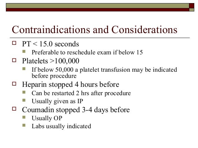
How long after spinal surgery can you stop taking medications?
They’ll be concerned if you are taking any medications that affect your ability to clot normally. You may need to stop taking any of the following medications both before (for 48 hours) and after (for 24 to 48 hours) your procedure:
How long does it take to get a CT scan of your spine?
This procedure typically takes about an hour and may cause some discomfort or a minor headache.
How long do you have to stop taking clot medications?
You may need to stop taking any of the following medications both before (for 48 hours) and after (for 24 to 48 hours) your procedure: