How much does an X ray machine cost?
The X-Ray systems come bundled from the manufactures into the most typically used systems. These systems range from $15,000 for the most basic chiropractic style package up to 60,000+ depending on the options, generator type selected, and specific needs of your practice. Examples of X-Ray Rooms Smaller Chiropractic X Ray Room with CR:
How much does a medical X ray machine cost?
GE XQi. $28,000 - $32,000. GE XRd. $21,750 - $28,250. Siemens Multix. $15,250 - $16,750. If you want to see more about X-ray equipment service, support, and parts you might also want to check out these related articles: 3 Key Elements of Effective X-Ray Preventive Maintenance.
What does an X ray machine look like?
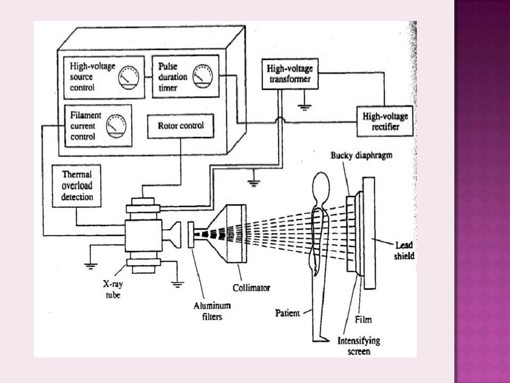
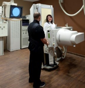
What does the equipment look like? The equipment typically used for bone x-rays consists of an x-ray tube suspended over a table on which the patient lies. A drawer under the table holds the x-ray film or image recording plate. Sometimes the x-ray is taken with the patient standing upright, as in cases of knee x-rays.
What is the X-ray machine used for?
X-Rays Uses Medical Use: They are used for medical purposes to detect the breakage in human bones. Security: They are used as a scanner to scan the luggage of passengers in airports, rail terminals, and other places. Astronomy: It is emitted by celestial objects and are studied to understand the environment. More items...

Are there different types of X ray machines?
X-ray machines come in a variety of styles, and each one has its separate strengths. The number of options is staggering so, to make the selection process more approachable, we'll break it down into three general categories: portables, floor-mounted, and ceiling-mounted.
How many types of X-ray are there?
The 5 different types of X-rays exist because each is used for a particular reason.
What are the 2 types of X-rays?
There are two types of X-ray generated: characteristic radiation and bremsstrahlung radiation.
What is the name of an X ray machine?
Medical x-ray systems. X-ray fluorescence analyzers (portable and bench-top) X-ray photoelectron spectrometers. Electron beam evaporators.
What are the five types of radiology?
The most common types of diagnostic radiology exams include:Computed tomography (CT), also known as a computerized axial tomography (CAT) scan, including CT angiography.Fluoroscopy, including upper GI and barium enema.Magnetic resonance imaging (MRI) and magnetic resonance angiography (MRA)Mammography.More items...•
Where are X-rays performed?
They normally are performed in a hospital radiology department, a dentist’s office, or a clinic that specializes in diagnostic ...
What is a conventional X-ray?
Conventional radiology is primarily used for viewing bones, bone fractures, tissues dense in calcium, dental X-rays, and the chest.
What does a 3D breast scan show?
Tumors will show up as irregular shaped white masses. Today’s mammography has advanced to include 3D images that show the entire breast. This new technology helps to increase the likelihood of early detection of breast cancers.
What is the procedure used to examine arteries, veins, and organs?
Angiography. This technique is used to examine arteries, veins, and organs in order to diagnose and treat blockages or other problems within the blood vessels. A thin tube known as a catheter is inserted into an artery or vein from the groin or arm.
What does a CT scan show?
A CT scan can show organs, the skeleton, tissues, and any abnormalities within these systems. This type of image can show tumors and lesions in the abdomen. A CT scan may also be used to look at the following:
What is the name of the X-ray that shows the breast?
Most women are familiar with this special X-ray known as a mammogram. It creates detailed images of the breast to be used both as a screening tool to detect cancer at an early stage, or diagnose breast disease from symptoms like pain, a lump, or discharge from the nipple.
What is a computerized tomography scan?
Computerized tomography combines a traditional X-ray with computer processing to create a better resolution. A CT scan creates a series of cross-sectional images or slices to form a 3D image. This allows Southwest Diagnostic Imaging to view different parts of the body from different angles.
When should X-rays be performed?
They should be performed only when the referring physician judges them to be necessary to answer a clinical question or to guide treatment of a disease.
Why are X-rays important?
X-ray imaging exams are recognized as a valuable medical tool for a wide variety of examinations and procedures. They are used to: noninvasively and painlessly help to diagnose disease and monitor therapy; support medical and surgical treatment planning; and.
Why should ionizing radiation be used in X-rays?
Therefore, all examinations using ionizing radiation should be performed only when necessary to answer a medical question, treat a disease, or guide a procedure. The clinical indication and patient medical history should be carefully considered before referring a patient for any X-ray examination.
What is the purpose of a medical imaging exam?
1. determining if the examination is needed to answer a clinical question ; 2. considering alternative examinations that use less or no radiation exposure, such as ultrasound or MRI, if medically appropriate; and. 3. checking the patient's medical imaging history to avoid duplicate examinations.
What is medical imaging?
Description. Medical imaging has led to improvements in the diagnosis and treatment of numerous medical conditions in children and adults. There are many types - or modalities - of medical imaging procedures, each of which uses different technologies and techniques. Computed tomography (CT), fluoroscopy, and radiography ...
What is the risk of X-rays?
Another risk of X-ray imaging is possible reactions associated with an intravenously injected contrast agent, or “dye”, that is sometimes used to improve visualization.
Does CT scan cause tissue damage?
tissue effects such as cataracts, skin reddening, and hair loss, which occur at relatively high levels of radiation exposure and are rare for many types of imaging exams. For example, the typical use of a CT scanner or conventional radiography equipment should not result in tissue effects, but the dose to the skin from some long, complex interventional fluoroscopy procedures might, in some circumstances, be high enough to result in such effects.
Intraoral X-ray Sensors
Although Renew Digital does not market or sell intraoral X-ray sensors, these devices have replaced film X-rays in many modern dental practices. They are faster and easier for capturing bitewings, FMX series, and other intraoral images and require much less radiation than their film counterparts.
Digital Panoramic X-rays
Many of today’s digital dental X-ray machines for panoramic imaging include additional features like extraoral bitewings and TMJ projections. Today’s digital panoramic X-ray machines are designed to enhance practice efficiency and streamline diagnoses. Some digital panoramic models are upgradeable to cephalometric and cone beam imaging as well.
Dental Cone Beam Systems
Cone beam computed tomography, or CBCT imaging, is quickly becoming the standard of care for many dental and dental specialty practices. It allows practitioners and their patients to have the most comprehensive view of the anatomy with 3D imaging to aid in diagnoses, treatment planning, and communicating with the patient.
Phosphor Plate X-ray Systems
Renew Digital does not market or sell phosphor plate X-ray systems, but they are still widely used in many dental practices. These allow for practices to use film equipment without upgrading to digital dental X-ray machines.
Consider Digital Dental X-Ray Machines
If your practice is still using traditional film or phosphor plate X-ray systems, we encourage you to consider the direct digital options. Search for a system that makes your existing imaging tasks more efficient but also allows you to add new options for diagnostics, patient education, and treatment planning.
Contact Us
At prices up to 30-50% less than the cost of new digital dental X-ray machines, it makes sense to shop with Renew Digital when considering upgrading your practice technology. With today’s current challenges to increase patient safety and practice efficiency, it’s never been a better time to go digital.
What are the different types of x-rays?
Here are the different types of commonly ordered medical x-rays and the reasons why they are performed. 1. Chest X-Ray. If you’re having trouble breathing, experiencing a persistent cough or feeling pain in your chest, your doctor may order a chest x-ray . A chest x-ray takes an image of your upper body and the bones and organs inside, ...
What Is an X-Ray?
An x-ray is a type of electromagnetic radiation. The light we see is also a form of electromagnetic radiation. However, unlike visible light, x-rays have much more energy, so they can pass through the human body. When x-rays pass through the part of your body under examination, they produce an image on film.
What is the purpose of x-rays for urinary tract?
With this type of x-ray, your doctor can evaluate your urinary tract and gain insight into the shape, size and position of your kidneys, ureters and bladder. They may determine the presence of kidney or ureteral stones or other reasons for your symptoms.
Why do doctors order x-rays?
It can feel scary when your doctor orders an x-ray, and no one wants to hear they need further testing. However, x-rays are quick, painless procedures that help your doctor identify the cause of your symptoms, so you can get the treatment you need. Doctors use different types of x-rays to diagnose everything from minor bone fractures to potentially life-threatening diseases. Overall, x-rays are a critical tool that helps save lives.
How to stand in front of xray machine?
During the imaging procedure, you’ll stand in front of the x-ray machine and face forward for one image. Usually, the x-ray technologist will then ask you to stand sideways for a second image. You’ll be asked to hold your breath for a few seconds as the x-ray is taken.
How to do a hand xray?
During a hand x-ray, you’ll be asked to place your hand flat on a table and keep it still as the image is taken. You may need to change the position of your hand a few times to provide the necessary images. The technologist will do everything they can to complete the x-ray quickly.
Why are x-rays important?
Overall, x-rays are a critical tool that helps save lives. We’ll explore the different types of x-rays doctors commonly order to help their patients get the proper care. If you have any questions about the x-ray process, reach out to us at Envision Imaging.
What is the purpose of X-rays?
This X-ray test is used to determine if there are obstructions in the coronary arteries, and may be conducted if a patient is experiencing chest pain. Contrast material is injected into one of the arteries of the heart.
How long does an MRI last?
It also requires the body to be placed in a special machine containing a strong magnet. There are different types of MRI machines and each procedure can last anywhere from 30 minutes to two hours. About 30 million MRI scans are performed in the U.S. each year.
What is CAT scan?
A CAT scan, or computed axial tomography, is a noninvasive test used to create multiple X-ray images, which are then used to create a 3D image of the patient's body structures. It may be performed alone, or with a contrast medium.
What is extremity MRI?
Extremity MRI: This is a diagnostic imaging procedure that uses a closed MRI machine to look at the tissues in the arms and legs. Unlike a traditional MRI procedure that uses a large tube-shaped device, an extremity MRI uses a smaller scanner designed specifically for the body's extremities. This eliminates the potential for claustrophobia, which some patients experience when enclosed in a full-body MRI machine. A traditional MRI requires you to lie completely still, but an extremity MRI won't limit your body movements quite as much. One may undergo an extremity MRI to diagnose any of the following conditions in the arms, legs, hands, and feet:#N##N#Arthritis#N#Fractures#N#Bone infections#N#Tumors of the bone or soft tissue#N#Nerve-related issues#N#Stress injuries or injuries related to torsion or heavy impact 1 Arthritis 2 Fractures 3 Bone infections 4 Tumors of the bone or soft tissue 5 Nerve-related issues 6 Stress injuries or injuries related to torsion or heavy impact
What is MRI in medical?
RAI Marketing. Physicians often use magnetic resonance imaging (MRI) to diagnose and treat medical conditions that can be seen only by use of x-ray or magnetic fields and radio frequencies. An MRI machine produces detailed pictures of internal body structures. This radiology imaging technique has improved by great leaps and bounds over the years, ...
What type of MRI is used to detect aneurysms?
3 Tesla MRI: This type of closed MRI uses magnetic fields that have double the strength of a traditional MRI machine, thus producing an even more detailed image in less time. It is commonly used to identify the signs of stroke, tumors, or aneurysms in the brain; to examine the heart and circulatory system for damage resulting from a heart attack or heart disease, or blockages in the blood vessels; to look for conditions such as arthritis, disc disease, or bone infections in the bones and joints; or to analyze the state of internal organs like the liver, kidneys, uterus, ovaries, or prostate.
Why is an open MRI considered an alternative to a closed MRI?
An open MRI was created to offer an alternative to patients who either have symptoms of anxiety or suffer from claustrophobia or obesity due to the enclosed-nature and duration of study normally attributed to closed MRIs. While the open MRI excels at providing comfort, it does not provide the same level of detail as a closed MRI at this time. See an example of a closed MRI on the left and an open MRI on the right.
Can you lie still during an MRI?
A traditional MRI requires you to lie completely still, but an extremity MRI won't limit your body movements quite as much. One may undergo an extremity MRI to diagnose any of the following conditions in the arms, legs, hands, and feet: . Arthritis.
