Correspondingly, how often should vital signs be taken during labor? Every 4 hours during the latent phase of labour. Every 2 hours during the active phase of labour.
What are the early signs of Labor?
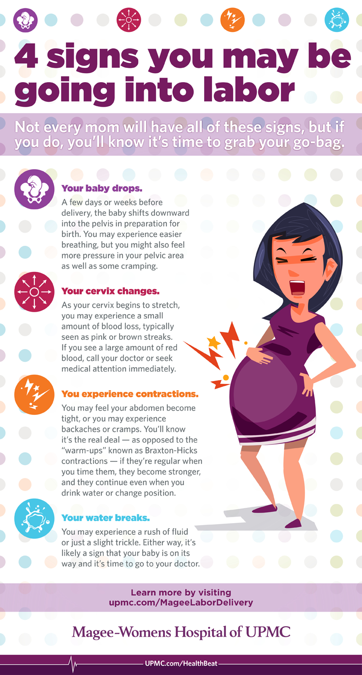
The appearance of bloody show (the discharge of a small amount of blood and mucous from the cervix), which may happen as early as three days before labor starts Irregular contractions of varying intensity.
How do you monitor the progress of Labor?
Inform patient on progress of her labor. Assist patient with pant-blow breathing. Monitor maternal vital signs and fetal heart rate every 30 minutes -1 hour, or depending on the doctor’s order. Contraction monitoring is also continued. When perineal bulging is noticeable, prepare for delivery.
How long does labor last during pregnancy?
If it is the first time that a woman is having a baby, labor usually lasts between 12 and 14 hours; in pregnancies that follow, labor is often shorter, averaging between six and eight hours. A woman should talk to her doctor about what the signs of the start of labor are. These include:
What does a nurse assess during labor and delivery?
In addition, the nurse assesses the following: vital signs, physical exam, contraction pattern (frequency, interval, duration, and intensity), intactness of membranes through vaginal exam, and fetal well-being through fetal heart rate, characteristic of amniotic fluid, and contractions.

What are normal vital signs during labor?
Check her vital signs Maternal pulse rate: normal range is 80-100 beats/minute, but should not be greater than 110 beats/minute in a woman in labour. Temperature: average 37oC; if it is between 37.5-38.4oC the woman has a low grade fever; if it is 38.5oC or above, she has a high grade fever.
What is the frequency of vital signs monitoring during postpartum?
Nursing guidelines in the United States recommend the assessment of maternal HR and BP every 15 minutes for two hours in the immediate postpartum period and more frequently if complications are encountered [6].
How often should the mother's blood pressure and pulse rate be checked during oxytocin infusion?
Assess blood pressure, pulse, respiratory rate every hour. Assess intake and output every 4 hours.
How often are OBS done in hospital?
According to the hospital escalation protocol, patients are considered at high risk when their NEWS score is 6 or above, requiring observations at least once every 4 hours.
How often should a nurse check on a patient?
With this in mind, one simple recommendation is to perform vital signs reassessment every 2 hours for monitored patients and every 4 hours for patients who aren't on a cardiac monitor.
At what time interval should BP be checked in a woman who has just delivered a baby?
Earlier postpartum visits Six weeks is too long to wait for a blood pressure check. By that time, the problem could reach dangerous levels. So, ACOG recommends that patients with hypertension during pregnancy see their Ob/Gyn specialist three days after discharge from the hospital.
When is the best time to assess FHR and maternal BP during labor?
Half-hourly during the active phase of labour. At least every 15 minutes if fetal distress is suspected. Directly after every bout of maternal pushing during the second stage of labour.
How do you monitor labor progress?
Cervical dilatation, descent of the fetal head and uterine contractions are used in assessing the progress of labour. About 1 cm/hour cervical dilatation and 1 cm descent in four hours indicate good progress in the active first stage.
What assessments are vital for the nurse to perform on the postpartum patient?
BREASTS. The breasts are assessed for: ... UTERUS. The fundus is assessed for: ... BOWEL. Assessment of the bowel is important in all postpartum patients. ... BLADDER. Assessment of urination and bladder function includes: ... LOCHIA. ... EPISIOTOMY/PERINEUM. ... LOWER EXTREMITIES. ... CESAREAN SECTION ISSUES.More items...
What is normal blood pressure postpartum?
Normal: A normal, healthy blood pressure reading is 120/80. Once we creep over that, the brain's ability to regulate blood flow is impacted at varying levels, depending on the patient. High: During pregnancy and the few weeks after, your blood pressure may be 140/90.
What are normal vital signs for a newborn?
Able to maintain stable body temperature of 97.0°F to 98.6°F (36.1°C to 37°C) in normal room environment. Heartbeat. Normally 120 to 160 beats per minute. It may be much slower when an infant sleeps.
What is postnatal assessment?
First 24 hours after birth: All postpartum women should have regular assessment of vaginal bleeding, uterine contraction, fundal height, temperature and heart rate (pulse) routinely during the first 24 hours starting from the first hour after birth. Blood pressure should be measured shortly after birth.
Where are vital records located?from cdc.gov
states, New York City, and the District of Columbia; Arizona, Colorado, Delaware, Florida, Georgia, Hawaii, Illinois, Mississippi, North Carolina, Ohio, South Carolina, Tennessee, and Utah maternal mortality review committees; Building U.S. Capacity to Review and Prevent Maternal Deaths Team, CDC; CDC Foundation; Association of Maternal and Child Health Programs. CDC developed this report using information from state maternal mortality review committees and is responsible for the conclusions in the report.
How to determine timing of death in pregnancy?from cdc.gov
Otherwise, timing of death in relation to the end of pregnancy was determined by comparing date of death on the death certificate with date of live birth or fetal death on linked birth or fetal death certificates. The specific timing of postpartum deaths was classified as unknown if there was no linked birth or fetal death certificate.
What is PMSS in pregnancy?from cdc.gov
PMSS was established in 1986 by CDC and the American College of Obstetricians and Gynecologists (ACOG) to evaluate the causes of death and risk factors associated with pregnancy-related deaths. PMSS methodology has been described previously ( 2 ); CDC’s Division of Reproductive Health requests that all states, the District of Columbia, and New York City send death certificates, linked live birth or fetal death certificates, and additional data when available, on deaths that occurred during pregnancy or within 1 year after delivery. Information on individual deaths are reviewed by medically trained epidemiologists to determine the pregnancy-relatedness and cause ( 4 ). A death is determined to be pregnancy-related if the death was caused by a pregnancy complication, a chain of events initiated by pregnancy, or the aggravation of an unrelated condition by the physiologic effects of pregnancy. Cause of death coding includes the following 10 mutually exclusive categories: hemorrhage; infection; amniotic fluid embolism; thrombotic pulmonary or other embolism (i.e., air, septic, or fat); hypertensive disorders of pregnancy (i.e., preeclampsia or eclampsia)*; anesthesia complications; cerebrovascular accidents †; cardiomyopathy; other cardiovascular conditions (e.g., congenital heart disease, ischemic heart disease, cardiac valvular disease, hypertensive heart disease, and congestive heart failure); and other noncardiovascular medical conditions (e.g., endocrine, hematologic, immunologic, and renal).
What is the leading cause of death during the first 6 weeks of pregnancy?from cdc.gov
Hemorrhage, hypertensive disorders of pregnancy, and infection were leading causes of death during the first 6 days postpartum. From 6 weeks postpartum (43 days) through the end of the first year (365 days), cardiomyopathy was the leading cause of death.
How many days postpartum do black women die?from cdc.gov
Timing of deaths did not significantly differ between black and white women for most periods; however, a greater proportion of deaths among black women (14.9%) occurred 43–365 days postpartum compared to the proportion of deaths among white women (10.2%) that occurred during the same period (p<0.01).
What is the most common cause of death in the late postpartum period?from cdc.gov
Cardiomyopathy was the most common cause of death in the late postpartum period (43–365 days postpartum). The higher proportion of pregnancy-related deaths in the late postpartum period among black women is likely attributable to higher proportion of pregnancy-related deaths due to cardiomyopathy among these women ( 8 ).
How many days postpartum do you die from cardiomyopathy?from cdc.gov
Deaths caused by cardiomyopathy most commonly occurred 43–365 days postpartum; deaths caused by other cardiovascular conditions occurred most commonly during pregnancy and within 42 days postpartum. The leading causes of death also varied by time relative to the end of pregnancy.
What are the vital signs of a woman in labor?
When women first present to the labor and delivery unit, vital signs, including temperature, heart rate, oxygen saturation, respiratory rate, and blood pressure, should be obtained and reviewed for any abnormalities. The patient should be placed on continuous cardiotocographic monitoring to ensure fetal wellbeing.
How does labor start?
The first stage of labor begins when labor starts and ends with full cervical dilation to 10 centimeters. Labor often begins spontaneously or may be induced medically for a variety of maternal or fetal indications. Methods of inducing labor include cervical ripening with prostaglandins, membrane stripping, amniotomy, and intravenous oxytocin. Although precisely determining when labor starts may be inexact, labor is generally defined as beginning when contractions become strong and regularly spaced at approximately 3 to 5 minutes apart. Women may experience painful contractions throughout pregnancy that do not lead to cervical dilation or effacement, referred to as false labor. Thus, defining the onset of labor often relies on retrospective or subjective data. Friedman et al. were some of the first to study labor progress and defined the beginning of labor as starting when women felt significant and regular contractions. He graphed cervical dilation over time and determined that normal labor has a sigmoidal shape. Based on the analysis from his labor graphs, he proposed that labor has three divisions. First, a preparatory stage marked by slow cervical dilation, with large biochemical and structural changes. This is also known as the latent phase of the first stage of labor. Second, a much shorter and rapid dilational phase is also known as the active phase of the first stage of labor. Third, a pelvic division phase, which takes place during the second stage of labor.
How many centimeters does it take for a cervical dilation to occur?
The cervix changes more rapidly and predictably in the active phase until it reaches 10 centimeters and cervical dilation and effacement are complete. Active labor with more rapid cervical dilation generally starts around 6 centimeters of dilation.
How to manage low risk labor?
Labor is a natural process, but it can suffer interruption by complicating factors, which at times necessitate clinical intervention. The management of low-risk labor is a delicate balance between allowing the natural process to proceed while limiting any potential complications. During labor, cardiotocographic monitoring is often employed to monitor uterine contractions and fetal heart rate over time. Clinicians monitor fetal heart tracings to evaluate for any signs of fetal distress that would warrant intervention as well as the adequacy or inadequacy of contractions. Vital signs of the mother are taken at regular intervals and whenever concerns arise regarding a clinical status change. Laboratory testing often includes the hemoglobin, hematocrit, and platelet count and is sometimes repeated following delivery if significant blood loss occurs. Cervical exams are usually performed every 2 to 3 hours unless concerns arise and warrant more frequent exams. Frequent cervical exams are associated with a higher risk of infection, especially if a rupture of membranes has occurred. Women should be allowed to ambulated freely and change positions if desired. An intravenous catheter is typically inserted in case it is necessary to administer medications or fluids. Oral intake should not be withheld. If the patient remains without food or drink for a prolonged period of time, intravenous fluids should be considered to help replace losses but do not need to be used continuously on all laboring patients. Analgesia is offered in the form of intravenous opioids, inhaled nitrous oxide, and neuraxial analgesia in those who are appropriate candidates. Amniotomy is considered on an as-needed basis for fetal scalp monitoring or labor augmentation, but its routine use should be discouraged. Oxytocin may be initiated to augment contractions found to be inadequate.
What is the process of delivery of a fetus and placenta?
Labor is the process through which a fetus and placenta are delivered from the uterus through the vagina. Human labor divides into three stages. The first stage is further divided into two phases. Successful labor involves three factors: maternal efforts and uterine contractions, fetal characteristi ….
What are the stages of labor?
Stages of Labor. Labor is the process through which a fetus and placenta are delivered from the uterus through the vagina. Human labor divides into three stages. The first stage is further divided into two phases. Successful labor involves three factors: maternal efforts and uterine contractions, fetal characteristi ….
What are the common complaints of a labor patient?
Common chief complaints include painful contractions, vaginal bleeding/bloody show, and fluid leakage from the vagina. It is up to the clinician to determine if the patient is in labor, defined as regular, clinically significant contractions with an objective change in cervical dilation and/or effacement.
What are the vital signs of a woman in labor?
When women first present to the labor and delivery unit, vital signs, including temperature, heart rate, oxygen saturation, respiratory rate, and blood pressure, should be obtained and reviewed for any abnormalities. The patient should be placed on continuous cardiotocographic monitoring to ensure fetal wellbeing.
How many stages of labor are there?
Labor is the process through which a fetus and placenta are delivered from the uterus through the vagina.[1] . Human labor divides into three stages. The first stage is further divided into two phases.
How long does the second stage of labor last?
The second stage of labor commences with complete cervical dilation to 10 centimeters and ends with the delivery of the neonate. This was also defined as the pelvic division phase by Friedman. After cervical dilation is complete, the fetus descends into the vaginal canal with or without maternal pushing efforts. The fetus passes through the birth canal via 7 movements known as the cardinal movements. These include engagement, descent, flexion, internal rotation, extension, external rotation, and expulsion.[1] In women who have delivered vaginally previously, whose bodies have acclimated to delivering a fetus, the second stage may only require a brief trial, whereas a longer duration may be required for a nulliparous female. In parturients without neuraxial anesthesia, the second stage of labor typically lasts less than three hours in nulliparous women and less than two hours in multiparous women. In women who receive neuraxial anesthesia, the second stage of labor typically lasts less than four hours in nulliparous women and less than three hours in multiparous women.[1] If the second stage of labor lasts longer than these parameters, then the second stage is considered prolonged. Several elements may influence the duration of the second stage of labor, including fetal factors such as fetal size and position, or maternal factors such as pelvis shape, the magnitude of expulsive efforts, comorbidities such as hypertension or diabetes, age, and history of previous deliveries. [8]
How does labor start?
The first stage of labor begins when labor starts and ends with full cervical dilation to 10 centimeters.[1] Labor often begins spontaneously or may be induced medically for a variety of maternal or fetal indications.[5] Methods of inducing labor include cervical ripening with prostaglandins, membrane stripping, amniotomy, and intravenous oxytocin.[5] Although precisely determining when labor starts may be inexact, labor is generally defined as beginning when contractions become strong and regularly spaced at approximately 3 to 5 minutes apart.[1] Women may experience painful contractions throughout pregnancy that do not lead to cervical dilation or effacement, referred to as false labor. Thus, defining the onset of labor often relies on retrospective or subjective data. Friedman et al. were some of the first to study labor progress and defined the beginning of labor as starting when women felt significant and regular contractions.[6] He graphed cervical dilation over time and determined that normal labor has a sigmoidal shape. Based on the analysis from his labor graphs, he proposed that labor has three divisions. First, a preparatory stage marked by slow cervical dilation, with large biochemical and structural changes. This is also known as the latent phase of the first stage of labor. Second, a much shorter and rapid dilational phase is also known as the active phase of the first stage of labor. Third, a pelvic division phase, which takes place during the second stage of labor. [1]
How is labor monitored?
Clinicians monitor fetal heart tracings to evaluate for any signs of fetal distress that would warrant intervention as well as the adequacy or inadequacy of contractions. Vital signs of the mother are taken at regular intervals and whenever concerns arise regarding a clinical status change. Laboratory testing often includes the hemoglobin, hematocrit, and platelet count and is sometimes repeated following delivery if significant blood loss occurs. Cervical exams are usually performed every 2 to 3 hours unless concerns arise and warrant more frequent exams. Frequent cervical exams are associated with a higher risk of infection, especially if a rupture of membranes has occurred. Women should be allowed to ambulated freely and change positions if desired.[3] An intravenous catheter is typically inserted in case it is necessary to administer medications or fluids. Oral intake should not be withheld. If the patient remains without food or drink for a prolonged period of time, intravenous fluids should be considered to help replace losses but do not need to be used continuously on all laboring patients.[3] Analgesia is offered in the form of intravenous opioids, inhaled nitrous oxide, and neuraxial analgesia in those who are appropriate candidates.[4] Amniotomy is considered on an as-needed basis for fetal scalp monitoring or labor augmentation, but its routine use should be discouraged.[3] Oxytocin may be initiated to augment contractions found to be inadequate.
What is the first stage of labor?
Labor is a process that subdivides into three stages. The first stage starts when labor begins and ends with full cervical dilation and effacement. The second stage commences with complete cervical dilation and ends with the delivery of the fetus. The third stage initiates after the fetus is delivered and ends when the placenta is delivered.
What is the presenting part of labor?
The presenting fetal part also begins the process of engagement into the pelvis during the first stage. Throughout the first stage of labor, serial cervical exams are done to determine the position of the fetus, cervical dilation, and cervical effacement.
How do you know if you are in labor?
Symptoms. A woman should talk to her doctor about what the signs of the start of labor are. These include: Back pain. The appearance of bloody show (the discharge of a small amount of blood and mucous from the cervix), which may happen as early as three days before labor starts.
How long does labor take?
It usually begins within two weeks before or two weeks after a woman's estimated due date. If it is the first time that a woman is having a baby, labor usually lasts between 12 and 14 hours; in pregnancies that follow, labor is often shorter, averaging between six and eight hours.
What is the best anesthesia for labor?
The usual forms of anesthesia given during labor are: 1 Pudendal block, in which a local anesthetic is injected through the vaginal wall. This is useful for uncomplicated deliveries and ones where the mother wants to push during delivery. 2 Regional anesthesia. The most often used for labor and delivery is an injection of a local anesthetic in the back. 3 General anesthesia. This is not used for routine deliveries because it can depress both the mother's and the fetus's vital signs.
How long does it take for amniotic fluid to leak through the cervix?
When this happens a woman should call her doctor right away. Between 80 and 90% of women with ruptured membranes go into labor within 24 hours. If this doesn't happen and the baby is due, labor is induced to reduce the risk of infection.
Why is it important to monitor heart rate during labor?
Between a third to half of the babies who develop fetal distress or die during delivery do so without any significant signs beforehand. Electronically monitoring the baby can help pinpoint fetal distress before it becomes dire.
What does a doctor do when a baby is in labor?
If labor is active and the baby is due, a labor-and-delivery nurse or a doctor will do an internal examination with a gloved hand to evaluate how the labor is going.
What happens if a baby is not born in labor?
If this doesn't happen and the baby is due, labor is induced to reduce the risk of infection. When a woman in labor arrives at the hospital, her blood pressure, heart and breathing rates, temperature and weight are recorded. She will be checked for signs of swelling. Blood and urine tests will be given.
How many phases are there in labor?
The progression of labor is traditionally divided into three phases, and each phase deals with different concerns and considerations. Having gained mastery of this, nurses are able to implement nursing interventions to safeguard the welfare of both the mother and the baby. Establishing Therapeutic Relationship.
What information is taken during admission to labor?
When a patient arrives at the labor floor, pertinent information about the pregnant woman’s health history is taken during admission. These include personal data (e.g. blood type, allergies, etc.), previous illness, pregnancy complications, preferences for labor and delivery, and childbirth preparations. Standard obstetric, medical, and social history taking is also done.
What is the transition phase of cervical dilation?
Transition Phase starts from 8 cm cervical dilatation to 10 cm (full) cervical dilatation and full cervical effacement. During this time, patient may be exhausted and withdrawn or aggressive and restless. Patient’s urge to push is noticeable. Here are nursing responsibilities in this phase:
Why is it important to determine when a patient last voided?
Determine when patient last voided because a full bladder can hinder fast labor progress.
What is the best position for a woman during labor?
Allow patient to be continually active. Upright maternal positions are recommended for women on the first stage of labor. Patients without pregnancy complications can still walk around and make necessary birth preparations.
Why is the first 4 hours of labor called the 4th stage?
The first four hours after birth is sometimes referred to as the fourth stage of labor because this is the most critical period for the mother. The nurse is set to perform nursing interventions that would prevent the patient from infection and hemorrhage.
When is admission to labor room done?
Admission into labor room is only done when the patient is in active labor.
What is fetal heart monitoring?
The Association of Women’s Health, Obstetric and Neonatal Nurses (AWHONN) asserts that the availability of registered nurses (RNs) and other health care professionals who are skilled in fetal heart monitoring (FHM) techniques, including auscultation and electronic fetal monitoring (EFM), is essential to maternal and fetal well-being during antepartum care, labor, and birth. Fetal heart monitoring requires advanced assessment and clinical judgment skills and should not be delegated to unlicensed assistive personnel or others who do not possess the appropriate licensure, education, and skills validation. A woman’s preferences and clinical presentation should guide selection of FHM techniques with consideration given to use of the least invasive methods. In general, the least invasive method of monitoring is preferred in order to promote physiologic labor and birth. Labor is dynamic; therefore, consideration of maternal preferences and identification of risk factors should occur upon admission to the birth setting and should be ongoing throughout labor.
How often do you use auscultation?
The range of frequency of assessment using auscultation in these studies varied from every 15-30 minutes during the first stage of labor to every 5-15 minutes during the second stage of labor. In most studies, a 1:1 nurse to patient ratio was used for auscultation protocols. These classic studies included low risk and/or high risk patient populations. Specific dilatation parameters for stages of labor generally were not defined in these studies, with the exception of
What is the purpose of intrapartum fetal surveillance?
The intent of intrapartum fetal surveillance is to assess uterine activity, fetal well-being, and the fetal heart rate (FHR) response to labor in order to make appropriate, physiologically based clinical decisions (
Is electronic fetal heart monitoring a substitute for nursing care?
Electronic fetal heart monitoring is not a substitute for appropriate professional nursing care and support of women in labor. Perinatal nurses are most often the primary health care professionals responsible for FHM. AWHONN’s Guidelines for Professional Registered Nurse Staffing for Perinatal Units (
How often should you reassess your vital signs?from nursingcenter.com
Patients with abnormal vital signs should be reassessed no less frequently than every 2 hours for the first 4 hours, then every 4 hours if clinically stable. * ESI Level 4: Vital signs should be reassessed per acuity and clinical assessment, but no less frequently than every 4 hours. * ESI Level 5: Upon discharge.
What is the frequency of vital signs reassessment?from nursingcenter.com
A review of the Agency for Healthcare Research and Quality's Emergency Severity Index (ESI) showed that there's no mention of the frequency of vital signs reassessment. On the contrary, vital signs assessment in triage is only required for patients who meet Level 3 criteria (patients who are predicted to require two or more resources). 4 The ESI consists of a five-level triage algorithm designed for EDs that places patients into five groups from ESI 1 (most urgent) to ESI 5 (least urgent) on the basis of acuity and the amount of resources required. 4 A review of the Emergency Nurses Association guidelines showed that they don't directly provide a recommendation for reassessing vital signs. 5 Thus, it's up to the facility to determine the frequency of vital signs reassessment based on the cliental served. Vital signs reassessment policies were reviewed from various facilities and all varied.
Why is it important to have vital signs reassessed?from nursingcenter.com
Numerous errors can occur in the ED, including missing signs and symptoms of deteriorating patient condition. Vital signs assessment serves as an early warning of a change in patient condition, playing an important role in assisting the healthcare professional to prevent adverse events. That's why it's necessary to ensure that vital signs are reassessed accordingly after triage.
What happens if a nurse doesn't sign a note?from nursingcenter.com
In some EHR systems, if the nurse doesn't sign the note, then the physician won't have access to the information, which can create a delay in patient treatment. One study found inconsistencies regarding documentation of ED patients' vital signs in the appropriate EHR fields. 1.
Where to record vital signs?from nursingcenter.com
The best practice is to record all vital signs in the vital signs section of the cover sheet or designated location based on the type of electronic health record (EHR) system being used. The vital signs section of the EHR was developed to allow healthcare providers quick access to the information. In some EHR systems, if the nurse doesn't sign the note, then the physician won't have access to the information, which can create a delay in patient treatment. One study found inconsistencies regarding documentation of ED patients' vital signs in the appropriate EHR fields. 1
How long does it take to get BP in the ED?from nursingcenter.com
In one exploratory study reviewing over 43,232 patient visits to 94 different EDs, the median time between documentation of BP in the ED was every 2.3 hours for all patients. 1 In a retrospective chart review of 202 randomly selected adult ED patients, it was concluded that a greater time between vital signs assessment can lead to errors by not detecting changes in patient condition. 2
What are the three things to look for in a woman in labor?
In order to memorise what aspects to inspect on the abdomen of a woman in labour, you can take the initial ‘S’ letters of the three points to look out for: size, shape and scars.
How to check fetal heart rate?
Use a fetoscope or stethoscope to listen to the fetal heart rate immediately after a contraction. Listening to sounds inside the abdomen is called auscultation. Count the number of fetal heartbeats for a full minute at least once every 30 minutes during the active phase first stage of labour and every 5 minutes during the second stage. If there are fetal heart rate abnormalities (less than 120 or more than 160 beats per minute, sustained for 10 minutes), suspect fetal distress and refer urgently to a health facility, unless the labour is progressing fast and the baby is about to be born. (You will learn about fetal distress in Study Session 4.)
What is the function of a vaginal exam?
The functions of a vaginal examination are to: Determine if true labour has begun and the stage it has reached, based on measuring the dilatation of the cervix. Assess the progress of labour in terms of the rate of increase in cervical dilatation and the descent of the fetus down the birth canal.
How to get a vaginal exam done?
Wash your hands thoroughly with soap and clean water for two full minutes. Then put on new sterile gloves. Tell the mother what you are going to do. Vaginal examination is done using two gloved fingers. Try to collect all the information you need before withdrawing from the vagina, because once you have withdrawn your fingers you should not put them back in again.
What are the dangers of vaginal bleeding?
Danger symptoms include vaginal bleeding (heavier than show), persistent headache, blurring of vision, convulsions, loss of consciousness, epigastric or severe abdominal pain, fever, leakage of amniotic fluid before the onset of labour, and abnormal vaginal discharge.
What is a sign in a health care?
(A symptom is something that a person experiences and can tell you about; a sign is something that only a trained health worker will notice, or can discover from an examination or test. )
How many live babies can a woman have at 40 weeks?
A woman comes to your Health Post in labour at full term. She tells you that she has previously given birth to two live babies (both at the gestational age of 40 weeks), and one dead baby (stillbirth) at 32 weeks. She also had a spontaneous miscarriage at 26 weeks. Record the gravidity and parity of this woman.
How often should you check vital signs during a blood transfusion?from iv-therapy.net
What is your standard for frequency of vital signs during a blood transfusion? Most references I have looked at state "vital signs according to your institution's policy". Transfusion Services offered no standard in their regulations either. We currently check VS at 15 minutes, then every 30 minutes thereafter. There is some interest in going to 15 minutes after starting, then hourly thereafter.
How long after a transfusion should you have vitals?from iv-therapy.net
Evidence Based recommendations, and the Joint Commission standard, is for vitals signs pre-transfusion, 15 minutes into the transfusion, and within 1 hour after end of the transfusion (no routine vitals are recommended at any set interval during the transfusion).