
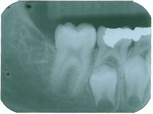
Periapical radiography Periapical radiography describes intraoral techniques designed to show individual teeth and the tissues around the apices. Each image usually shows two to four teeth and provides detailed information about the teeth and the surrounding alveolar bone.
What does periapical mean?
peri· api· cal | -ˈā-pi-kəl also -ˈap-i-kəl Medical Definition of periapical : of, relating to, occurring in, affecting, or being the tissues surrounding the apex of the root of a tooth periapical infection a periapical abscess Learn More About periapical Dictionary Entries Near periapical periaortic periapical periaqueductal
What are Intraoral periapical images?
Intraoral images can be divided into three categories: Periapical projections, which show the entire length of the tooth and the surrounding bone Bitewing projections, which show only the crowns of teeth and the adjacent alveolar crests Occlusal projections, which show an area of teeth and bone larger than periapical images.
What is an associated periapical radiolucency?
Periapical radiolucency is characterized by chronic or acute inflammatory lesions or lacerations around the apex of your tooth’s root. It is usually triggered by bacterial invasion of the dental pulp and its presence is often an indication of poor oral health status.
What is periapical pathology?
Periapical lesions resulting from necrotic dental pulp are among the most common pathologic conditions within alveolar bone, the majority being periapical cysts or granulomas. Diagnosis of these lesions frequently depends on conventional radiography and this is important in determining the treatment plan.
How would you describe dental radiographic findings?
In general, findings in dental radiographs are classified as radiolucent, radiopaque or mixed density, depending on their appearance when compared to the adjacent bone. One of the most common clinical observations seen among radiolucent lesions is the pericoronal radiolucency around unerupted teeth.
How do you read a periapical radiograph?
13:0324:47How to Read Dental X-Rays - YouTubeYouTubeStart of suggested clipEnd of suggested clipAnd here is actually one of the maxillary sinuses above the roots of the teeth. So here in thisMoreAnd here is actually one of the maxillary sinuses above the roots of the teeth. So here in this periapical. We can actually see a small triangular radial lucency forming on the side of this tooth.
What is the purpose of periapical film?
The film shows the entire crown and root of the teeth which provides vital information to aid in the diagnosis of the most common dental diseases; specifically tooth decay, tooth abscesses and periodontal bone loss or gum disease.
What is intraoral periapical radiograph?
Intraoral periapical radiographs (IOPAR) are widely used for the preoperative planning and evaluation for most minor oral surgical procedures owing to it simplicity, significantly lower cost, less radiation exposure and easy availability in a dental clinical set-up.
What appears radiopaque on a dental radiograph?
Radiolucent structures appear dark or black in the radiographic image. Radiopaque – Refers to structures that are dense and resist the passage of x-rays. Radiopaque structures appear light or white in a radiographic image.
What are the limitations of periapical radiography?
The periapical radiography is nowadays the main radiographic investigations used but presents some limits as 3D anatomic alteration, geometric compression, and possible anatomical structures overlapping that can obscure the area of interest.
What is the difference between bitewing and a periapical image?
What is the difference between a bitewing and a periapical image? Bitewing is upper and lower teeth in occlusion. Periapical is entire tooth from crown to 2-3mm past the apex of the root to show periapical bone.
What are the techniques for periapical radiography?
Two types of exposure techniques may be used for intraoral periapical radiography: the paralleling and the bisecting angle technique (Figures 1 and 2). With the paralleling technique, the tooth and the sensor are both kept on a parallel planes.
How do you write a radiographic interpretation?
1:386:07Radiographic interpretation made easy case 8 Solved examplesYouTubeStart of suggested clipEnd of suggested clipBecause generally between the premolar roots you would see a faint radiolucency. And that is what isMoreBecause generally between the premolar roots you would see a faint radiolucency. And that is what is the outline of the mental foramen. Which is here pointed out by the red dotted.
What are the three types of dental images?
There are three types of diagnostic radiographs taken in today's dental offices -- periapical (also known as intraoral or wall-mounted), panoramic, and cephalometric. Periapical radiographs are probably the most familiar, with images of a few teeth at a time captured on small film cards inserted in the mouth.
What does it mean intraoral periapical first film?
This dental procedure code, refers to a type of X-ray known as periapical. This term is used because these X-rays capture the entire tooth all the way down to the tissues at the tip of the tooth root – an area referred to as the periapical area.
How do you read a radiograph?
How to interpret the radiograph?Describe the location of the lesion.Describe the internal structure of the lesion: radiopaque or radiolucent.Describe the size, shape and border of the lesion.Describe the effect of the lesion to the surrounding structures.
How do you read an occlusal radiograph?
7:4512:56Occlusal Radiography - YouTubeYouTubeStart of suggested clipEnd of suggested clipThe top edge of the x-ray cone is lined up parallel to the plane of the film. And the verticalMoreThe top edge of the x-ray cone is lined up parallel to the plane of the film. And the vertical angulation is noted on the vertical angulation scale.
What are the techniques for periapical radiography?
Two types of exposure techniques may be used for intraoral periapical radiography: the paralleling and the bisecting angle technique (Figures 1 and 2). With the paralleling technique, the tooth and the sensor are both kept on a parallel planes.
How do you read a vet radiograph?
2:3426:07VET Talks - How to Read Radiographs - YouTubeYouTubeStart of suggested clipEnd of suggested clipSo whatever you see could be anywhere. From left right. When you do event reduce or dorsoventral toMoreSo whatever you see could be anywhere. From left right. When you do event reduce or dorsoventral to flatten it the other way around or algebra.
What is a periapical radiograph?
PERIAPICAL RADIOGRAPH. 2. DENTAL X-RAYS X-rays are produced by “boiling off” electrons from a filament (the cathode)and accelerating the el to the target at the anode. The accelerated x-rays are decelerated by the target material, resulting in bremsstrahlung. 3. History of X-Ray DISCOVEY Wilhelm Conrad Roentgen, ...
How are horizontal and vertical angulations of the X-ray tubehead determined?
26. The horizontal and vertical angulations of the X-ray tubehead are automatically determined by the positioning devices if placed correctly. The X-ray beam is aimed accurately at the centre of the film — all areas of the film are irradiated and there is no coning off or cone cutting. Reproducible radiographs are possible at different visits and with different operators.
How are dental x-rays produced?
2. DENTAL X-RAYS X-rays are produced by “boiling off” electrons from a filament (the cathode)and accelerating the el to the target at the anode. The accelerated x-rays are decelerated by the target material, resulting in bremsstrahlung.
Who invented the x-ray?
3. History of X-Ray DISCOVEY Wilhelm Conrad Roentgen, Bavarian physicist, discovered the x-ray on 1895. In 1895, German dentist Otto Walkhoff made the 1st dental radiograph. In 1895, New York physician made the 1st dental radiograph in the united states using the skull.
What is the most important diagnostic tool in dentistry?
6. Radiograph in dentistry X-rays in dentistry serves as the most important diagnostic tool. Radiograph in dentistry are divided into two: 1. Intraoral radiograph. 2. Extraoral radiograph.
What is periapical radiograph?
If a suspected root fracture is not visible on a periapical radiograph, a second radiograph made with a different angulation may be helpful. A fracture that is not perpendicular to the beam may not be detectable. Thus a tooth with a history of trauma but no associated clinical finding should be monitored and evaluated radiographically on a periodic basis.
What should a dentist consider when evaluating a radiograph?
After concluding that a patient requires a radiograph, the dentist should consider which radiographic examination is most appropriate to meet all the patient's diagnostic and treatment planning needs. In choosing one, the dentist considers the anatomic relationships, the size of the field, and the radiation dose from each view. For example, a panoramic radiograph provides broad area coverage with moderate resolution. Intraoral films give more detailed information but a significantly higher radiation dose per unit area exposed. The clinician must use clinical judgment to weigh these factors.
What is the first step in ordering a radiograph?
Accordingly, the first step is a careful examination of the patient. The clinical examination provides indications as to the nature and extent of the radiographic examination appropriate to the situation. Transillumination of anterior teeth should be conducted to evaluate for interproximal decay. Posterior interproximal radiographs form the foundation of the ADA guidelines. Other intraoral or extraoral radiographs should be added as indicated by the clinical examination.
How often should you take a radiograph for caries?
The rate of caries varies from person to person and the length of intervals between exposures must be determined clinically. The rate of caries progression is more rapid in deciduous teeth. In carious prone patients radiograph should be taken every 6 - 12 months; in non-caries prone patients every 18 - 24 months is adequate.
What is occlusal view?
Occlusal views are intraoral radiographs in which the film is positioned in the occlusal plane. They are often used in children in place of periapical views because of the small size of the patient's mouth. In adults, occlusal radiographs may supplement periapical views, providing visualization of a greater area of teeth and bone. They are useful for demonstrating impacted or abnormally placed maxillary anterior teeth or visualizing the region of a palatal cleft. They may also demonstrate buccal or lingual expansion of bone.
