What is the difference between B cell receptor and antibody?
B cell receptor and antibody are two types of molecules that relate to B cells. The B cells are one of the two types of lymphocytes that the the bone marrow produce. What is an Antibody — Definition, Structure, Role 3. B cell receptor BCR is a type of receptor molecule that we can find on the surface of the B cells.
Do all B cells produce the same antibodies?
B Cells Make Antibodies as Both Cell-Surface Receptors and Secreted Molecules. As predicted by the clonal selection theory, all antibody molecules made by an individual B cell have the same antigen-binding site. The first antibodies made by a newly formed B cell are not secreted.
How do B cells react to antigens?
- B cells express multiple identical antigen binding receptors (BCRs) on their surface.
- When BCRs are shed into body fluids, they are called immunoglobulins or antibodies.
- BCRs consist of two heavy and two light chains bound together by disulfide bonds.
- B cells can recognize most antigens without prior processing. ...
What is one similarity between cell receptors and antibodies?
Similarities Between B Cell Receptor and Antibody. B cell receptor and antibody are two types of functional molecules that relate to B cells. Both are immunoglobulin molecules. Therefore, they contain two heavy (H) polypeptide chains and two light (L) chains.

Are B cell receptor antibodies?
Antibodies. These are cell surface receptors that are attached to the plasma membrane. The B-Cells produce protein structures called antibodies. The B-cell receptors attach to free moving antigens.
What type of antibody is B cell receptor?
The B cell receptor is composed of two parts: A membrane-bound immunoglobulin molecule of one isotype (IgD, IgM, IgA, IgG, or IgE). With the exception of the presence of an integral membrane domain, these are identical to a monomeric version of their secreted forms.
What are B cell receptors known as?
The B cell receptor (BCR) stands sentry on the front lines of the body's defenses against infection. Embedded in the surface of the B cell—one of the principal immune cells—its job is to bind foreign substances called antigens.
Is an antibody a receptor?
An antigen receptor is basically an antibody protein that is not secreted but is anchored to the B-cell membrane. …from the trillions of different antigen receptors that are produced by the B and T lymphocytes.
Is the BCR an antibody?
Membrane-bound immunoglobulin on the B-cell surface serves as the cell's receptor for antigen, and is known as the B-cell receptor (BCR). Immunoglobulin of the same antigen specificity is secreted as antibody by terminally differentiated B cells—the plasma cells.
What is the difference between BCR and TCR?
TCRs usually recognize antigenic peptides in complex with major histocompatibility complex (MHC) molecules, whereas BCRs and antibodies bind directly to the antigen surface.
What is B-cell receptor composed of?
B cell receptors are made up of four peptides – two light chains and two heavy chains – that comprise two antigen-binding regions. Light chains are classified as either kappa or lambda, while the heavy chains can be IgG, IgA, IgM, IgD, or IgE isotypes.
How are B cell receptors made?
B-cell receptors (BCRs) are membrane-bound immunoglobulins that recognize and bind foreign proteins (antigens). BCRs are formed through random somatic changes of germline DNA, creating a vast repertoire of unique sequences that enable individuals to recognize a diverse range of antigens.
How many B cell receptors are there?
105Each B cell has approximately 105 such receptors in its plasma membrane. As we discuss later, each of these receptors is stably associated with a complex of transmembrane proteins that activate intracellular signaling pathways when antigen binds to the receptor.
What are the types of antibodies?
5 types of antibodies, each with a different function There are 5 types of heavy chain constant regions in antibodies (immunoglobulin) and according to these types, they are classified into IgG, IgM, IgA, IgD, and IgE. They are distributed and function differently in the body.
What is the difference between antibodies and antigen receptors?
The main difference between antigen and antibody is that an antigen is a substance that can trigger an immune response in the body whereas n antibody is the globin protein produced in response to a specific antigen. In order to elicit an immune response, an antigen should bind to an antibody or T-cell receptor.
What are antibodies examples?
An antibody is a protein produced by the body's immune system when it detects harmful substances, called antigens. Examples of antigens include microorganisms (bacteria, fungi, parasites, and viruses) and chemicals.
Is IgG on B cells?
memory B cell differentiation is immunoglobulin isotype. B cells that have switched to IgG, IgE, or IgA are more prone to differentiate to plasma cells than memory B cells (54–58).
Which antibody classes can function as the antigen receptor for naive B cells?
Of the five antibody classes, notice that only two can function as the antigen receptor for naïve B cells: IgM and IgD (Figure 2). Mature B cells that leave the bone marrow express both IgM and IgD, but both antibodies have the same antigen specificity.
What class of antibody has Pentameric?
Natural immunoglobulin M (IgM) antibodiesNatural immunoglobulin M (IgM) antibodies are pentameric or hexameric macro-immunoglobulins and have been highly conserved during evolution. IgMs are initially expressed during B cell ontogeny and are the first antibodies secreted following exposure to foreign antigens.
How do B cells make IgG?
The production of antibodies by B lymphocytes is initiated on recognition of extracellular antigen by clonotypic antigen receptors expressed on the B-cell surface. On secondary antigen encounter, antibody responses are more vigorous and dominated by the production of immunoglobulin G (IgG) antibodies.
What are the differences between B-Cell receptors and T-Cell receptors?
T-cell receptors can not identify free antigens. These T-cell receptors can recognize an antigen if it is associated with the MHC (Major Histocompa...
What are immunoglobulins?
Immunoglobulins are glycoproteins that are found in five different variables. IgA, IgD, IgE, IgM and IgG are those variants. They specifically bind...
Is antibody also a receptor?
The initial antibodies made by the B cells are embedded equally into the plasma layer. Here, they act as receptors for antigen.
What is the difference between B cell receptor and antibody?
Another difference between B cell receptor and antibody is that the B cell receptors bind with a specific antigen to activate the B cell while antibodies can bind to the antigen and elicit immune responses through the complement pathway and recruit other immune cells to destroy the pathogen.
What is the B cell receptor?
B cell receptor is the type of immunoglobulin that a particular clone of B cells produce in response to a particular pathogen. These immunoglobulins are not secreted into the circulation but, they are inserted into the cell membrane. They bind to their specific antigen and the antigen-bound B cell receptors are processed and presented again to ...
What are the antibodies that are not secreted into the circulation called?
The antibodies that are not secreted into the circulation are called immunoglobulins . Hence, BCRs are such immunoglobulins on the surface of the B cells. Figure 1: B Cell Receptor.
What is the BCR?
B cell receptor (BCR) is a type of receptor molecule that we can find on the surface of the B cells. T helper cells induce B cells to proliferate and produce specific antibodies against a particular pathogen. Furthermore, a clone of B cells produces only one type of antibodies. A typical B cell may contain around 10 5 of such antibodies. Moreover, the initial antibodies produced by the B cells are not secreted to the circulation but are inserted into the cell membrane to serve as BCRs. The antibodies that are not secreted into the circulation are called immunoglobulins. Hence, BCRs are such immunoglobulins on the surface of the B cells.
How many antibodies can a B cell produce?
The antibody-secreting B cells are called the plasma B cells and a matured plasma B cell can produce around 2000 antibodies per second. An antibody is made up of four polypeptide chains: two heavy (H) chains and two light (L) chains held together by both covalent and non-covalent bonds.
What is an antibody clone?
An antibody is a protein molecule that the B cells produce in response to a particular pathogen. A particular antibody clone is specific to that particular pathogen. Also, the T helper cells present the antigens of the pathogen for B cells for activation.
What are the two types of molecules that relate to B cells?
B cell receptor and antibody are two types of molecules that relate to B cells. The B cells are one of the two types of lymphocytes that the the bone marrow produce.
What is the bridge between the B cell receptor and the immunoglobulin isotype?
Disulfide bridges connect the immunoglobulin isotype and the signal transduction region. The B cell receptor is composed of two parts: A membrane-bound immunoglobulin molecule of one isotype (IgD, IgM, IgA, IgG, or IgE).
How does a B cell activate?
A B cell is activated by its first encounter with an antigen (its "cognate antigen") that binds to its receptor, resulting in cell proliferation and differentiation to generate a population of antibody-secreting plasma B cells and memory B cells. The B cell receptor (BCR) has two crucial functions upon interaction with the antigen.
How does the BCR signaling pathway work?
The BCR signaling pathway is initiated when the mIg subunits of the BCR bind a specific antigen. The initial triggering of the B cell receptor is similar for all receptors of the non-catalytic tyrosine-phosphorylated receptor family. The binding event allows phosphorylation of immunoreceptor tyrosine-based activation motifs (ITAMs) in the associated Igα/Igβ heterodimer subunits by the tyrosine kinases of the Src family, including Blk, Lyn, and Fyn . Multiple models have been proposed how BCR-antigen binding induces phosphorylation, including conformational change of the receptor and aggregation of multiple receptors upon antigen binding. Tyrosine kinase Syk binds to and is activated by phosphorylated ITAMs and in turn phosphorylates scaffold protein BLNK on multiple sites. After phosphorylation, downstream signalling molecules are recruited to BLNK, which results in their activation and the transduction of the signal to the interior.
How does the BCR work?
Through biochemical signaling and by physically acquiring antigens from the immune synapses, the BCR controls the activation of the B cell. B cells are able to gather and grab antigens by engaging biochemical modules for receptor clustering, cell spreading, generation of pulling forces, and receptor transport, which eventually culminates in ...
What are heterodimers in B cells?
Heterodimers may exist in the B cells as either an association or combination with another pre B cell-specific proteins or alone, thereby replacing the mIgM molecule. Within the BCR, the part that recognizes antigens is composed of three distinct genetic regions, referred to as V, D, and J. All these regions are recombined and spliced at the genetic level in a combinatorial process that is exceptional to the immune system. There are a number of genes that encode each of these regions in the genome and can be joined in various ways to generate a wide range of receptor molecules. The production of this variety is crucial since the body may encounter many more antigens than the available genes. Through this process, the body finds a way of producing multiple different combinations of antigen-recognizing receptor molecules. Heavy chain rearrangement of the BCR entails the initial steps in the development of B cell. The short J H (joining) and D H (diversity) regions are recombined first in early pro-B cells in a process that is dependent on the enzymes RAG2 and RAG1. After the recombination of the D and J regions, the cell is now referred to as a “late pro-B” cell and the short DJ region can now be recombined with a longer segment of the V H gene.
What is the role of pulling forces in the BCR?
On the other hand, pulling forces delinks the antigen from the BCR, thus testing the quality of antigen binding. The receptor's binding moiety is composed of a membrane-bound antibody that, like all antibodies, has two identical paratopes that are unique and randomly determined. The BCR for an antigen is a significant sensor ...
What is the BCR?
The B cell receptor (BCR) is a transmembrane protein on the surface of a B cell. A B cell receptor includes both CD79 and the immunoglobulin. The plasma membrane of a B cell is indicated by the green phospholipids. The B cell receptor extends both outside the cell (above the plasma membrane) and inside the cell (below the membrane).
What type of antibody does a B cell produce?
Such cells make and secrete large amounts of soluble (rather than membrane-bound) antibody, which has the same unique antigen-binding site as the cell-surface antibody that served earlier as the antigen receptor(Figure 24-17). Effector B cells can begin secreting antibody while they are still small lymphocytes, but the end stage of their maturation pathway is a large plasma cell(see Figure 24-7B), which continuously secretes antibodies at the astonishing rate of about 2000 molecules per second. Plasma cells seem to have committed so much of their protein-synthesizing machinery to making antibody that they are incapable of further growth and division. Although many die after several days, some survive in the bone marrow for months or years and continue to secrete antibodies into the blood.
Where are antibodies made in a B cell?
The first antibodies made by a newly formed B cell are not secreted. Instead, they are inserted into the plasma membrane, where they serve as receptors for antigen. Each B cell has approximately 105such receptors in its plasma membrane.
What are the two sites of antibody binding?
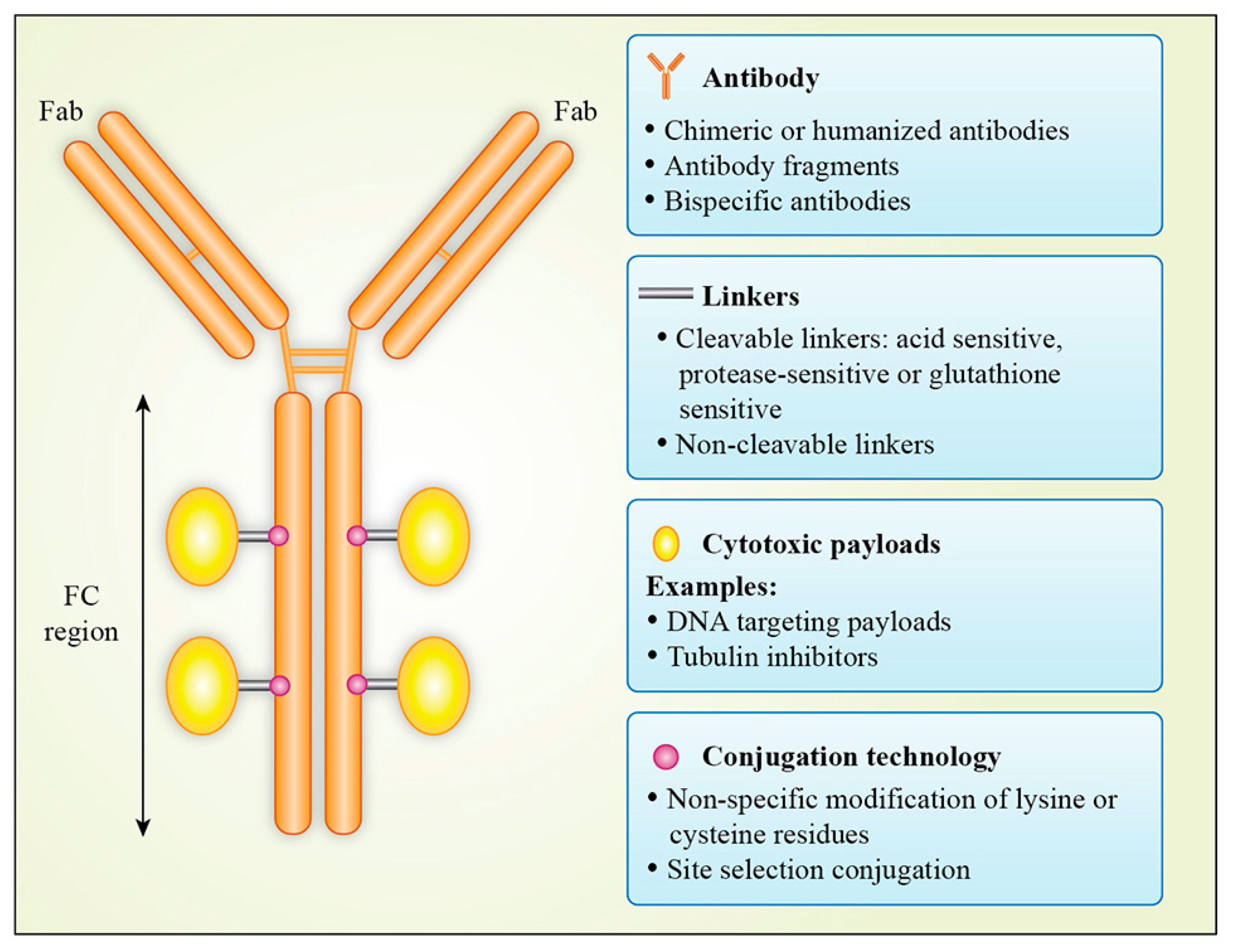
The simplest antibodies are Y-shaped molecules with two identical antigen-binding sites, one at the tip of each arm of the Y (Figure 24-18). Because of their two antigen-binding sites, they are described as bivalent. As long as an antigen has three or more antigenic determinants, bivalent antibody molecules can cross-link it into a large lattice (Figure 24-19). This lattice can be rapidly phagocytosed and degraded by macrophages. The efficiency of antigen binding and cross-linking is greatly increased by a flexible hinge regionin most antibodies, which allows the distance between the two antigen-binding sites to vary (Figure 24-20).
How many classes of antibodies do mammals make?
Mammals make five classes of antibodies, each of which mediates a characteristic biological response following antigen binding. In this section, we discuss the structure and function of antibodies and how they interact with antigen. B Cells Make Antibodies as Both Cell-Surface Receptors and Secreted Molecules.
Why are antibodies protective?
The protective effect of antibodies is not due simply to their ability to bind antigen. They engage in a variety of activities that are mediated by the tail of the Y-shaped molecule. As we discuss later, antibodies with the same antigen-binding sites can have any one of several different tail regions. Each type of tail region gives the antibody different functional properties, such as the ability to activate the complement system, to bind to phagocytic cells, or to cross the placenta from mother to fetus.
Which antibodies can pass from mother to fetus?
The binding of the antibody-coated bacterium (more...) IgG molecules are the only antibodies that can pass from mother to fetus via the placenta. Cells of the placenta that are in contact with maternal blood have Fc receptors that bind blood-borne IgG molecules and direct their passage to the fetus.
What happens when a B cell is activated?
B cell activation. When naïve or memory B cells are activated by antigen (and helper T cells—not shown), they proliferate and differentiate into effector cells. The effector cells produce and secrete antibodies with a unique antigen-binding (more...)
What is the B cell receptor?
The B-cell receptor is a complex of surface immunoglobulin with the accessory molecules Igα and Igβ. Following receptor cross-linking by binding of antigen to the BCR, a complex cascade of signaling molecules becomes involved in transducing the signal from the BCR to eventually result in B-cell activation and proliferation, or anergy and death.
What are the differences between B cells and antibodies?
B cell receptors for antigen are almost identical in structure to secreted antibodies. The only structural difference is that the C-terminal region of the heavy chains contain a short hydrophobic stretch which spans the lipid bilayer of the membrane. In addition to the antibody module that recognizes antigen, B cell receptors have short transmembrane chains (Ig-α and Ig-β) that are involved in signal transduction (Cambier et al., 1994; Pleiman et al., 1994; Reth, 1994). Membrane IgM may also be associated with prohibitin and a prohibitin-related protein (Terashima et al., 1994); membrane IgD may also be associated with two other as yet unidentified proteins (Kim et al., 1994). The precise pathways of signalling from the receptors to the interior of the cell are not yet fully understood but involve associated protein kinases (Pleiman et al., 1994), and lead to rapid cleavage of phosphatidyl inositol and mobilization of calcium (reviewed by Weiss and Littman, 1994; Cambier et al., 1993, 1994; Peaker, 1994 ).
What are the signals of BCR?
BCR signals are transduced within seconds, and positive regulators such as CD19 and negative regulators of these signals such as CD22 and FcγRIIB either expand or dampen them. CD19 is a 95-KDa glycoprotein that is upregulated at the pre-B-cell stage and remains on the B-cell surface until B cells differentiate into plasma cells. The cytoplasmic domain of CD19 is phosphorylated by Lyn and through recruitment of PI3K amplifies the signals emanating from antigen binding to BCR. In the absence of CD19, B cells are unable to respond to membrane-bound antigen but can sense soluble antigen in a comparable manner to normal, WT B cells ( Depoil et al., 2008 ). Association of CD19 with surface CD21 (complement receptor type 2 that binds C3 complement components), the tetraspanin CD81, and leu13 molecules ( Tedder, Inaoki, & Sato, 1997) provides a means of connecting the BCR complex with the complement receptors in response to antigens coupled to C3d. In fact, BCR responses to membrane-bound antigen require CD81, demonstrating that CD19–CD81 complexes are important for these responses ( Mattila et al., 2013 ).
How do BCRs form?
BCR microclusters form simultaneously with BCR binding to antigen and continue to grow by recruitment of both antigen and BCR molecules. Although multivalency of the antigen likely contributes to BCR microclustering, it is not an absolute requirement ( Fleire et al., 2006; Tolar, Hanna, et al., 2009 ). Instead, BCR microclusters form by a surprisingly complex process that involves several mechanisms ( Pierce & Liu, 2010; Tolar, 2011 ). Initially, membrane curvature at the contact sites can lead to diffusional confinement of antigen-engaged BCRs. The engaged BCRs then become trapped in a tightly packed cluster via oligomerization ( Tolar, Hanna, et al., 2009; Tolar & Pierce, 2010; Tolar, Sohn, Liu, & Pierce, 2009 ). These microclusters are also corralled by the actin cytoskeleton ( Treanor et al., 2011 ), which increases the rebinding rate of dissociated BCRs. Further growth of the microclusters is possible through continuous trapping of diffusing BCRs and also by active movement of BCR clusters centripetally, which merges small microclusters together.
What is the role of BCR in the activation of B lymphocytes?
Interaction of the BCR with its specific antigen is a necessary first step in the activation of B lymphocytes. Investigators noted that the BCR includes a short intracytoplasmic tail, consisting of three amino acid residues that is too short to interact with second messengers in the cytoplasm involved in transmitting a signal to the nucleus. As described in Chapter 17, in 1990, Kerry Campbell and John Cambier at the University of Colorado Medical School sought and found accessory molecules associated with the BCR that serve this transduction function. Two other groups of investigators, one led by Joachim Hombach in Freiburg, Germany ( Hombach et al., 1990 ), and a second headed up by Carel van Noesel in the Netherlands, reported independently in 1990 the isolation of these same molecules. Originally termed Ig-α and Ig-β, these chains have been characterized by monoclonal antibodies and are now called, respectively, CD79a and CD79b.
How do B lymphocytes become self-reactive?
B lymphocytes expressing potentially self-reactive BCRs can be deleted by the induction of apoptosis or can be rendered unresponsive by induction of anergy. A third process for regulating B lymphocytes reactive to self comprises replacing the gene coding for the V region of one of the chains of the BCR of the developing B lymphocyte. This process is termed receptor editing and was demonstrated experimentally in the early 1990s by two groups of investigators, both of which used transgenic mice.
What is the function of BCR?
The BCR is essential for the humoral immune response. Membrane Ig-based BCR, generated through gene recombination, have a vast repertoire that can recognize an incredible number of antigens in various biological, chemical, and physical forms. Antigen-binding of BCRs triggers multiple signaling cascades by inducing BCR clustering and the formation of the BCR SMAC in lipid rafts. BCR signaling determines B-cells’ fate, death or survival. BCR signaling is tightly regulated by both internal and external signals to control B-cell responses. The BCR is also responsible for antigen capture, internalization, and intracellular transportation for antigen processing and presentation. The ability of BCRs to capture and internalize antigen determines if B-cells can acquire the essential signals from T-cells for proliferation and differentiation into long-lived memory B-cells and plasma cells, which are responsible for long-term humoral protection.
What happens when a B cell receptor binds its cognate antigen?
When a B-cell receptor binds its cognate antigen,it undergoes changes and can secrete a soluble form of that receptor.This soluble form of that receptor is known as antibody.The antibody is specific for that antigen it encountered on the original B cell receptor.This antibody has an Fc region on its heavy chain that can be recognized by innate immune cells (macrophages) and these cells rid the body of the pathogen.
What is the B cell in resting state?
When B cell is in resting state The receptor it has in its surface (in its plasma membrane) is BCR (B-cell receptor). But when this BCR binds to its cognate antigen then that leads to differentiation of the B-cell to 2 types of cell: antibody-secreting plasma cell and memory cell (which stays in the body for quick recognition and response to the future attack by the same antigen).
What happens when antibodies are binding to the surface of the cell?
Antigen binding to the surface of antibodies of the cell cause rapid division. The cells would proliferate and its progeny differentiate into two kinds of cells- effector cells and memory cells.
What is the function of antibodies?
Such as opsonization ,ADCC, complement activation etc to eliminate antigens from the circulations.
Where is the BCR located?
B cell receptor is found on the membrane of the B lymphoctyes. It contains surface bound immunoglobulins IgM and IgD as the binding receptor along with some co receptor proteins.the function of BCR is to bind specific antigen to stimulate B cell to process and present that antigen which can be recognized by T Helper cel
Which cell has receptors that help in receptor cross linking or binds to the antigens?
The plasma membrane of B cell contains BCR or B cell receptors that help in receptor cross linking or binds to the antigens , a series of complex signalling cascade occurs and activate the B cell , eventually it proliferate and produce two kinds of cells :
What is the role of T-helper cells in the MHC II complex?
T-helper cells identify and bind to the MHC-II complex. The association would activate the cells to convert into effector cells which would secrete various growth factors such as cytokines. These secreted cytokines would play an important role in activating B-cell, T-cells, and macrophages required for immune response.
Where are antibodies made in a B cell?from ncbi.nlm.nih.gov
The first antibodies made by a newly formed B cell are not secreted. Instead, they are inserted into the plasma membrane, where they serve as receptors for antigen. Each B cell has approximately 105such receptors in its plasma membrane.
Which cell makes antibodies?from ncbi.nlm.nih.gov
B Cells Make Antibodies as Both Cell-Surface Receptors and Secreted Molecules
What are the IgG4 antibodies?from ncbi.nlm.nih.gov
IgG4 antibodies are often formed following repeated or long-term exposure to antigen in a non-infectious setting and may become the dominant subclass. Examples are long-term bee keepers and allergic individuals that underwent immune therapy (8, 29–31). In immunotherapy, relief of symptoms appears to correlate with IgG4 induction. Switching to IgG4 may be modulated by IL10, linking this subclass downregulation of immune responses or tolerance induction (8, 32). IgG4 may also represent the dominant antibody subclass in immune responses to therapeutic proteins, such as factor VIII and IX (33–35) and at least some recombinant antibodies such as adalimumab (36). Furthermore, helminth or filarial parasite infections may result in the formation of IgG4 antibodies (37, 38), and high IgG4 titers can be associated with an asymptomatic infection (39).
How does affinity of an antibody relate to the antigenic determinant?from ncbi.nlm.nih.gov
The affinityof an antibody for an antigenic determinant describes the strength of binding of a single copy of the antigenic determinant to a single antigen-binding site, and it is independent of the number of sites. When, however, a polyvalent antigen, carrying multiple copies of the same antigenic determinant, combines with a polyvalent antibody, the binding strength is greatly increased because all of the antigen-antibody bonds must be broken simultaneously before the antigen and antibody can dissociate. As a result, a typical IgG moleculecan bind at least 100 times more strongly to a polyvalent antigen if both antigen-binding sites are engaged than if only one site is engaged. The total binding strength of a polyvalent antibody with a polyvalent antigen is referred to as the avidityof the interaction.
What is an IgG subclass?from microbenotes.com
IgG Subclasses. Functions of Immunoglobulin G (IgG) References. Antibodies, or ‘immunoglobulins’, are glycoproteins that bind antigens with high specificity and affinity. In humans there are five chemically and physically distinct classes of antibodies (IgG, IgA, IgM, IgD, IgE). Immunoglobulin G (IgG), the most abundant type of antibody, ...
Why are antibodies bivalent?from ncbi.nlm.nih.gov
Because of their two antigen-binding sites, they are described as bivalent. As long as an antigen has three or more antigenic determinants, bivalent antibody molecules can cross-link it into a large lattice (Figure 24-19). This lattice can be rapidly phagocytosed and degraded by macrophages.
Why do antibodies cross link?from ncbi.nlm.nih.gov
Antibody-antigen interactions. Because antibodies have two identical antigen-binding sites, they can cross-link antigens. The types of antibody-antigen complexes that form depend on the number of antigenic determinants on the antigen. Here a single species (more...)

Overview
The B cell receptor (BCR) is a transmembrane protein on the surface of a B cell. A B cell receptor is composed of a membrane-bound immunoglobulin molecule and a signal transduction moiety. The former forms a type 1 transmembrane receptor protein, and is typically located on the outer surface of these lymphocyte cells. Through biochemical signaling and by physically acquiring antigens from …
Development and structure of the B cell receptor
The first checkpoint in the development of a B cell is the production of a functional pre-BCR, which is composed of two surrogate light chains and two immunoglobulin heavy chains, which are normally linked to Ig-α (or CD79A) and Ig-β (or CD79B) signaling molecules. Each B cell, produced in the bone marrow, is highly specific to an antigen. The BCR can be found in a number of identical co…
Signaling pathways of the B cell receptor
There are several signaling pathways that the B cell receptor can follow through. The physiology of B cells is intimately connected with the function of their B cell receptor. The BCR signaling pathway is initiated when the mIg subunits of the BCR bind a specific antigen. The initial triggering of the B cell receptor is similar for all receptors of the non-catalytic tyrosine-phosphorylated receptor family. T…
The B cell receptor in malignancy
The B cell receptor has been shown to be involved in the pathogenesis of various B cell derived lymphoid cancers. Although it may be possible that stimulation by antigen binding contributes to the proliferation of malignant B cells, increasing evidence implicates antigen-independent self-association of BCRs as a key feature in a growing number of B cell neoplasias. B cell receptor signalling is currently a therapeutic target in various lymphoid neoplasms. It has been shown tha…
See also
• Co-stimulation
• T-cell receptor
External links
• B-Cell+Antigen+Receptors at the US National Library of Medicine Medical Subject Headings (MeSH)