Full Answer
What is a dicrotic notch?
The dicrotic notch is caused by the aortic valve closing which causes a change in pressure that can be seen on the waveform. This marks the end of systole and the start of diastole. How the notch looks on the downward waveform changes with age and cardiac health. The less distinct the notch, the great probability of coronary heart disease.
What causes a dicrotic notch in the heart?
The dicrotic notch is caused by the aortic valve closing which causes a change in pressure that can be seen on the waveform. This marks the end of systole and the start of diastole.
What does no dicrotic notch in PVR indicate?
Dicrotic notch must be present on the diastolic limb of the PVR waveforms for a normal healthy artery. Absence of the dicrotic notch is suggestive of a non-compliant artery. decrease of 20mmHg in pressure between adjacent levels of the ipsilateral extremity denotes disease.
What is the difference between anacrotic and dicrotic?
The waveform can be separated into an anacrotic (upstroke) and dicrotic (downstroke) limbs. The origin of the term is from Greek dikrotos, which means "beating twice" ( krotos meaning "stroke"); anacrotic having been abbreviated from anadicrotic.
What does a Dicrotic notch represent?
The dicrotic notch The notch represents the nadir point that occurs immediately after the closure of the aortic valves and precedes the secondary dicrotic wave. The notch is frequently used as a marker for the end of the ventricular ejection period.
What causes the Dicrotic notch in the blood pressure waveform?
The dicrotic notch and the dicrotic wave that follow it are thought to be due to a reflected pressure wave. The depth of the dicrotic notch appears to increase following infusion of vasodilators, as demonstrated by the below waveform that was recorded after infusion of hydralazine.
What is the significance of the Dicrotic notch in the cardiac cycle?
When pressure within the ventricles drops below pressure in both the pulmonary trunk and aorta, blood flows back toward the heart, producing the dicrotic notch (small dip) seen in blood pressure tracings. The semilunar valves close to prevent backflow into the heart.
What does a good arterial waveform look like?
1:4916:05The Arterial Line Waveform EXPLAINED! - YouTubeYouTubeStart of suggested clipEnd of suggested clipIncrease in arterial blood pressure as the heart is ejecting blood where the waveform begins itsMoreIncrease in arterial blood pressure as the heart is ejecting blood where the waveform begins its rise is where the aortic valve actually opens and marks the beginning of systole.
How do you get Dicrotic notch?
Detection of dicrotic notch For dicrotic notch detection, the algorithm utilizes the first-order difference of the filtered BP pulse. The algorithm first computes the first-order difference of the filtered BP pulse. The first-order difference time series is then smoothed using Savitzky–Golay filter.
What factors would affect the shape or position of the Dicrotic notch?
The change in shape and position of the dicrotic wave is due to it being caused by reflections of the arterial pressure wave rather than aortic valve closure.
Why is the Dicrotic notch important quizlet?
Why is the dicrotic notch important? Review each of the pressure line graphs in Focus Figure 17.2. As ventricular pressure decreases below aortic pressure, the aortic semilunar valve closes. In response, blood rebounds against this closed valve, producing a slight increase in the aortic pressure.
Which valve causes Dicrotic notch?
the aortic valve closureSince most medical textbooks explain the origin of the dicrotic notch as caused by the aortic valve closure itself, this is commonly transmitted in medical physiology courses.
What is a normal waveform?
Normal cerebrovascular waveforms. Normal flow waveforms in the cerebral arteries show a rapid systolic upstroke, reflecting normal proximal vessels and cardiac function, but the characteristics of the diastolic portion of the wave- form are determined by the resistance of the distal vascular bed.
What is a normal arterial line pressure?
Normal Ranges: Systolic Blood Pressure: 90 – 120 mm Hg. Diastolic Blood Pressure: 50 – 80 mm Hg. Mean Arterial Pressure: 70 – 100 mm Hg.
What is the Dicrotic notch and why does it follow the T wave quizlet?
The dicrotic notch in the pulse waveform follows the second heart sound. The dicrotic notch is a short-lived decrease in pressure in the ascending aorta, which occurs following closure of the aortic valve.
What is the Dicrotic notch quizlet?
dicrotic notch. a small plateau or dip in the pressure wave caused by the closure of the aortic valve. end diastolic volume. total volume of blood in the ventricles at the end of diastole. stroke volume.
Why is the Dicrotic notch important quizlet?
Why is the dicrotic notch important? Review each of the pressure line graphs in Focus Figure 17.2. As ventricular pressure decreases below aortic pressure, the aortic semilunar valve closes. In response, blood rebounds against this closed valve, producing a slight increase in the aortic pressure.
What is a Dicrotic wave?
dicrotic wave the second portion of the tracing of a sphygmograph of the arterial pulse or arterial pressure after the dicrotic notch, attributed to the reflected impulse of closure of the aortic valves. Called also recoil wave.
What is the notch in the aorta?
In perfect circumstances, when measured in the aorta, this notch is very sharp and it actually does represent the closing of the aortic valve. In fact, when it is measured in the aorta the notch is called the incisura, because it cuts into the waveform. However, further down the arterial tree the incisura disappears. It is replaced by the dicrotic notch, a mutant offspring of several reflected waves, only vaguely related to the behaviour of the aortic valve.
Why is the incisura soft?
As you move further out into the peripheral circulation, the incisura ends up being slurred and softened. It is generally believed that the peripheral dicrotic notch owes more of its shape to the vascular resistance of peripheral vessels than to the closing of the aortic valve. This is best illustrated in this image, modified from O'Rourke via McDonalds Blood Flow In Arteries (6th ed).
What is the dicrotic notch?
The notch represents the nadir point that occurs immediately after the closure of the aortic valves and precedes the secondary dicrotic wave.
What does the solid arrow on the dicrotic notch mean?
The solid arrow indicates where one would expect to see the notch (approximately one-third the way down the descending limb ) and the dashed arrow shows where the notch was actually seen.
What wave shows the dicrotic notch?
Arterial pressure wave showing the dicrotic notch. Adapted from: Wikimedia Commons with acknowledgement to the originial uploader of this image Benutzer:Lupino under the Creative Commons Attribution/Share-Alike License.
What is the notch used for?
The notch is frequently used as a marker for the end of the ventricular ejection period. The secondary dicrotic wave occurs as the elastic recoil of the arterial tree clamps down against a closed aortic valve causing a secondary pressure increase.
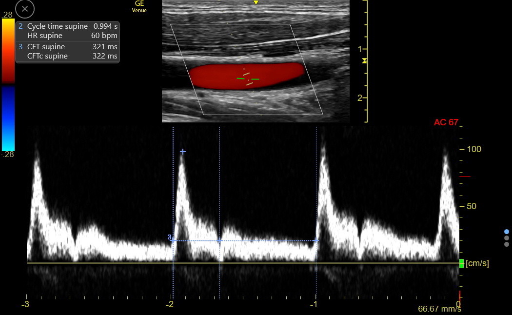
What is the dicrotic notch in systole?
Arterial Waveform: Systole begins during the sharp rise from the baseline (troughs). Diastole begins on the down slope where the bulge appears. The bulge is referred to as the dicrotic notch. This signifies the closure of the aortic valve at the end of systole. The peaks average the systolic pressure.
Where does diastole begin?
Systole begins during the sharp upstroke from the trough. Diastole begins at the dicrotic notch on the backside of the waveform. This is an arterial waveform, but it is for the pulmonary artery. Pay attention to the scale at the left.
What happens when diastole comes to an end?
Diastole in a ventricle becomes very close to zero. Also, as diastole comes to an end, the pressure begins to rise revealing a little hill with what is referred to as the end-diastolic pressure–this is associated with ventricular filling of blood prior to ventricular contraction.
When does notching disappear in the uterus?
The presence of an early diastolic notch can however be a normal finding in a non-pregnant uterus and even in a gravid uterus, at least up to 16 weeks, but notching typically begins to disappear in the gravid uterus by 13 weeks with clearly established diastolic flow by 20 weeks 7.
What is uterine flow notching?
Uterine artery flow notching refers to a phenomenon observed in uterine arterial Doppler ultrasound assessment.
Which side of the placenta is bilateral notching?
Bilateral notching is more concerning. Unilateral notching of the uterine artery on the ipsilateral side of the placenta, if the placenta is along one lateral wall (right or left) carries the same significance as bilateral notching.
Is bilateral notching the same as unilateral notching?
Bilateral notching is more concerning. Unilateral notching of the uterine artery on the ipsilateral side of the placenta, if the placenta is along one lateral wall (right or left) carries the same significance as bilateral notching. The presence of an early diastolic notch can however be a normal finding in a non-pregnant uterus and even in a gravid uterus, at least up to 16 weeks, but notching typically begins to disappear in the gravid uterus by 13 weeks with clearly established diastolic flow by 20 weeks 7.
A few ground rules about Pulse Volume Recording Interpretation
Pressures trump tracings. This means that if the pressures are normal but the tracings are not, believe the pressures. However, in general these two pieces of information should align. So before scratching off a discrepancy, think.
Step 1 – Look at the Ankle Brachial Index
Starting the analysis of pulse volume recordings and segmental pressure measurements by looking at the ankle brachial index makes a lot of sense. The ankle brachial index offers very quick information about the following points: