
Is green fluorescent protein hydrophobic?
Green fluorescent protein is extremely hydrophobic compared to bacterial proteins. Unique characteristics of GFP enable it to be purified from bacterial cell proteins using HIC columns.
How is GFP different from other bacterial proteins?
Moreover, GFP is much more ? hydrophobic amino acids, which collectively make the entire protein hydrophobic. hydrophobic than most of the other bacterial proteins.
How do you purify GFP from bacterial cells?
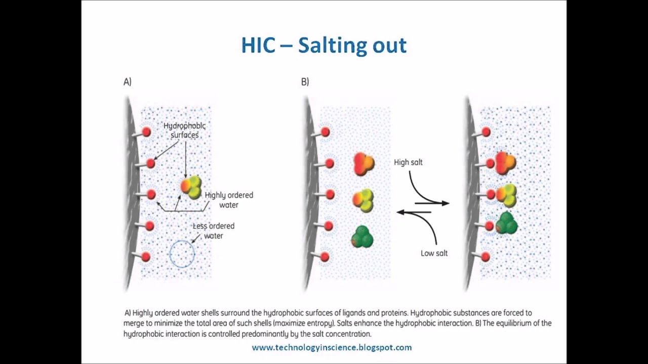
Unique characteristics of GFP enable it to be purified from bacterial cell proteins using HIC columns. When placed in a buffer containing a high concentration of salt, the HIC matrix selectively binds hydrophobic GFP molecules while allowing the bacterial proteins to pass through the column.
Why use GFP as a model protein?
GFP serves as a useful model protein, due to its stability, unique light absorbance peak at 397 nm, and fluorescence when exposed to UV light (5).
See more
Is GFP soluble?
The engineered GFP 1–10 is about 50% soluble, and the inclusion body fraction is processed to take advantage of the enrichment and partial purification afforded by using inclusion bodies.
What are the properties of GFP?
GFP is a fluorescent protein that can be expressed in vivo. If GFP is exposed to light, it emits a green fluorescent signal. This property has had an enormous impact on cell biology by enabling the imaging of almost any protein, in transcription studies by working as a reporter gene, and in biochemical applications.
What are the disadvantages of GFP?
Disadvantage of GFP: It is necessary that each fusion protein be tested for its functionality in vivo because GFP-tag is so relatively large that affect the function of fused protein of interest. The GFP signal can not be amplified in a controlled manner, possibility preventing detection of low expression levels.
What type of molecule is GFP?
Green fluorescent protein (GFP) is a protein in the jellyfish Aequorea Victoria that exhibits green fluorescence when exposed to light. The protein has 238 amino acids, three of them (Numbers 65 to 67) form a structure that emits visible green fluorescent light.
Is GFP weakly hydrophobic?
Green Fluorescent Protein (GFP) is extremely hydrophobic compared to bacterial proteins. This unique characteristic of GFP enables the purification of GFP from bacterial cell proteins using hydrophobic interaction chromatography (HIC).
What is a major advantage of using green fluorescent protein GFP in cell biology?
The chief advantage of GFP/S65T is that it is excited primarily by blue light, making it more useful as a label.
How long does GFP fluorescence last?
The half-life of unmodified GFP is approximately 26 h;8 thus, it takes several days for the passively transferred protein to degrade leading to an overestimation of transduction achieved at early time points.
Is GFP toxic to humans?
GFP is cytotoxic by a variety of mechanisms in addition to immunogenicity. Initiation of the apoptosis cascade has been postulated as a possible mechanism for the toxicity of GFP and cellular death.
What are the advantages of having a GFP tag?
The primary advantage of the chimeric GFP tag having an internal hexa-histidine sequence is that such a tag allows maximum flexibility for protein or peptide fusions since both N- and C-terminal ends of the GFP are available for fusion.
What causes GFP to glow?
Scientists knew that GFP glows because three of its amino acids form a fluorophore, a chemical group that absorbs and emits light.
What is GFP made of?
Green fluorescent protein (GFP) was originally derived from the jellyfish Aequorea victoria (Prendergast and Mann, 1978). It has 238 amino acid residues and a green fluorophore, which is comprised of only three amino acids: Ser65-Tyr66-Gly67.
What is the difference between GFP and EGFP?
The main difference between GFP and EGFP is that the GFP (stands for Green Fluorescent Protein) is a protein that exhibits bright green fluorescence when exposed to blue light whereas the EGFP (stands for Enhanced Green Fluorescence Protein) exhibits stronger fluorescence than GFP.
Why is GFP special?
GFP is amazingly useful for studying living cells, and scientists are making it even more useful. They are engineering GFP molecules that fluoresce different colors. Scientists can now make blue fluorescent proteins, and yellow fluorescent proteins, and a host of others.
Where do the fluorescent properties of GFP come from?
Abstract. Green fluorescent protein (GFP) is a protein produced by the jellyfish Aequorea victoria, that emits bioluminescence in the green zone of the visible spectrum. The GFP gene has been cloned and is used in molecular biology as a marker.
What are the applications of GFP?
Overall, GFP can be used to visualize specific cell types in intact animals, organs, and tissues. This prospect is significantly useful in fields such as immunology, neurobiology, development, and carcinogenesis.
What makes GFP glow?
Scientists knew that GFP glows because three of its amino acids form a fluorophore, a chemical group that absorbs and emits light.
What is the unique feature of GFP?
Unique characteristics of GFP enable it to be purified from bacterial cell proteins using HIC columns. When placed in a buffer containing a high concentration of salt, the HIC matrix selectively binds hydrophobic GFP molecules while allowing the bacterial proteins to pass through the column.
How is GFP purified?
GFP is purified from the bacterial lysate using hydrophobic interaction chromatography (HIC) columns provided in this kit. Green fluorescent protein is extremely hydrophobic compared to bacterial proteins. Unique characteristics of GFP enable it to be purified from bacterial cell proteins using HIC columns.
What is green fluorescent protein?
Green Fluorescent Protein (GFP) is extremely hydrophobic compared to bacterial proteins. This unique characteristic of GFP enables the purification of GFP from bacterial cell proteins using hydrophobic interaction chromatography (HIC). When placed in a buffer containing a high concentration of salt, the HIC matrix selectively binds hydrophobic GFP ...
What is the chromophore of GFP?
The GFP chromophore can exist as anionic or neutral, which explains the spectroscopic observation of two coincident absorbance and excitation bands [Fig. 3(b)]. The neutral protonated or A form absorbs in the ultraviolet (UV) at about 395 nm, whereas the anionic or B form absorbs at about 475 nm. Curiously, in wild-type GFP both bands are present at equilibrium in an A:B intensity ratio of about 6:1. This ratio is surprisingly insensitive to the environmental pH and salt concentration21implying that in wild type, some as yet poorly understood mechanism protects the chromophore from changes in the solvent environment.
What is the reaction scheme for ESPT in wild type GFP?
Proposed reaction scheme for ESPT in wild-type GFP. An ∼ 390 nm wavelength photon excites the system (upper right) and causes a series of proton transfer events to form the excited state anion (I*) , left center, which subsequently emits a green photon (∼ 510 nm). A low frequency event is transformation of the ground state I to a metastable ground state anion, B (lower right).
What is the advantage of GFP/S65T?
The chief advantage of GFP/S65T is that it is excited primarily by blue light, making it more useful as a label.4However at low pH, fluorescence is quenched and ESPT is not observed. Shortly after determining the structure of GFP/S65T, we initiated a collaboration with Titia Sixma's group to compare results with their structure of wild-type GFP, and proposed an atomic mechanism for the process. In this proposal, wild-type GFP has a built-in proton wire from the chromophore OH, through a water molecule and the hydroxyl of serine 205 to the acceptor Glu 222 (Fig. 4).33Stoner-Ma et al.30subsequently used ultrafast vibrational spectroscopy to confirm that Glu222 is the proton acceptor. Remarkably, the observed rate of protonation at Glu222 (t∼ 15 ps) approximately matches the decay of blue fluorescence, suggesting that the proton transfer process is concerted (i.e., all three protons in Fig. 4move simultaneously). Alternatively, proton transfer may be stepwise but the intermediate steps may take place too quickly for these techniques to resolve. Further studies of the proton transfer pathways in GFP are currently being conducted by us34and by others,35using a combination of mutagenesis, high resolution crystallography, and ultrafast spectroscopic techniques. GFP has turned out to be an outstanding system for study of the geometric factors controlling the rate of proton transfer, one of the most elementary reactions in biological chemistry.
What is the function of protein folding?
Protein folding is essential for both chromophore formation and for fluorescence; see reviews.4,9,10As is now well known, three amino acids within the central helix (serine 65, tyrosine 66, and glycine 67) rearrange covalently during the folding reaction, and in the presence of molecular oxygen become oxidized to form the chromophore. Denatured GFP is nonfluorescent, but fluorescence is regained upon renaturation.11The isolated chromophore is also not fluorescent, either as the naked molecule12or within the isolated hexapeptide.13From these observations, it is clear that in addition to promoting the chemistry of fluorophore formation, a primary function of the polypeptide fold is to tightly restrain the chromophore. In this way, excitation energy is dissipated as light, rather than through nonradiative processes. Nature can have it both ways, though and in a later section, I'll discuss what makes “chromoproteins” nonfluorescent (although their purpose in Nature remains a mystery).
What is the emission of anionic chromophore?
Emission from the anionic chromophore is rather simply described as a transition from the first sin-glet excited state to ground (that is, a transition between the lowest unoccupied and highest occupied molecular orbital, see for example a recent study24). In most fluorescent proteins, protonation of the chromophore quenches fluorescence. The reason for this behavior is not completely understood, however in wild-type GFP and a few of its relatives, the chromophore is fluorescent in either protonation state (that is, upon either A or B band excitation). Comparison of the GFP chromophore with model compounds suggests that the excited state of the protonated chromophore (A*) should emit in the blue but instead, green emission is observed.21The Boxer group25used ultrafast fluorescence upconversion spectroscopy to show that upon excitation by 397-nm light, blue emission at 460 nm is observed as expected, but the blue emission decays with biphasic time constants of about 3 and 12 ps. The blue decay is matched by the rise in 510-nm green emission. Both rates are slowed by a factor of 5-6 upon substitution of exchangeable hydrogens with deuterium, suggesting that the A species is converted into an excited anionic intermediate I* by proton transfer. The I* state subsequently emits a green photon.
How does light excitation affect chromophore?
Light excitation promotes electrons to nonbonding orbitals, so in the minimum energy configuration of the first excited state of the chromophore, the two rings are approximately perpendicular.15If such rearrangements are permitted by the environment, energy is usually dissipated by thermal rather than radiative processes. Consequently, the chromophore configuration and overall planarity are rigidly enforced by the surrounding protein matrix using a combination of hydrogen bonds and hydrophobic side chains, which in turn determine the shape of the enclosing cavity. Experimental evidence for the importance of a close cavity fit was provided quite early on by mutants at the Tyr 66 position (e.g., Phe, His, and Trp), all of which have substantially reduced brightness.16However, brightness in these mutants can be improved by further mutagenesis of protein side chains lining the chromophore cavity. This result is consistent with the theoretical predictions that a loose cavity permits energy dissipation via internal conversion.17,18
How many strands are in a GFP/S65T crystal?
2), which seems remarkably well suited to its multiple tasks. The polypeptide backbone folds into a previously unobserved motif: a β-barrel of eleven strands, surrounding a central helix. Short distorted helical segments cap the barrel ends and help isolate the internal chromophore from solvent. An all-atom representation appears as a nearly perfect cylinder about 25 Å in diameter and 40 Å tall.
What is GFP bind?
Binding - a solution containing the cell contents, including GFP, is run through the column in high salt. GFP binds via the hydrophobic effect; most other molecules don’t. Those that don’t stick flow through the column and are discarded.
What happens when you heat up GFP?
Different proteins denature at different temperatures depending on the particular non-covalent bonds that give them their shape. When GFP denatures, the chromophore is exposed to the surrounding water and it is no longer fluorescent.
Why are proteins not fluorescent?
Most proteins are not fluorescent because, in order to be fluorescent, a molecule must have particular structural elements that are not normally found in proteins. However, the jellyfish Aequoria victoria produces a protein, called Green Fluorescent Protein (GFP), that absorbs ultraviolet (UV) light and gives off longer wavelength green light. This makes GFP an interesting protein for study. GFP has been used in a wide variety of experiments exploring protein structure and folding, gene expression, development, etc. In this lab, we will use GFP as an example of a ‘typical’ protein for biochemical study.
What is affinity chromatography?
Chromatography is a term that describes a huge and diverse set of methods for separating molecules based on their different properties. We are using affinity chromatography today. In it’s simplest form, it consists of three steps:
How to purify GFP?
The bacteria need to be ruptured in order to release the GFP, which can then be purified using column chromatography.
How to isolate a single fluorescent green colony of bacteria?
One can isolate a single fluorescent green colony of bacteria and grow large amounts of the bacteria in a liquid growth media. Bacteria in liquid media can be concentrated by centrifugation. After the bacterial cells are lysed to release the cancer curing protein, the protein can be isolated by passage through a chromatography column which has an affinity for the cancer-curing protein.
Why does the LB plate glow green?
The LB/amp/ara plate glowed green primarily because of arabinose, which explains why the other plate did not glow. Describe how you might recover a cancer-curing protein from the bacterial cells: One can isolate a single fluorescent green colony of bacteria and grow large amounts of the bacteria in a liquid growth media.
Does a supernatant contain GFP?
The supernatant contains the bacterial growth media and does not contain the desired GFP.
Is a bacterial protein hydrophobic?
hydrophobic than most of the other bacterial proteins.
