Is osteoid osteoma rare?
Osteoid osteoma (OO), a rare bone-producing tumor, can occur anywhere in the skeleton, [1, 2] and in the spine it is frequently located in the posterior elements involving the lamina, pedicles, or the transverse and spinous processes [3, 4].
Should I worry about osteoma?
While osteomas are not cancerous, they can sometimes cause headaches, sinus infections, hearing issues or vision problems – however, many benign osteomas don't require treatment at all. If treatment is needed, your doctor may prescribe surgery, pain relievers, or other minimally invasive techniques to provide relief.
How common are osteomas?
They are seen commonly associated with the nose and the paranasal sinuses, the commonest being the frontal sinus. The incidence of osteoma of frontal bone and frontal sinus ranges from 37-80% in the reported cases. [2] But isolated cases of osteoma of the forehead, without involvement of the sinus, are rare.
Will osteoid osteoma go away?
Osteoid osteomas may go away on their own. But these tumors may not disappear for years. You can treat an osteoid osteoma with NSAIDs. NSAIDs can be nonprescription (aspirin, ibuprofen or naproxen) or prescription.
Can osteoid osteoma turn cancerous?
An osteoid osteoma is a benign bone-forming tumor that has no potential to become malignant. It classically causes severe pain at night relieved with nonsteroidal anti-inflammatory medications. Occasionally, it resolves without treatment.
Can osteomas shrink?
Osteoid osteomas may shrink on their own. But that often takes years. Some people get pain relief from nonsteroidal anti-inflammatory drugs (NSAIDs).
How fast does osteoma grow?
The mean linear growth rate of osteomas was estimated to be 0.117 mm/yr (95% CI, 0.004, 0.230) in maximal dimension, assuming linear growth.
How long does it take to recover from osteoid osteoma?
After surgery, patients may spend up to a week in the hospital, and may require up to 6 months before they can return to normal activities.
Is osteoma surgery safe?
The authors have found that excision of forehead osteoma using an endoscope coupled with proper instruments to be very safe and effective with minimal morbidity.
What causes an osteoma to grow?
What causes osteoid osteoma? An osteoid osteoma occurs when certain cells divide uncontrollably, forming a small mass of bone and other tissue. This growing tumor replaces healthy bone tissue with abnormal, hard bone tissue. No one knows exactly why this occurs.
Is osteoma removal painful?
How painful is osteoma surgery? Most of Dr. Fishman's osteoma treatment patients describe a bruised or sore sensation, or a headache, after surgery, with most discomfort fading away within 1-2 days after treatment.
Can osteoid osteoma be seen on xray?
Plain films may suggest the diagnosis, but in complex areas such as the spine, pelvis, wrist, and foot, additional imaging modalities are often required. On plain radiography and CT, a typical nidus surrounded by sclerosis or cortical thickening characterizes osteoid osteoma.
Why do osteomas develop?
Most osteomas have no identifiable cause. A small percentage are present or develop soon after birth. Others are believed to result from inflammation or trauma. In rare cases osteomas may be a component of an underlying hereditary disorder.
Is osteoma surgery safe?
The authors have found that excision of forehead osteoma using an endoscope coupled with proper instruments to be very safe and effective with minimal morbidity.
How do you get rid of osteoma?
The most common treatment option for osteomas is surgery on the skull base. Osteomas of the skull base may be approached directly using endoscopic sinus surgery. This minimally invasive approach allows surgeons to access the tumor through the natural corridor of the nose, without making an open incision.
What is the cause of osteoma?
Arising from the normal bony walls of the sinus cavities, osteomas are the most common tumor involving the paranasal sinuses. Causes of osteoma development that have been theorized include congenital, inflammatory, or traumatic factors, but in most cases the cause of the osteoma is unknown.
When is osteoid osteoma more common?
While osteoid osteoma can occur at any age, it is more common between the ages of 5 and 25 , and nearly three times as likely to affect boys as girls. Osteoid osteoma is often confused with other diseases such as osteomyelitis, osteoblastoma, Langerhans cell histiocytosis, or a stress fracture. Your child’s doctor may need to perform specific ...
What is osteoid osteoma?
Osteoid osteoma is a common benign tumor that usually develops in the long bones of the leg — the femur (thigh bone) and tibia (shin bone) – but can occur in any bone. In 7-20 percent of cases, osteoid osteoma occurs in the spine.
What is the best treatment for osteoid osteoma?
The gold standard for treating osteoid osteoma is CT-guided radiofrequency ablation (RFA). This minimally-invasive, outpatient procedure is performed in the Interventional Radiology Suite at Children’s Hospital ...
How long does it take for osteoid osteomas to heal?
Many osteoid osteomas will resolve on their own, but it may take several years. If your child is experiencing pain, he may benefit from nonsteroidal anti-inflammatory medications or regular analgesics such as aspirin, Tylenol® or ibuprofen.
Do children with osteoid osteoma have long term outcomes?
Children with osteoid osteoma have good long-term outcomes.
Where is osteoid osteoma most commonly found?
Osteoid osteoma is a benign tumor of the bone. This tumor is most frequently found in the legs but may occur also at other bones in nearly any part of the body. Osteoid osteoma is a tumor of children and young adults, it is very rare in older adults over the age of 50. .
What is the best treatment for osteoid osteoma?
Treating the pain: The most significant symptom of osteoid osteoma is pain, this can be treated with aspirin, ibuprofen or other over-the-counter anti-inflammatory drugs. Some patients have relief from certain medications for a while, but then these medications stop working.
How far away from osteoid osteoma can you get radiofrequency?
If the tumor nidus is more than 1 cm away from these structures the procedure can usually be safely performed.
How long does it take to recover from osteoid surgery?
The operation usually entails a longer hospital stay of at least several days. Osteoid osteomas are frequently located in weight-bearing bones and during the recovery period from surgery a longer period of limited weight bearing is required, often with crutches for a number of weeks.
Can osteoma get worse?
Diagnosing an Osteoid Osteoma. Osteoid osteoma causes a lot of pain in almost all patients. This pain is most frequently in the night, and patients sometimes wake up from the pain. The pain may also occur during the day. Sometimes the pain gets worse over time. It may only be dull, but sometimes also very sharp and gets worse with activity.
Does osteoma have to be removed?
In rare instances this tumor even heals spontaneously. Therefore the tumor does not necessarily have to be removed.
What is the best test for osteoid osteoma?
The following tests may be used in the diagnosis of osteoid osteoma: X-ray: scan that uses invisible beams of electromagnetic energy to produce images of internal tissues, bones, and organs on film. Pain is generally present before the osteoid osteoma is visible on X-ray, so diagnosing these tumors often takes some time.
How big is an osteoblastoma?
Osteoid osteomas do not grow larger than 1.5-2 centimeters, or about .75 inches. A related tumor type, osteoblastoma, is very similar, but is usually larger than 2 cm.
What are the bones of the spine called?
The bones of the spine are called vertebrae, and osteoid osteomas tend to affect the posterior (rear) portions of the vertebrae . They can occur in vertebrae at any level: cervical (neck), thoracic (upper- and mid-back), lumbar (lower back), or sacral (base of the spine).
What is the term for a tumor that develops in the bones?
Osteoid Osteoma. Osteoid = a substance produced by cells that make new bone. Osteoma = a type of tumor that develops in bones. Osteoid osteomas are small, benign bone tumors. Osteoid osteomas most commonly occur in the legs, hands, fingers, and spine.
Is osteoid osteoma a bone tumor?
The causes of osteoid osteoma are not yet understood. It is a relatively common bone tumor. It is more common in males than females, and equally common across races. Most people diagnosed with osteoid osteoma are in the first, second, and third decades of life.
Can a MR scan confirm osteoid osteoma?
MR scans may be able to rule out other diagnoses, but these scans alone are not useful in diagnosing osteoid osteoma. Biopsy: a procedure in which tumor tissue is removed for study in the laboratory. A biopsy can confirm the diagnosis of osteoid osteoma.
Can osteoid osteoma cause scoliosis?
Spinal osteoid osteoma may cause muscle spasms that produce scoliosis, or a bending and twisting of the spine. This is especially common with osteoid osteomas in the lumbar region. Scoliosis caused by muscle spasm is generally painful. Some osteoid osteomas, however, cause no symptoms at all.
How to treat osteoid osteoma?
One option for surgical treatment of an osteoid osteoma is to scrape or scoop out the entire tumor, particularly the nidus—or central core. Your doctor will take great care to ensure that the entire tumor is removed; otherwise, it may grow back.
How old are osteomas?
They are also found in the hands, fingers, and spine. Osteoid osteomas may occur at any age, but are most common between the ages of 4 and 25 years old. Males are affected approximately three times more often than females. Osteoid osteomas are benign (noncancerous).
How long does it take for osteoid osteomas to go away?
Most osteoid osteomas will disappear on their own over several years. For some patients, regular use of over-the-counter nonsteroidal anti-inflammatory drugs (NSAIDs), such as aspirin, ibuprofen, and naproxen, provides pain relief.
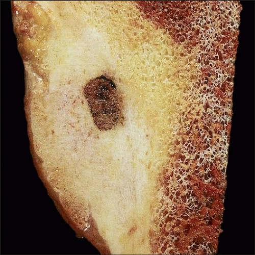
What is the abnormal bone that forms around tumor cells?
They do, however, typically cause reactive bone to form around them. They also make a new type of abnormal bone material called osteoid bone. This osteo id bone, along with the tumor cells, forms the nidus of the tumor, which is a clear spot seen on x-rays. Osteoid osteomas may occur in any bone in the body, but are most often found in the bones ...
What is the procedure to diagnose osteoid osteoma?
Biopsy . A biopsy may be necessary to confirm the diagnosis of osteoid osteoma. In a biopsy, a tissue sample of the tumor is taken and examined under a microscope. Your doctor may give you a local anesthetic to numb the area and take a sample using a needle.
What is the best imaging for osteoid osteoma?
Imaging Studies. X-rays. X-rays create clear pictures of dense structures such as bone and are helpful in diagnosing an osteoid osteoma. An x-ray of the painful area may reveal thickened bone surrounding a small central core of lower density—a distinctive characteristic of the tumor. Computerized tomography (CT) scan.
Is osteoid osteoma a tumor?
An osteoid osteoma is a benign (noncancerous) bone tumor that usually develops in the long bones of the body, such as the femur (thighbone) and tibia (shinbone). Although osteoid osteomas can cause pain and discomfort, they do not spread throughout the body.
Is osteoma a benign bone tumor?
Osteoid osteoma is a painful, benign and common bone tumor that is prevalent in young adults. The typical clinical presentation consists of pain that becomes worse at night and is relieved by nonsteroidal anti-inflammatory drugs.
Is osteoid osteoma a multifaceted pathology?
However, osteoid osteoma is a multifaceted pathology that can have unusual presentations, such as intraarticular osteoid osteoma, epiphyseal location, lesions at the extremities and multicentric nidi, and frequently present atypical clinical and radiological manifestations.
Where is osteoid osteoma found?
♦ Males are affected approximately three times more than females. ♦ 50 percent of osteoid osteoma lesions are found in the fibula or tibia. ♦ The cortex of long bones is the most commonplace for osteoid osteoma tumors. ♦ Osteoid osteoma makes up 12 percent of all skeletal neoplasms.
What is the most common symptom associated with osteoid osteoma?
The most common symptom associated with an osteoid osteoma is a dull, aching pain that gets worse, especially at night. The pain is not typically related to any movement, and in many cases, it appears for no apparent reason.
How long does it take for osteoid osteomas to go away?
In most cases, osteoid osteomas will go away without treatment, although this can take several years. Using over-the-counter pain medications and anti-inflammatory drugs can help bring relief. This type of medication may not work for long, so it may be necessary to try several anti-inflammatory medications.
What happens when osteoblasts break down old bone?
In bones, osteoblasts that create new bone multiply quickly, while osteoclasts that break old bone down, are not. The osteoclast becomes part of the osteoma, and as it hardens into a tumor, pressure on the bone increases, causing pain.
Can osteoid osteoma cause bone swelling?
Complications of Osteoid Osteoma. These tumors can be very painful, and sometimes, complications can occur as a result of bone swelling. Some of the more serious complications include: You should seek immediate medical attention if you experience bone pain, along with any of the following symptoms:
Can you have surgery for osteoid osteoma?
Taking anti-inflammatory medications for an extended period of time can do damage to your stomach, which makes surgery an option for an osteoid osteoma. The two most common surgical procedures for osteoid osteoma are:
Is osteoid osteoma a tumor?
An osteoid osteoma is a non-cancerous bone tumor. As a benign tumor, these can cause pain and discomfort, but will not spread to other parts of the body.
Where do osteoid osteomas occur?
Most osteoid osteomas occur in long tubular bones of the limbs (especially the proximal femur), but any bone may be involved. Furthermore, osteoid osteomas are usually cortical lesions but they can occur anywhere within the bone including medullary, subperiosteal (most commonly in the talus), and intracapsular 2.
What age group is osteoid osteomas?
Epidemiology. Osteoid osteomas are usually found in children, adolescents, and young adults, between the ages of 10 and 35 years 2 . They account for ~10% of all benign bone lesions and there is a male predilection (M:F 2-4:1) 2.
What are the three concentric parts of osteoid osteoma?
An osteoid osteoma is composed of three concentric parts 1: The nidus releases prostaglandins (via the enzymes cyclo-oxygenase-1 and cyclo-oxygenase-2) which in turn result in pain 2.
How long does it take for osteoid osteoma to resolve?
The average time to resolution is 33 months.
How old are osteoid osteomas?
Osteoid osteomas are usually found in children, adolescents, and young adults, between the ages of 10 and 35 years 2 .
What is the name of the benign bone forming tumor that typically occurs in children?
Osteoid osteoma. Osteoid osteomas are benign bone-forming tumors that typically occur in children (particularly adolescents). They have a characteristic lucent nidus less than 1.5 or 2 cm and surrounding osteosclerotic reaction, which classically causes night pain that is relieved by the use of salicylate analgesia, e.g. aspirin.
Where are osteoid osteomas found?
Osteoid osteomas are most commonly found in the metaphyseal and diaphyseal regions of the tibia and femur (2). However, they can also be found in the spine, hands, pelvis, sacrum, and feet (2). Intra-articular osteoid osteomas differ from intra-cortical osteoid osteomas and are recognized as separate entities (11). They are a benign tumor and do not metastasize or have the capacity to become malignant (5). They are typically categorized based on their location within bone (11). This includes either the cortical portion, subperiosteal, or medullary portion (11).
What is the most common presentation of osteoid osteoma?
The most common presentation of an osteoid osteoma is a dull and local pain that increases at night (4). The area of abnormal osteoid, known as the nidus, is innervated by sensory nerve fibers that produce prostaglandins (2). Levels of prostaglandins as high as 100 to 1000 times normal have been documented within the nidus of an osteoid osteoma (7). In a survey of patients with hip osteoid osteomas, they found that 90% of the affected population had pain at nighttime and 88% felt that the pain was relieved with NSAIDs (2).
How to treat osteoid osteomas?
Currently, the gold standard for symptomatic osteoid osteomas is thermal radiofrequency ablation under CT guidance (3) . The procedure is performed under a general anesthetic (10). Under CT guidance, a drill bit is inserted into the cortex of the bone (10). Once within the nidus, a radiofrequency probe is inserted into the nidus and heated to 90 degrees Celsius for 4 minutes (10). Research finds that the procedure is typically successful in 90% of the cases (3). In a study done by Rosenthal in the Journal of Bone & Joint Surgery, there was no difference in the rate of recurrence when comparing open excision to radiofrequency ablation (9).
What is the osteoid of bone?
The osteoid of bone is a matrix of Type I collagen that is embedded in a glycosaminoglycan gel that contains proteins that bind strongly to calcium (1). The osteoid is made by the osteoblast cells (1). Bone is made up of the combination of osteoid, osteoblasts, osteoclasts, and mineral salts (1).
Can a CT scan show osteoid osteomas?
First line imaging for osteoid osteomas is a plain radiograph (5). The nidus is seen as an oval radiolucency and is surrounded by bony sclerosis (5). When evaluating, second line imaging CT scans have been found to be more accurate in diagnosing these tumors than MRIs (5). In a study comparing the two modalities, CT scans identified the tumor in 67% of the cases and MRI only identified them in 3% of the cases (6). However, due to the small size of the osteoid osteomas, 1mm slices on a CT would be required and this can lead to large radiation doses for the patient (10). The small size of the tumors is also the reason why an MRI may miss the nidus of the tumor (10). Frequently an MRI will only identify an area of non-specific edema within bone, which can be misinterpreted as a stress fracture (10).
Can osteoid osteoma cause swelling in knee?
Physical exam findings can sometimes reveal a knee effusion due to the osteoid osteoma’s proximity to the joint (5). There can also be local bony tenderness and soft tissue swelling (5). Physicians should also evaluate for limb-length discrepancy, which could be the result of a femoral osteoid osteoma (5).
Is osteoid osteomas a benign tumor?
Osteoid osteomas are one of the most common benign bone tumors found. They have a characteristic presentation of nocturnal pain relieved with NSAIDs. Diagnosis can sometimes be difficult due to their location or small size on radiographs. In the future, treatment options will continue to shift towards minimally invasive options, such as arthroscopic excision.
