What structures are retroperitoneal?
The primary retroperitoneal structures are the adrenal glands, kidneys, ureters, inferior vena cava, and the rectum. Secondary retroperitoneal structures include the duodenum (except for the proximal first segment), the pancreas (head, neck and body), ascending colon, and descending colon.
Which organ is retroperitoneal?
Retroperitoneal organs lie behind the posterior sheath of the peritoneum and include the aorta, esophagus, second and third parts of the duodenum, ascending and descending colon, pancreas, kidneys, ureters, and adrenal glands.
What part of the duodenum is retroperitoneal?
The secondary retroperitoneal organs, which were initially intraperitoneal and became retroperitoneal structures during embryologic development due to the regression of peritoneal tissue lying on the posterior wall of the abdominal cavity (the mesentery of these structures fuse with the posterior abdominal wall), are the ascending and descending colon, duodenum except the bulbus part (first half of duodenum segment 1) and pancreas.
What organs are retroperitoneal Quizlet?
retroperitoneal space Area posterior to peritoneum and anterior to muscular body wall Contains pancreas, kidneys, ureters, and parts of the digestive tract sandwich sign represents the anterior and posterior node masses surrounding mesenteric vessels sex hormones

Retroperitoneal Organs
The abdominal cavity is the large space that is vertically enclosed by the vertebral column and abdominal muscles, and horizontally by the diaphragm and pelvis. The peritoneum is a serous membrane that lines the abdominal cavity and supports the abdominal organs.
Where is the Retroperitoneal Cavity?
The intraperitoneal space is the part of the abdominal cavity that is surrounded by the peritoneum, and houses organs such as the stomach and the intestines. The retroperitoneal cavity is the space located behind the intraperitoneal space where the kidneys are located.
A Mnemonic for Retroperitoneal Organs
The retroperitoneal organs mnemonic SAD PUCKER can be used to summarize retroperitoneal organs.
The Structure of Retroperitoneal Organs
The retroperitoneum is divided into three main spaces anatomically, which house different retroperitoneal structures:
What is the retroperitoneum?
The retroperitoneum is an anatomical space located behind the abdominal or peritoneal cavity. Abdominal organs that are not suspended by the mesentery and lie between the abdominal wall and parietal peritoneum are said to lie within the retroperitoneum. Several individual spaces make up the retroperitoneum. These spaces are the anterior pararenal space, posterior pararenal space, and the perirenal space. Each of these spaces contains parts of various organs and structures. These structures include organs that contribute to several systems in the body, including the urinary, adrenal, circulatory, gastrointestinal, and endocrine systems.[1] This article will discuss the structure, function, embryology, and anatomy of the retroperitoneum, and will also include discussion of its clinical significance and specific surgical considerations.
What are the three spaces of the retroperitoneum?
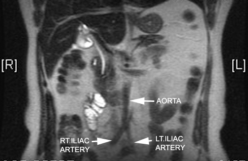
The retroperitoneum divides into three main anatomical spaces: the anterior pararenal space, perirenal space, and posterior pararenal space . The anterior para renal space contains the head, neck, and body of the pancreas (the tail of the pancreas is within the splenorenal ligament), ascending and descending colon, and the duodenum (except for the proximal first segment). Structures contained within the perirenal space include the adrenal gland, kidney, ureters, and renal vessels. The posterior pararenal space, which is surrounded by the posterior leaf of the renal fascia and muscles of the posterior abdominal wall, contains no major organs and is composed primarily of fat, blood vessels, and lymphatics.[2] There is also a fourth, less well-defined space known as the great vessel space. It lies anterior to the vertebral bodies and psoas muscles and contains the aorta, inferior vena cava, and surrounding fat. [3]
What is retroperitoneal fibrosis?
Retroperitoneal fibrosis is an uncommon collagen vascular disorder. It is the result of a fibrotic reaction within the retroperitoneum, and its cause is not well understood. It has correlations with both benign and malignant conditions, certain medications, and idiopathic cases, which have also been described. Patients will often present initially with symptoms of ureteric obstruction. Reportedly CT or MRI are of equal value in diagnosis. Imaging typically shows contrast-enhancing fibrosis encasing the structures of the retroperitoneum resulting in obstruction and displacement of the ureters or vascular structures. An underlying cause remains unfound in over 70% of cases. Treatment and outcomes vary and are dependent upon etiology and the degree of obstruction. [13]
Which muscle is located on the posterior margin of the retroperitoneum?
Muscles within the retroperitoneum can be organized based on their location. Muscles contributing to the posterior margin of the retroperitoneal space consist largely of the transverse abdominal, psoas, quadratus lumborum, and iliacus. The paraspinous muscles contribute to the medial boundary on either side of the spine, and the abdominal musculature forms the lateral margin. The superior border is formed in part by the diaphragm, while the iliopsoas muscle is the primary muscle contributing to the inferior border. [8]
Can retroperitoneum be a site of bleeding?
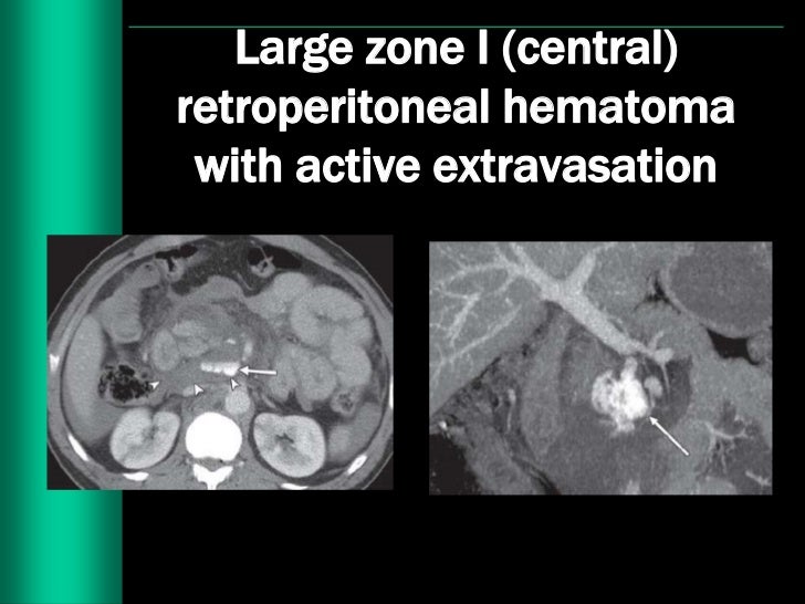
The retroperitoneum can occasionally be a site of significant bleeding , usually after trauma, surgical intervention, or even spontaneously in patients with vascular lesions (e.g., abdominal aortic aneurysm) or those treated with anticoagulation therapy. Presentation varies based on etiology. Symptoms can include hypotension, tachycardia, ecchymoses in the affected areas, fatigue, hematuria, and flank or back pain. Computed tomography of the abdomen is the diagnostic imaging of choice. Management of retroperitoneal hematoma is almost always MEDICAL, with resuscitation, blood transfusions, and reversal of anticoagulation as necessary. [14]
What age does retroperitoneal fibrosis occur?
Trusted Source. , it occurs most often between the ages of 40 and 60. However, it can develop at any age. The condition occurs twice as often in men as in women. Specific conditions linked with retroperitoneal fibrosis can include: smoking. exposure to asbestos.
What are the symptoms of retroperitoneal fibrosis?
Initially, your body reacts to the reduced blood flow. Symptoms that occur in the early stages of this condition include: dull pain in the abdomen or back that may be hard to pinpoint.
Why wrap the affected ureter in fat tissue from the intestines?
wrap the affected ureter in fat tissue from the intestines to protect it from fibrosis regrowth
What is the term for the condition where the stomach is surrounded by fibrous tissue?
It occurs when excess fibrous tissue develops in the space behind your stomach and intestine called the retroperitoneal area.
Can retroperitoneal fibrosis be prevented?
How to prevent retroperitoneal fibrosis. The majority of cases cannot be linked to any specific cause, so prevention may not be possible. However, the condition is associated with the use of some medications to treat high blood pressure and medications to treat migraines called ergotamines.
Can retroperitoneal fibrosis cause kidney failure?
This may result in chronic kidney failure and long-term blockage of the ureters, which can cause urine backup and kidney swelling. Untreated retroperitoneal fibrosis can also lead to the cutting off of the blood supply to the legs, which in turn can lead to further dangerous complications.
What is the abdominal aortic aneurysm associated with?
Figure 2: Abdominal aortic aneurysm associated with the right common iliac artery aneurysm. The access to the bifurcation of the right iliac artery will be difficult in RP approach due to the right lateral position of the patient, therefore, unsuitable for RP approach
Why are open aortic surgeries so challenging?
Patients undergoing open surgery these days are inevitably either young or have challenging anatomy, and as a consequence, the open procedures are often more challenging due to obesity, calcified vessels , and poor-quality necks requiring supra/intrarenal clamps.
What is the best way to repair an aneurysm?
Most Vascular Surgical units are familiar with the transperitoneal (T P) approach to AAA. Another useful approach to AAA is retroperitoneal (RP). Our unit has found RP a more suitable approach for repairing the AAAs with challenging anatomy and in patients with poor physiological function. RP approach provides excellent access to the juxta/supra renal aorta. In addition, patients undergoing RP have less post-operative morbidity and shorter hospital stay compared to those undergoing TP repair. Our unit recommends RP approach as the preferred option to repair AAAs unsuitable for endovascular repair. Given the relative lack of familiarity with this technique as it is not widely practised, we have described the technique/our experience and its advantages over conventional open surgery.
When did Cooley and De Bakey repair the aorta?
In the early 1950s, Cooley and De Bakey [1] resected saccular aneurysms and repaired the arterial walls by lateral suture. Since then, there have been significant advances in aortic surgery. Various approaches to the aorta have been described which broadly can be classified as transperitoneal (TP) or retroperitoneal (RP).
Where is the incision for a rectus abdominis?
The incision starts at the lateral edge of the rectus abdominis approximately midway between the umbilicus and the pubic symphysis and then extends posterolaterally toward the 11 th and the 12 th rib [Figure 4]. These ribs can be excised if further exposure is required. Diathermy is then used to dissect through the abdominal musculature. A plane is then developed between the posterior abdominal wall and the posterolateral peritoneum displacing the left kidney, the ureter, tail of the pancreas, and the spleen anteriorly. This is done by a combination of sharp and blunt dissection by gently sweeping the tissue in front of the posterior abdominal wall using a pack and cauterizing bleeding vessels with diathermy. Any breach in the peritoneum at this stage is sutured with vicryl. Exposure of the aorta and retraction of the abdominal wall and abdominal contents is greatly aided using self-retaining retraction systems. Increased exposure is facilitated by retraction of the abdominal musculature outward and upward. The dissection is then continued anterior and medial to the psoas muscle to reach the side wall of the aorta. This dissection is continued cranially to the neck and caudally to the aortic bifurcation. The periaortic fat is vascular and is carefully dissected to ensure good hemostasis. The left gonadal vein needs ligation to expose the left renal artery and thus the neck of the aneurysm [Figure 5] and [Figure 6].
Is transperitoneal aneurysm repair good?
The transperitoneal approach is most familiar to surgeons and provides good intraperitoneal access. It has been the standard approach to open aneurysm repair. The RP approach is practiced in fewer centers, and consequently, expertise in the technique is limited. With the decreasing number of aneurysms in the UK population and restriction of training hours, this expertise is likely to diminish even further. Our unit has been performing RP AAA repair for more than two decades and is one of the few centers in the UK that offers RP repair to patients with AAAs.
Is an aneurysm repair an open surgery?
Despite the development of endovascular techniques for abdominal aortic aneurysm (AAA) repair, open surgical repair remains necessary to treat anatomically complex an eurysms and may be beneficial for younger patients and those unable to comply with long-term surveillance. Not all aneurysms have a favorable anatomy for a simple EVAR procedure and may require a complex endovascular procedure or open surgery. RP access is especially suited for challenging necks as the approach facilitates better access to the suprarenal aorta. The evidence also suggests that the RP approach is better in patients with compromised respiratory function as it reduces postoperative morbidity and shortens postoperative stay when compared with transperitoneal repair. [2]
Why is the retroperitoneal space entered posterolaterally?
The retroperitoneal space is entered posterolaterally to avoid tearing the parietal peritoneum (Figure 2). The posterior peritoneum, posterior layers of Gerota’s fascia, and the left kidney are retracted anteriomedially and cephalad to expose the left psoas muscle and periaortic tissue. The fascia remains intact on the psoas, which minimizes dissection-related injury of the genitofemoral nerve and bleeding from the iliopsoas muscle. Exposure is maintained with a self-retaining retractor. Care must be taken to avoid vigorous retraction of the anterior and cephalad margin of the incision because this can result in injury to the spleen or kidney.
What is retroperitoneal exposure?
A traditional retroperitoneal exposure is the preferred approach to an AAA repair in the hostile abdomen, which includes patients with prior intraabdominal surgery resulting in adhesions, the presence of stomas, peritoneal dialysis, reoperative aortic surgery, prior abdominopelvic radiation treatment, inflammatory aneurysm, and morbid obesity. Relative contraindications to the retroperitoneal approach include limited exposure of the right renal and iliac arteries as well as the inability to fully examine the intraabdominal cavity for additional pathology at the time of surgical intervention. Most recently, this approach has been used for complex aortic reconstructions owing to the excellent exposure of the suprarenal and infradiaphragmatic aorta. It is also useful when treating pararenal aortic aneurysms as well as explanting failed endografts.
What are the landmarks of the infrarenal neck of the aortic aneurysm?
The landmarks for the infrarenal neck of the aortic aneurysm are the origin of the left crus of the diaphragm, the lumbar branch of the left renal vein, and the left renal artery ( Figure 4 ). The lumbar branch of the left renal vein, which crosses the aorta in a posterior and perpendicular fashion and caudad to the renal artery, ...
Which muscle is divided to the lateral border of the rectus abdominis?
The muscle layers of the lateral abdominal wall are divided to the lateral border of the rectus abdominis. This includes the external oblique, internal oblique, transversus abdominis, and transversalis fascia, respectively. The transversus abdominis is initially divided laterally and then medially to separate the peritoneum, which is usually thicker and more discrete laterally, from the underlying muscle. The intercostal muscles are divided on the superior margin of the underlying rib.
What is the purpose of distal arterial control?
Distal arterial control is obtained first to prevent embolization. If there is extensive right iliac artery disease, then a right suprainguinal counterincision is made to obtain extraperitoneal exposure of these vessels. Right iliac artery control can also be obtained with a balloon occlusion catheter at the time the aneurysm sac is incised. Alternatively, vertical groin incisions can be made to access the femoral vessels for distal arterial control.
What causes retroperitoneal inflammation?
Retroperitoneal inflammation can happen when harmful bacteria come in contact with the organs in the retroperitoneal space or the lining that encloses your abdominal cavity . Possible causes of retroperitoneal inflammation include:
What is the term for inflammation of the retroperitoneum?
Retroperitoneal inflammation is also known as retroperitonitis.
What is the space between the intestines and the back?
In less complicated terms, it’s the space in your abdomen between your abdominal cavity (the area where your intestines are) and your back. It houses several major organs, including: kidneys. bladder. abdominal aorta. adrenal glands. Inflammation often happens in response to an infection.
What tests are done to check for retroperitoneal space?
Your doctor will assess your symptoms. Then they will typically order an ultrasound, abdominal X-ray, CT scan, or MRI. These imaging tests will help reveal any abnormalities in the retroperitoneal space. This will allow your doctor to assess your condition.
What can cause bacteria to enter the retroperitoneal space?
A ruptured appendix, stomach ulcers, or a perforated colon can allow bacteria into your retroperitoneal space .
Is retroperitoneal space a serious condition?
It has a high mortality rate. However, early diagnosis and treatment can improve your outlook. The retroperitoneal space is the space between your peritoneum and your posterior abdominal wall.
Is retroperitoneal inflammation a long term condition?
Your hospital stay may be lengthy. Retroperitoneal inflammation is a serious condition that can have life-threatening consequences.