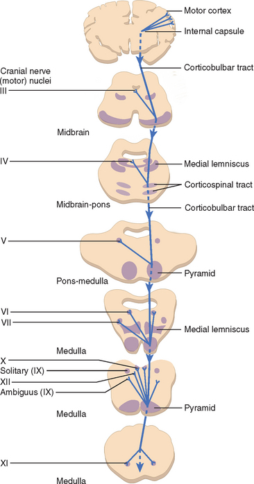
The facial muscles are innervated peripherally (infranuclear innervation) by the ipsilateral 7th cranial nerve and centrally (supranuclear innervation) by the contralateral cerebral cortex. Central innervation tends to be bilateral for the upper face (eg, forehead muscles) and unilateral for the lower face.
Which side of the brain controls the facial nerve?
The lower branches of the facial nerve that supply muscles in the lower two-thirds of the face are controlled by messages from only one side of the brain (the contralateral or opposite side). The upper branches of the facial nerve, which control the upper part of the face, receive messages from both sides of the brain.
What is the facial nerve?
The facial nerve is the seventh cranial nerve (CN VII). It arises from the brain stem and extends posteriorly to the abducens nerve and anteriorly to the vestibulocochlear nerve.
What is the difference between Central and peripheral facial nerve palsy?
In a central lesion, the forehead should lift symmetrically, due to bilateral cortical innervation of the frontalis muscle. However, in a peripheral lesion, the patient will be unable to wrinkle their forehead on one side, or have fewer wrinkles on that side. Asymmetry in forehead wrinkles is a sign of peripheral facial nerve palsy.
What is facial nerve decompression?
Facial nerve decompression surgery is also sometimes carried out in certain cases of facial nerve compression. Voluntary facial movements, such as wrinkling the brow, showing teeth, frowning, closing the eyes tightly (inability to do so is called lagophthalmos ), pursing the lips and puffing out the cheeks, all test the facial nerve.
See more
Is facial nerve contralateral or ipsilateral?
The fibers that control the lower face travel from the cortex down to the brainstem. In the brainstem, these fibers cross over to the opposite, or contralateral, facial nerve.
Is facial nerve palsy contralateral?
In central facial palsy, paralysis is contralateral to the lesion, and eyelid and forehead muscles are not affected!
Is the facial nerve bilaterally innervated?
The supranuclear innervation is bilateral to the muscles of the forehead and eyes but only contralateral to the muscles of the lower part of the face. This accounts for the sparing of the upper facial muscles with a contralateral cortical lesion. Figure 62.1 shows the anatomy of the facial nerve.
Is cranial nerve 7 ipsilateral or contralateral?
The main function of each of the two 7th cranial nerves is facial movement on the same side (ipsilateral). Left sided forehead wrinkle, left eyelid closure, and movement of the left half of the face is stimulated by the left 7th cranial nerve.
Does the facial nerve Decussate?
Answer and Explanation: Facial nerve donot decussate. There are only 2 cranial nerves that decussate, and these nerves are the second and fourth cranial nerves.
Is facial droop on same side as stroke?
A stroke occurs due to the blockage of a blood vessel in the brain. Motor neurons traveling from the cortex of either brain hemisphere stimulate facial muscles on the opposite side of the body. Thus, when a stroke impacts one hemisphere of the brain, it will cause facial weakness in the opposite side of the face.
Which cranial nerve is contralateral?
Contralateral and Unilateral InnervationNerveInnervationFacial (VII)Mixed bilateral symmetry and contralateral innervationGlossopharyngeal (IX)Neither bilateral symmetry or contralateral innervation*Vagus (X)Bilateral symmetrySpinal accessory (XI)Contralateral innervation2 more rows
Is the trigeminal nerve contralateral?
The spinothalamic tract from the contralateral half of the body is near the trigeminal tract and nucleus. Therefore it follows that at these levels there can be contralateral loss of body pain and temperature associated with an ipsilateral loss of facial pain and temperature if the lesion is sufficiently large.
Which cranial nerves are bilaterally innervated?
CN IX is innervated bilaterally and has sensory, parasympathetic, and motor components. The sensory division receives general sensory fibers from the tonsils, pharynx, middle ear, and the posterior one-third of the tongue, as well as taste fibers from the posterior third of the tongue.
Is cranial nerve 3 contralateral?
Saccade movement originates in the frontal eye fields and initiates the CONTRALATERAL CN III (vertical movement) or VI (horizontal eye movement). Pursuit eye movement originates at the top of the brainstem and initiates CN III (vertical) or CN VI (horizontal) on the IPSILATERAL side.
How can you differentiate bilateral UMN and LMN facial palsy?
If the forehead is not affected (i.e. the patient is able to raise fully the eyebrow on the affected side) then the facial palsy is likely to be an upper motor neuron (UMN) lesion. Paralysis which includes the forehead, such that the patient is unable to raise the affected eyebrow, is a lower motor neuron (LMN) lesion.
How can you differentiate upper and lower motor facial nerve palsy?
A lower motor neurone lesion causes weakness of all the muscles of facial expression. The angle of the mouth falls. Weakness of frontalis occurs, and eye closure is weak. With an upper motor neurone lesion frontalis is spared, normal furrowing of the brow is preserved, and eye closure and blinking are not affected.
Which side is affected in facial palsy?
Bell's palsy is a condition that causes sudden weakness in the muscles on one side of the face. In most cases, the weakness is temporary and significantly improves over weeks. The weakness makes half of the face appear to droop. Smiles are one-sided, and the eye on the affected side resists closing.
Does Bell's palsy affect both sides of the face?
Generally, Bell's palsy affects only one side of the face; however, in rare cases, it can affect both sides. Symptoms appear suddenly over a 48 - 72-hour period and generally start to improve with or without treatment after a few weeks, with recovery of some or all facial function within six months.
How can you differentiate between bilateral UMN and LMN facial palsy?
If the forehead is not affected (i.e. the patient is able to raise fully the eyebrow on the affected side) then the facial palsy is likely to be an upper motor neuron (UMN) lesion. Paralysis which includes the forehead, such that the patient is unable to raise the affected eyebrow, is a lower motor neuron (LMN) lesion.
How can you differentiate upper and lower motor facial nerve palsy?
A lower motor neurone lesion causes weakness of all the muscles of facial expression. The angle of the mouth falls. Weakness of frontalis occurs, and eye closure is weak. With an upper motor neurone lesion frontalis is spared, normal furrowing of the brow is preserved, and eye closure and blinking are not affected.
Which facial nerve controls the frontalis muscle?
Six of the facial nerve branches control facial movement. The temporal nerve controls the frontalis muscle. The zygomatic nerve controls the orbicularis oculi. The buccal nerve controls the buccinator and orbucularis oris muscles. The mandibular nerve controls the mentalis muscle. The cervical nerve controls the platysma, and the posterior auricular nerve controls the occipitalis muscle.
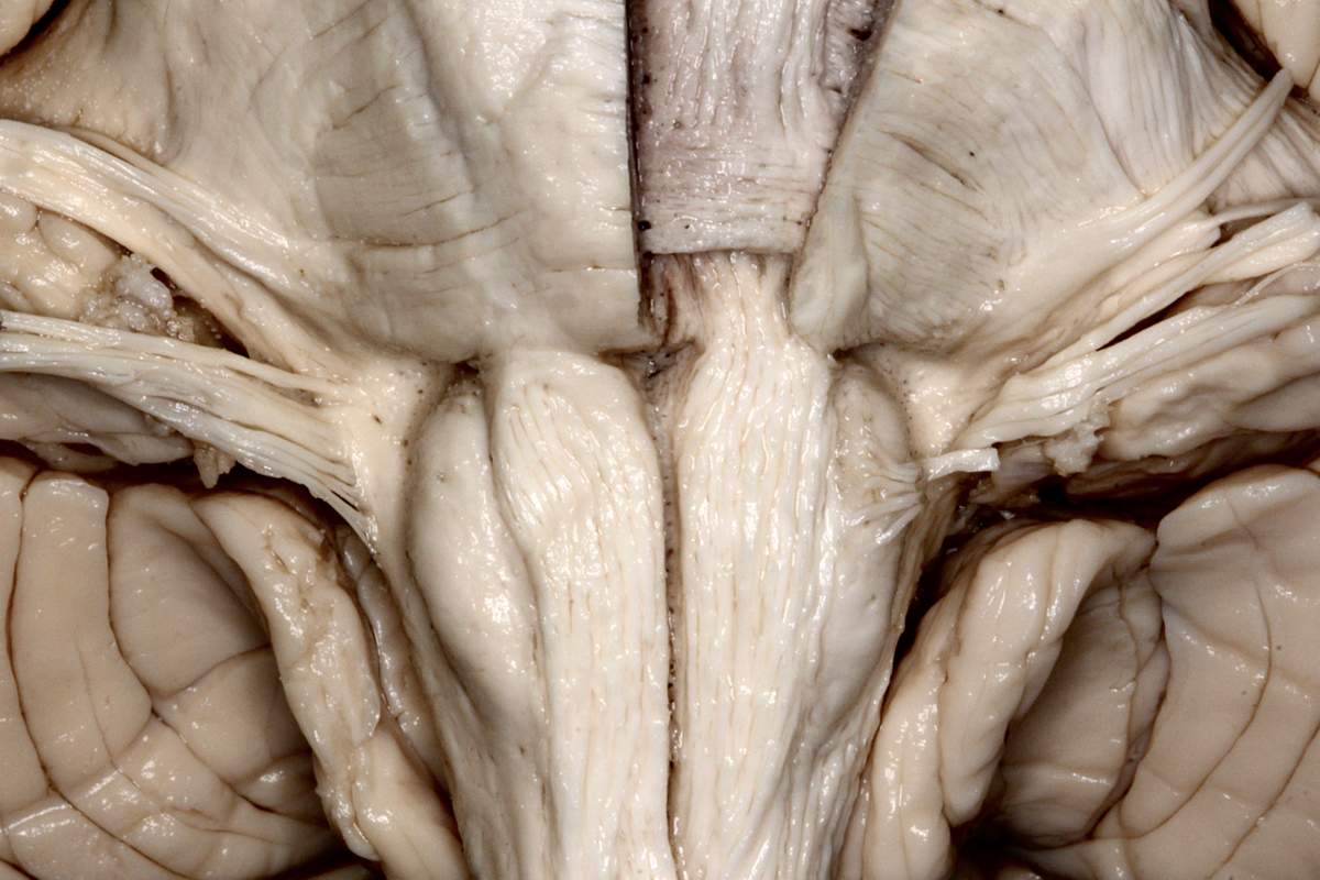
Where is the facial nerve located?
It is one of the longest cranial nerves, extending from the brainstem to the terminal (end) branches, which are located throughout the face. Several structures of the facial nerve—described as nuclei, segments, and branches—produce the four components of facial nerve function. 1
What are the branches of the facial nerve?
Most of the branches of the facial nerve are motor branches that stimulate the movement of the facial muscles. These muscles include: 1 the stapedius muscle in the ear, which controls the vibration of a bone in the ear to help moderate hearing 2 the stylohyoid muscle in the neck, which is involved with swallowing 3 the posterior belly of the digastric muscle, which is involved with movements of chewing, swallowing, talking, and breathing 4 the muscles of facial expression are controlled by the facial nerve 5 the frontalis muscle moves the forehead and eyebrows 6 the orbiculus oculi, which controls the muscles of the eyelids 7 the buccinator muscle, which moves the mouth and cheek 8 the orbicularis oris, which controls movements of the mouth and lips 9 the platysma, which is a large muscle in the neck that controls movements of the neck and jaw 10 the occipitalis muscle, which is located in the back of the head and moves the scalp skin posteriorly.
How many sections does the facial nerve have?
The facial nerve has: six major sections (described as segments) along the pathway from the brainstem to the terminal branches in the face. divisions and subdivisions (also called branches), which are small nerves in and around the face that merge along the segments into the main facial nerve.
How to recover from facial nerve damage?
If you have had any type of facial nerve disease or injury, recovery includes physical therapy , which can help your face and mouth muscles regain at least some of their strength. 10 The extent of recovery depends on the type and severity of the damage, how much of the nerve was involved, and the type of disease.
Why does the forehead move?
The fascinating thing about this redundancy is that if the facial nerve can’t function properly due to a problem in the brain, the muscles of the forehead can still move. When the area of the brain that controls the face becomes damaged, only the lower two-thirds of the face becomes weak.
What neurotransmitter is released by the facial nerve?
The motor branches of the facial nerve activate muscles to move by releasing acetylcholine, a neurotransmitter that binds to the surface of muscle cells. Activated muscles respond by contracting (becoming shorter in length), pulling or twisting nearby joints and bones, and ultimately producing movement of the face.
What is the facial nerve?
Introduction. The facial nerve is the seventh cranial nerve (CN VII). It arises from the brain stem and extends posteriorly to the abducens nerve and anteriorly to the vestibulocochlear nerve. It courses through the facial canal in the temporal bone and exits through the stylomastoid foramen after which it divides into terminal branches at ...
What are the two parts of the facial nerve?
It consists of two parts: a proper facial nerve and the intermediate nerve. The proper facial nervecontains only a motor component and a very small general somatic afferent component, whereas the intermediate nervecarries sensory and parasympathetic visceromotor components.
What nerve innervates the facial muscles?
As stated, the facial nerve innervates the following: 1 The muscles of facial expression – responsible for the expression of emotions by changing facial expression 2 The stylohyoid muscle – draws the hyoid bone backward, which initiates a swallowing action and elevates the tongue 3 The posterior belly of the digastric muscle – together with the anterior belly of the digastric muscle, elevates the hyoid bone and is involved in any complex movements involving the jaw 4 The stapedius muscle of the middle ear – stabilizes the stapes, preventing excessive movement in response to loud sounds
Which muscle is innervated by the second branchial arch?
The second branchial arch also produces the muscles of the face, the occipitofrontalis muscle, the platysma, the stylohyoid muscle, the posterior belly of the digastric muscle, the stapedius muscle , and the auricular muscles, all of which are innervated by CN VII. [1] Nerves. The facial nerve exits the brain stem from its ventrolateral surface ...
How is the corneal reflex tested?
The corneal reflexis tested by stimulating the cornea with a wisp of cotton. It results in the reflex closure of both eyelids. The afferent limb of this reflex is mediated by the trigeminal nerve, and the efferent limb is mediated by the facial nerve.
Which nerve carries descending parasympathetic GVE fibers from the superior salivatory nucleus?
The intermediate nerve carries descending parasympathetic GVE fibers from the superior salivatory nucleus and ascending GVA, GSA, and SVA fibers from the geniculate ganglion.
Which nerve is responsible for facial expression?
The facial nerve provides motor innervation of facial muscles that are responsible for facial expression, parasympathetic innervation of the glands of the oral cavity and the lacrimal gland, and sensory innervation of the anterior two-thirds of the tongue. The facial nerve is the seventh cranial nerve (CN VII).
What is the facial nerve?
The facial nerve is one of a group of nerves called the cranial nerves (CN), twelve pairs of nerves that , with the exception of the spinal accessory nerve (CN XI), originate in the brain and contribute to the peripheral nervous system (PNS).
What is the function of the facial nerve?
While it is indeed responsible for innervating the muscles of facial expression, the facial nerve is a complex structure containing many fiber types with a variety of functions, including motor, sensory, and autonomic. The following article will discuss the importance and versatility facial nerve.
Where do facial nerve fibers travel?
The fibers travel towards the floor of IV ventricle and go around the abducens nucleus and descend. The facial nerve emerges from the lateral surface of brainstem at the pontine-medullary junction between the VI and VIII nerves.
What is the vascular damage of the facial nerve?
Vascular damage to the facial nerve usually occurs at the supranuclear, pontine, and (rarely) cerebellopontine angle. Upper motor neuron (UMN) lesions occur in strokes and can easily be differentiated with lower motor neuron (LMN) lesions by their presentation. A LMN lesion causes paralysis of the whole side of face, ...
What are the components of the facial nerve?
The facial nerve contains many different types of fibers, including general sensory (afferent) fibers, special sensory fibers, visceral/autonomic motor (efferent) fibers, and somatic motor fibers. General sensory fibers in the facial nerve are responsible for transmitting signals to the brain from ...
Which nerve is responsible for innervating the lacrimal gland, submandibular gland, sublingual?
Visceral/autonomic motor fibers in the facial nerve are responsible for innervating the lacrimal gland, submandibular gland, sublingual gland, and the mucous membranes of the nasal cavity and hard and soft palates, allowing for production of tears, saliva, etc., from these locations.
Which nerve contains motor fibers?
the hypoglossal nerve (CN XII) Some of these contain motor fibers, some contain autonomic fibers, some contain somatic sensory fibers, some contain special sensory fibers, and some contain combinations of a number of these aforementioned fiber types.
What is the name of the facial nerve?
The facial and intermediate nerves can be collectively referred to as the nervus intermediofacialis.
Which cranial nerve controls facial expression?
The facial nerve (the labyrinthine segment) is the seventh cranial nerve, or simply CN VII. It emerges from the pons of the brainstem, controls the muscles of facial expression, and functions in the conveyance of taste sensations from the anterior two-thirds of the tongue.
What is the function of parasympathetic innervation?
Parasympathetic innervation serves to increase the flow of saliva from these glands. It also supplies parasympathetic innervation to the nasal mucosa and the lacrimal gland via the pterygopalatine ganglion. The parasympathetic fibers that travel in the facial nerve originate in the superior salivatory nucleus .
How many segments are there in the facial nerve?
The path of the facial nerve can be divided into six segments: intracranial (cisternal) segment. meatal (canalicular) segment (within the internal auditory canal) labyrinthine segment (internal auditory canal to geniculate ganglion) tympanic segment (from geniculate ganglion to pyramidal eminence)
Where does the communicating branch of the otic ganglion arise?
The communicating branch to the otic ganglion arises at the geniculate ganglion and joins the lesser petrosal nerve to reach the otic ganglion.
Which nerve is located in the labyrinthine segment?
The labyrinthine segment is very short, and ends where the facial nerve forms a bend known as the geniculum of the facial nerve ( genu meaning knee), which contains the geniculate ganglion for sensory nerve bodies. The first branch of the facial nerve , the greater petrosal nerve, arises here from the geniculate ganglion. The greater petrosal nerve runs through the pterygoid canal and synapses at the pterygopalatine ganglion. Postsynaptic fibers of the greater petrosal nerve innervate the lacrimal gland .
Which nerve runs through the pterygoid canal?
The greater petrosal nerve runs through the pterygoid canal and synapses at the pterygopalatine ganglion. Postsynaptic fibers of the greater petrosal nerve innervate the lacrimal gland . In the tympanic segment, the facial nerve runs through the tympanic cavity, medial to the incus .
1. Introduction
The principle of selective reinnervation is to use a cross face reinnervation to power eye closure and the ipsilateral masseteric nerve to power the smile.
2. Cross face reinnervation to power eye closure
A facelift or parotidectomy type incision is performed on both the ipsilateral and the contralateral side, and the distal branches identified as they exit the parotid gland. Existing lacerations may also be used if present.
3. Masseteric nerve reinnervation
The masseteric nerve is identified within the body of the masseter muscle at the level of the sigmoid notch.
4. Reinnervation of the lower division of the facial nerve
The lower division can be antagonistic to commissure elevation when trying to smile. Two options exist:
5. Aftercare
Routine wound care is all that is necessary for the majority of the procedures.
What is the ipsilateral facial weakness?
Lesions that damage the facial nerve in the brainstem, or after it exits the brainstem, result in ipsilateral facial weakness involving both the upper and lower face. It doesn’t matter where the innervation is coming from; if the nerve is damaged, all the muscles on that side of the face are weak. These lesions are referred to as “peripheral lesions” because they affect the facial nerve as it exits the brainstem. Patients will be unable to wrinkle their forehead, tightly close their eye, or smile on the affected side. This distinction can aid in localizing the lesion to the appropriate place in the nervous system, thereby narrowing the differential diagnosis.
How to tell if facial weakness is peripheral or central?
Mouth: First, inspect the patient’s mouth. Look at the nasolabial fold–the wrinkle between the corner of their nose and the corner of their mouth.
What causes weakness of the lower face?
Lesions that damage the motor cortex, such as acute ischemic strokes, will result in contralateral facial weakness of the lower face only, with preservation of the muscles of the upper face on both sides, due to the dual innervation of the upper face.
What causes facial paralysis?
Facial Weakness. The two most common causes of acute facial paralysis are Bell’s palsy and ischemic stroke. 1 EMS providers are often faced with the challenge of differentiating between these two diagnoses.
How long does it take for facial nerves to heal?
It’s the most common cause of facial nerve injury. 3 Deficits accumulate over hours to days, and reach maximum severity within three weeks. The symptoms may also develop at night while the patient is sleeping, making them seem more acute. Facial weakness typically recovers–partially or fully–within six months.
What nerves do brainstem strokes affect?
Due to the vascular supply of the brainstem, brainstem strokes typically affect multiple cranial nerves in addition to either motor or sensory tracts traveling to the spinal cord. 2 Bell’s palsy, on the other hand, typically affects only the facial nerve, causing only peripheral facial weakness.
How to tell if one eye is closed more than the other?
Eyes: First, inspect the eyes at rest. Look at the palpebral fissure–the space between the eyelids–to determine if one eye is opened more widely than the other. This may be a subtle sign of eye closure weakness. Next, ask the patient to close their eyes tightly. Normally, patients should be able to squeeze their eyes so tightly that the eyelashes are no longer visible. Asymmetry in eyelid closure is a sign of peripheral facial nerve palsy.