Are sinus bones?
Paranasal sinuses are named after the bones that contain them: frontal (the lower forehead), maxillary (cheekbones), ethmoid (beside the upper nose), and sphenoid (behind the nose).
Which bones make up the maxillary sinus?
The medial wall or base of the maxillary sinus is formed by the maxilla, and by parts of the inferior concha and palatine bone that overlie the maxillary hiatus.
Are sinus cavities under bone?
Your cheekbones hold your maxillary sinuses (the largest). The low-center of your forehead is where your frontal sinuses are located. Between your eyes are your ethmoid sinuses. In bones behind your nose are your sphenoid sinuses.
What are the walls of the maxillary sinus?
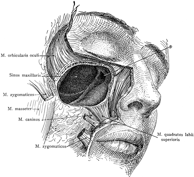
There are six maxillary sinus walls: the superior, anterior, lateral and medial walls are broad, with narrow posterior and inferior walls. Superior: the thin superior wall (forming most of the orbital floor), separates the contents of the orbit from the maxillary sinus.
Is maxilla a single bone?
In humans, the upper jaw includes the hard palate in the front of the mouth. The two maxillary bones are fused at the intermaxillary suture, forming the anterior nasal spine. This is similar to the mandible (lower jaw), which is also a fusion of two mandibular bones at the mandibular symphysis.
What type of bone is maxilla?
The answer to the question, “What type of bone is the maxilla bone?” is simple – it is an irregular facial bone. You can refer to the maxilla bone as a single unit or as two paired but fused bones.
Are your sinuses under your skull?
The frontal sinuses are positioned behind the forehead, while the maxillary sinuses are behind the cheeks. The sphenoid and ethmoid sinuses are deeper in the skull behind the eyes and maxillary sinuses. The sinuses are lined by mucous-secreting cells. Air enters the sinuses through small opening in bone called ostia.
Why does my maxillary sinus hurt?
Maxillary Sinusitis Sinusitis may be due to either a bacterial infection or an allergen. With an acute infectious maxillary sinus, there will usually be an acute ache in the dentition in close proximity or contact with the sinus floor. Percussion tenderness is common on all teeth in a specific quadrant.
How do you drain maxillary sinuses?
Maxillary Sinus MassagePlace each of your index and middle fingers on either side of your nose, just between your cheekbones and upper jaw. Try using your thumbs instead of your index fingers for stronger pressure.Gently massage this area using a circular motion.Repeat for around 30 seconds to a minute.
What is maxillary sinus fracture?
Maxillary sinus fractures (MSFs) are most commonly caused by blunt force trauma to the face. Depending on the magnitude and location of the direct injury, MSFs can vary in appearance and symptomatology.
Is maxillary sinus part of maxilla?
Maxillary sinuses The medial wall or base of the maxillary sinus is formed by the maxilla, and by parts of the inferior concha and palatine bone that overlie the maxillary hiatus.
How big is your maxillary sinus?
The ostium of the maxillary sinus is high up on the medial wall and on average is 2.4 mm in diameter; with a mean volume of about 10 ml.
Does the frontal bone contain a sinus?
There are four paired sinuses (named for the skull bones in which they are located) in the human head: Frontal sinuses: The right and left frontal sinuses are located near the center of the forehead (frontal bone) just above each eye.
Does the ethmoid bone have a sinus?
The ethmoid sinuses are within the ethmoid bone and are divided into two compartments, the anterior and posterior. The anterior ethmoid sinus drains into the middle meatus, and the posterior ethmoids drain into the sphenoethmoidal recess.
Where are the palatine bones?
The palatine bones contribute to the posterior part of the roof of the mouth and floor and lateral walls of the nose, the medial wall of the maxillary sinuses and the orbital floors. Each bone (Fig. 5-66) consists of horizontal and perpendicular plates (laminae) set at right angles to each other.
What does the maxillary sinus drain into?
The maxillary sinus drains into the middle meatus, with the ostium of the sinus opening into the nose on the superior aspect of the medial wall of the sinus, which may explain the high incidence of maxillary sinusitis.
Where is the maxillary sinus?
The maxillary sinus can normally be seen above the level of the premolar and molar teeth in the upper jaw.
How thick is the bone in the maxillary sinus?
However, the bone can vary in thickness in different individuals, ranging from complete absence to 12mm thick.
What is the name of the inflammation of the maxillary sinuses?
Maxillary sinusitis is inflammation of the maxillary sinuses. The symptoms of sinusitis are headache, usually near the involved sinus, and foul-smelling nasal or pharyngeal discharge, possibly with some systemic signs of infection such as fever and weakness.
Why is my sinus opacified?
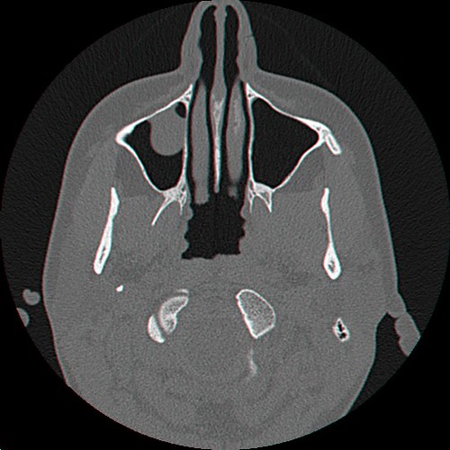
On radiographs, there is opacification (or cloudiness) of the usually translucent sinus due to retained mucus. Maxillary sinusitis is common due to the close anatomic relation of the frontal sinus, anterior ethmoidal sinus and the maxillary teeth, allowing for easy spread of infection.
What is the nasal wall of the maxillary sinus?
The nasal wall of the maxillary sinus, or base, presents, in the disarticulated bone, a large, irregular aperture, communicating with the nasal cavity. In the articulated skull this aperture is much reduced in size by the following bones: and a small part of the lacrimal above and in front.
What is the medial wall?
The medial wall is composed primarily of cartilage. The ostia for drainage are located high on the medial wall and open into the semilunar hiatus of the lateral nasal cavity; because of the position of the ostia, gravity cannot drain the maxillary sinus contents when the head is erect (see pathology).
How big is the first sinus?
It is the first sinus to appear as a shallow groove. At birth it measures about 7*4*4mm. It continues to develops throughout childhood at an annual rate of 2mm vertically and 3mm anteroposteriorly. It reaches its final size in the seventeenth to eighteenth year of life.
Where is the maxillary sinus located?
The maxillary sinus is one of the four paranasal sinuses found near the nose that drains into the middle meatus via the osteomeatal complex. Maxillary sinus.
What is the condition where the maxillary sinuses become inflamed?
Maxillary sinusitis is a condition where the maxillary sinuses become inflamed. Maxillary sinusitis can be caused by either a virus, bacteria or fungus, though acute sinusitis is most commonly caused by a bacterial infection, namely after a viral upper respiratory tract infection. The main culprits are usually Haemophilus, Streptococcus, Pneumococcus or Staphylococcus species. Maxillary sinusitis can also be caused by hay fever and other allergies.
What is the inferior wall of the maxillary sinus?
Inferior wall – formed by alveolar and palatine processes of the maxilla. The maxillary sinus also contains a medial wall, which is rectangular and made primarily of cartilage. It separates the sinus from the nasal cavity. Some of the walls have grooves in them to house nerves and blood vessels. Sinus and nasal cavity anatomy.
What is the largest sinus in the body?
Maxillary Sinus Anatomy. The maxillary sinus is the largest sinus in the body, and so the largest of the four paranasal sinuses. It contains three cavities: Alveolar recess – bounded by the alveolar process of the maxilla body, points downwards. Zygomatic recess – bounded by the zygomatic bone, points laterally.
Why does maxillary sinusitis persist?
Chronic maxillary sinusitis can often develop from an episode of acute sinusitis and may persist if there is poor drainage from the antrum to the nasal cavity. This reduction in drainage can be caused by a number of things, such as mechanical obstruction of the ostium or inhibited mucus clearance.
What causes maxillary sinusitis?
The main culprits are usually Haemophilus, Streptococcus, Pneumococcus or Staphylococcus species. Maxillary sinusitis can also be caused by hay fever and other allergies. The symptoms of acute maxillary sinusitis are: Maxillary sinus pain/pressure. Nasal congestion/discharge. Reduced smell perception.
Where is the ostium located?
The ostium is located on the medial wall near the roof of the maxillary sinus and is approximately 2.4mm in diameter. There are cilia near the maxillary ostium that beat towards it.
What is the maxillary sinus?
Maxillary sinus. The maxillary sinus (or antrum of Highmore) is a paired pyramid-shaped paranasal sinus within the maxillary bone which drains via the maxillary ostium into the infundibulum, then through hiatus semilunaris into the middle meatus. It is the largest of the paranasal sinuses.
Where are the sinuses located?
location: paired sinuses within the body of the maxilla. blood supply: small arteries from the facial, maxillary, infraorbital and greater palatine arteries. innervation: superior alveolar, greater palatine and infraorbital nerves.
Which nerve supplies the pre-molar teeth and overlying mucosa of the sinus?
posterior superior alveolar nerves : dental branches pierce the bone to supply the sinus mucosa. middle superior alveolar nerves : supply the pre-molar teeth and overlying mucosa of the sinus. anterior superior alveolar nerves : supplies canine and incisors and anterior wall of the sinus. greater palatine nerve.
Which sinuses form the anterior border of the pterygopalatine fossa?
The posterior wall forms the anterior border of the pterygopalatine fossa. There are several recesses of the maxillary sinus 5: infraorbital recess (superiorly) zygomatic recess (laterally) alveolar recess (inferiorly) palatine recess (variable extension of alveolar recess) Like the other paranasal air sinuses, these can vary in size.
What is the maxillary sinus?
The maxillary sinus is the paranasal sinus that impacts most on the work of the dentist as they will often be required to make a diagnosis in relation to orofacial pain that may be sinogenic in origin. Maxillary sinus disease is often coincidentally observed on radiographs, and dentists often have to make a diagnosis and plan treatment based on the interpretation of the image. This paper aims to guide the dental professional through some of the disease processes involving the paranasal sinuses and in particular the maxillary sinus. The outcome is to encourage comprehensive history taking and examination of the patient to facilitate an accurate diagnosis that will enable successful treatment.
What is the most common fungal disease in the maxillary sinus?
Most fungal disease of the maxillary sinus involves the organism Aspergillus which lives within moulds and spores and is regularly inhaled into the respiratory system. When infection occurs with Aspergillus in relation to dental foreign materials, the infection is normally contained within the confines of the maxillary sinus. 20 Foci of infection may lead to dystrophic calcification and the formation of rhinoliths, which may be seen on dental radiographs. ( Fig. 12) Large rhinoliths are known as fungal balls. Treatment is normally surgical with removal of any predisposing cause, and this is also increasingly being provided endoscopically with the aim of restoring normal mucociliary function.
What is the most common disease in the paranasal sinuses?
Inflammatory sinus disease is the most common disease process involving the paranasal sinuses. 3 When the maxillary sinus is involved, it is the disease entity where a dentist will most often be asked to make a differential diagnosis.
What is the direction of mucociliary activity in the maxillary sinus?
The direction of mucociliary activity in the maxillary sinus is in blue. Ethmoid polyps are in red, with antrochoanal polyps in green. Full size image. The ostium of the maxillary sinus is high up on the medial wall and on average is 2.4 mm in diameter.
What is the process of growth of the sinuses?
Growth of the sinus continues through life by a process called pneumonisation, such that the roots of maxillary teeth often project into the air space, and following loss of teeth, the sinus floor may be at a level below the nasal floor. The right and left sinuses are often of different dimensions.
What is the treatment for acute sinusitis?
Treatment of acute sinusitis is based on relief of symptoms and does not involve antibiotics unless the patient is pyrexial or there is evidence of spread of infection beyond the confines of the sinus. 8 Rather, treatment is based on topical nasal decongestants and saline irrigation of the nasal cavity.
Can maxillary sinuses be seen on radiographs?
Maxillary sinus disease is often coincidentally observed on radiographs, and dentists often have to make a diagnosis and plan treatment based on the interpretation of the image. This paper aims to guide the dental professional through some of the disease processes involving the paranasal sinuses and in particular the maxillary sinus.
What is sinusitis in the cheek?
Maxillary sinusitis is inflammation of the maxillary sinus which are air filled spaces inside inside the cheek bones. It is one pair of sinuses that is located above or at the level of roots of molar teeth. The maxillary sinuses are connected to the nasal cavity through an opening and therefore any nasal infections can trigger maxillary sinusitis. It is commonly seen after viral infections such as the cold and may be associated with certain dental procedures and problems.
What causes a deviated nasal septum?
A deviated nasal septum and nasal polyps are the conditions predisposing to maxillary sinusitis. It is also caused due to cysts and tumors of maxillary sinus. Dental causes of maxillary sinusitis include the spread of infection from roots of upper molar teeth to the floor of the sinus. The offending tooth is generally infected due ...
Can sinusitis be caused by a cold?
The maxillary sinuses are connected to the nasal cavity through an opening and therefore any nasal infections can trigger maxillary sinusitis. It is commonly seen after viral infections such as the cold and may be associated with certain dental procedures and problems.
Is sinusitis a chronic condition?
The feeling of being tired all the time and a low grade fever is commonly seen. Nasal discharge and headaches are more persistent in chronic forms. These symptoms are largely non-specific for maxillary sinusitis when compared to the acute form. Patients may therefore mistaken it for conditions such as hay fever (allergic rhinitis) and not seek appropriate treatment.
What are the sinuses in the skull?
What is a sinus? The sinuses are air-filled cavities inside of the skull that serve several purposes. There are 4 sets of sinuses (maxillary, ethmoid, frontal and sphenoid), and they are mainly distributed around the eyes. Besides making the skull lighter, sinuses also seem to have a protective role during trauma.
How to tell if you have a sinus fracture?
Fractures of the maxillary sinuses are usually coupled with cheekbone fractures. In cases of isolated maxillary sinus fractures, you may notice air under the skin of the cheek or notice bleeding during nose blowing.
What is a fracture of the ethmoid sinus?
Fractures of the ethmoid sinus are almost always associated with an orbital blowout fracture and would be diagnosed and treated accordingly. When a fracture involves the frontal sinus, you may notice a depression of the forehead. There may also be some bleeding from the nose.
What to do if a fracture does not involve the drainage pathway of the sinus and brain?
If the fracture does not involve the drainage pathway of the sinus and brain, then simple routine follow up with repeated CT scans are all that is required. However, if the fracture involves the drainage pathway or has caused CSF leakage, the fracture will need to be surgically reduced and plated.
Can a sinus fracture lead to a lifetime of sinus disease?
The nasal sinuses are usually lined with paper-thin bone and thus have a high tendency to sustain a fracture during trauma. Although not functionally as debilitating as other fractures of the facial bones, sinus fractures can lead to a lifetime of sinus disease. As such, sinus issues should be treated by a facial plastic surgeon ...
Can a sinus fracture be repaired?
The manner in which your sinus fracture is repaired is dependent on two factors, location and extent of damage. Ethmoid sinus fractures are typically not repaired. If any repair is needed, it is done in conjunction with an orbital blow out fracture repair. Similarly, maxillary sinus fractures may also not require repair.
Overview
Clinical significance
Maxillary sinusitis is inflammation of the maxillary sinuses. The symptoms of sinusitis are headache, usually near the involved sinus, and foul-smelling nasal or pharyngeal discharge, possibly with some systemic signs of infection such as fever and weakness. The skin over the involved sinus can be tender, hot, and even reddened due to the inflammatory process in the area. On radiographs, there is opacification (or cloudiness) of the usually translucent sinus due to retaine…
Structure
It is the largest air sinus in the body. Found in the body of the maxilla, this sinus has three recesses: an alveolar recess pointed inferiorly, bounded by the alveolar process of the maxilla; a zygomatic recess pointed laterally, bounded by the zygomatic bone; and an infraorbital recess pointed superiorly, bounded by the inferior orbital surface of the maxilla. The medial wall is composed primarily of cartilage. The ostia for drainage are located high on the medial wall and o…
History
The maxillary sinus was first discovered and illustrated by Leonardo da Vinci, but the earliest attribution of significance was given to Nathaniel Highmore, the British surgeon and anatomist who described it in detail in his 1651 treatise.
See also
• Ohngren's line
• Zygomatic complex fracture
External links
• Radiology image: Headneck:17Maxill from Radiology Atlas at SUNY Downstate Medical Center (need to enable Java)
• Cross section image: skull/x-front—Plastination Laboratory at the Medical University of Vienna
• lesson9 at The Anatomy Lesson by Wesley Norman (Georgetown University) (latnasalwall3, nasalcavitfrontsec)