What is the talus?
The talus is the bone at the top of the foot that serves as a perch for the tibia and holds the weight of the entire body. The talus is considered a short bone and is one of the main bones of the ankle.
What is the function of the talus cartilage?
This cartilage allows the talus to move smoothly against its neighbor bones. The talus bone sits between the bones of the lower leg and the calcaneus (heel bone). Fractures can occur in all parts of the talus bone.
Where is the neck of the talus bone?
The neck is between the "body" of the talus, under the tibia, and the "head" of the talus, located further down the foot. The talus often breaks in the mid-portion — or "neck" — of the bone.
Is it normal to walk and bear weight on a talus fracture?
Doctor said its okay I may start to use my ankle and weight bearing gradually and talus fracture takes a year to heal fully. Guys is it normal to walk and bear weight on my partly healed talus bone. Now I am cycling and walking with a one crutch.
Is the talus non weight bearing?
How Long is the Recovery after Talus Fracture Surgery? Following surgery to repair a fractured talus, a period of immobilization is necessary. The patient is typically non-weight bearing for 6-12 weeks.
Can you walk on a broken talus bone?
What is the long-term prognosis for a talus fracture? With or without surgery, your foot will be in a cast or splint. You won't be able to put any weight on it or walk on it. Depending on the complexity of your injury, you may have to wear the cast for eight to 12 weeks or more.
How important is the talus bone?
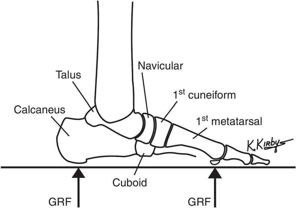
Where the talus meets the foot bones, it forms the subtler joint. This joint is important for walking on uneven ground. Besides connecting the foot to the leg and body, the talus helps transfer weight and pressure across the ankle joint.
What is the main function of the talus?
The talus is the second largest bone in the hindfoot region of the human body. Responsible for transmitting body weight and forces passing between the lower leg and the foot. Is a component of many multiple joints, including the talocrural (ankle), subtalar, and transverse tarsal joints.
How serious is a talus fracture?
Talus fractures are quite severe injuries and can lead to longstanding problems with the foot and ankle. There are early and late complications. Early complications most often are related to the significant swelling that can occur after these injuries, which can cause wound problems and infection.
How hard is it to break your talus?
The talus is a strong bone since it bears the entire body weight. To break the talus bone, it usually takes a lot of energy such as falling off of a ladder, a roof, or from a car accident. The most common area the talus fractures is in the mid portion in an area called the neck.
What happens if talus bone dies?
Avascular necrosis of the talus can be quite devastating and lead to total loss of the ankle joint with arthritis, deformity and pain. The development of AVN is determined to a large extent by the type of the talus fracture.
Why does my talus hurt when walking?
Osteochondral Lesions of the Talus (OLT) A sudden injury like a sprain can damage cartilage on your talus (heel bone) or cause fractures, blisters or sores in the bone underneath. You might notice a catch in your ankle, or it could lock up or still hurt months after a treated injury, which could be an OLT.
What is unique about the talus?
The talus is the second largest of the tarsal bones; it is also one of the bones in the human body with the highest percentage of its surface area covered by articular cartilage. It is also unusual in that it has a retrograde blood supply, i.e. arterial blood enters the bone at the distal end.
Is the tibia weight bearing?
The tibia is much thicker than the fibula. It is the main weight-bearing bone of the two. The fibula supports the tibia and helps stabilize the ankle and lower leg muscles.
How long does it take for a broken talus bone to heal?
Recovery can be prolonged. No weight or walking on the leg will be allowed for 8-12 weeks. Once the bone is healed, exercise and physical therapy is started to maximize the function of the ankle. The patient should expect some swelling about the foot for several months after the procedure.
Why does my talus hurt when walking?
Osteochondral Lesions of the Talus (OLT) A sudden injury like a sprain can damage cartilage on your talus (heel bone) or cause fractures, blisters or sores in the bone underneath. You might notice a catch in your ankle, or it could lock up or still hurt months after a treated injury, which could be an OLT.
How do you treat a fractured talus bone?
How do you fix a broken talus? Some talus fractures can be corrected by casting and rehabilitation. However, most talus fractures require surgery to correct the alignment of your bones. A talus fracture is a painful injury usually caused by a high-impact accident such as a car accident or fall.
How long does it take to recover from a fractured talus?
Recovery can be prolonged. No weight or walking on the leg will be allowed for 8-12 weeks. Once the bone is healed, exercise and physical therapy is started to maximize the function of the ankle. The patient should expect some swelling about the foot for several months after the procedure.
How do you pop a talus bone?
2:365:51How to SELF Adjust Your Ankles - YouTubeYouTubeStart of suggested clipEnd of suggested clipSo you want to do this a couple times a day if you are having talus. And pinch ment especially rightMoreSo you want to do this a couple times a day if you are having talus. And pinch ment especially right at the front it just helps loosen it up and you might find that it helps with pop.
What is the talus?
The talus is the second largest bone in the hindfoot region of the human body. Responsible for transmitting body weight and forces passing between the lower leg and the foot. The Talus (left talus shown in image) Is a component of many multiple joints, including the talocrural (ankle), subtalar, and transverse tarsal joints.
What is the structure of the talus?
Structure. The talus is an incredible bone; despite its small size, it transmits considerable force during the normal gait cycle and even more significant force during impact activities. The talus is shaped like a truncated cone and is wider anteriorly than posteriorly (it is an irregular saddle-shaped bone).
What is talar fracture?
Avascular Necrosis (AVN) Fractures of the talus have a high risk of developing AVN due to its inherently tenuous and limited blood supply. Talar fractures make up one percent of all foot and ankle fractures, and the most common (around 50%) talar fractures occur in the talar neck, the weakest part of the talus.
How many articular surfaces does the talus have?
Approximately two-thirds of its surface area is covered with articular cartilage and it has a tenuous blood supply, similar to the scaphoid. The talus has seven articular surfaces and is divided into the head, neck and body, and two processes, the posterior process and the lateral process.
What is the name of the joint that maintains the medial and lateral longitudinal arches?
Pes Planus. Through the subtalar joint, the talus maintains the medial and lateral longitudinal arches. Pes Planus occurs when there is an alteration of talus morphology of the articular facets, the biomechanics of the subtalar joint change, which can lead to pes planus.
What is the term for the pain in the posterior of the ankle and reduced plantarflexion?
Os Trigonum Syndrome. Os trigonum syndrome refers to pain posterior of the ankle and reduced plantarflexion caused by “the nutcracker-phenomenon”. When an os trigonum is present, this accessory ossicle together with surrounding soft tissues can become wedged between the tibia, talus and calcaneus.
What is the tarsal tunnel?
Tarsal Tunnel Syndrome occurs on the ankle's medial aspect, the tarsal tunnel forms a bony floor consisting of the tibia, talus, and calcaneus, and a fibrous roof consisting of the fibrous flexor retinaculum. The posterior tibial nerve, artery, vein, and posterior tibialis, flexor hallicus longus, and flexor digitorum longus tendons are inside of the tarsal tunnel, and the posterior tibial nerve has the potential to become entrapped within the space
What is the Talus Bone
Talus bone, alternatively known as talus, ankle bone, or astragalus, is the second-largest tarsal bone that connects the leg to the foot by forming the ankle joint.
Where is the Talus Located in the Foot
Talus is located in the hindfoot region, between the heel bone ( calcaneus) and the tibia and fibula of the lower leg.
Functions
The talus acts as the main connector between the foot and leg, forming the ankle joint.
Structure and Anatomy
As mentioned, this is a short, irregular, saddle-shaped bone, which can be divided into three parts: head, neck, and body. The talus also bears several articular surfaces and two protuberances on the back and side, the posterior and lateral processes. It is wider in front than the back.
What is vertical talus?
Vertical talus, also referred to as congenital convex pes valgus, is a congenital condition characterized by an irreducible and rigid dorsal dislocation of the navicular on the talus. This results in a rigid rocker bottom flatfoot. The calcaneus is in fixed equinus, and the Achilles tendon is very tight.
What is the talus parallel to?
Diagnostic Studies: Radiography reveals the talus parallel to the tibia, the calcaneus in an equinus position, and the navicular dislocated on the neck of the talus. The diagnosis is confirmed by a lateral view of the foot during maximum plantarflexion and maximum dorsiflexion.
What is the subtalar facet?
f. The subtalar (or calcaneal) facets on the inferior aspect of the talus are usually three in number and variable in shape. 1. The anterior subtalar (or calcaneal) facet is the most anterior facet on the plantar (inferior) surface of the talus, often somewhat continuous with the articular surface of the talar head. 2.
What is the difference between type 1 and type 2 talus?
Type 1 vertical talus is stiffer and is associated with a calcaneocuboid dislocation. Type 2 is not associated with such a dislocation. Treatment of type 1 deformities must focus on releasing the calcaneocuboid joint.
Why is the talus avascular?
This is because the talus has a precarious blood supply. As most surfaces of the talus contribute to joints they are lined by hyaline cartilage which is avascular. The only part where there is periosteum (and hence blood vessels) besides the nutrient vessels is the neck of the talus.
What is a congenital vertical talus?
Congenital vertical talus (CVT) is a rare deformity that goes by many names , including congenital convex pes valgus, rocker-bottom foot, and Persian slipper foot. CVT is a rare disorder, with the exact incidence unknown. It can be idiopathic or associated with a genetic or neuromuscular disorder. Physical examination reveals that the heel is in fixed equinus, with the forefoot dorsiflexed and everted. The arch is convex, and the head of the talus is prominent medially in the sole. Overall, the foot has a rocker-bottom appearance, and it is rigid and uncorrectable. A true CVT cannot be passively reduced with simple manipulation. If the talonavicular joint can be readily reduced and the ankle can be readily manipulated out of equinus, the deformity probably is either a calcaneovalgus positional deformity or the “oblique talus” of a severe flatfoot.
How to treat talus?
The first step in treating vertical talus is serial casting and manipulation. This stretches the skin, tendons, ligaments, and muscles. On very rare occasions this will resolve the problem; however, in almost all cases, reconstructive surgery is required. The surgery consists of lengthening the extensor hallucis longus and peroneus tertius. The talonavicular joint is then reduced and held with K-wire. Occasionally capsulotomies of the talonavicular and calcaneocuboid joints are required. The final phase of the surgery is to percutaneously lengthen the Achilles tendon.
Which bones are weight bearing?
The tibias present in the lower legs, below the knees are also weight bearing bones. The tibia is one of the most crucial, bones of the body that bear the weight and is most often broken. The tibia, is also known as the Shin bone, and connects the knee to the ankle joint. The tibia is connected to the knee joint, ...
What is weight bearing in orthopedics?
In orthopedic term, weight-bearing is described as the amount of weight a patient puts on the affected leg on which surgery has been performed. However, in general, weight bearing is the ability of the body part to support the weight of the body. Advertisement.
Why does my ankle hurt?
Ankle pain is often due to overstrain of the structures that keep the joint stable . Pain in the ankle can be due to traumatic or overuse injuries.
What are the joints that hold us up when we stand?
Weight-bearing joints are the joints that hold us up when we stand and carry the weight of our body. Ankles, knees and hip joints are the primary weight-bearing joint s of the body. Some other joints that bear the weigth of our body include, the joints of the feet, pelvis and the lower back and spine (especially the lower back).
Why should bones be in working condition?
Weight bearing bones of the body should be in working condition so as to enable the body to stand upright and walk. By understanding what bones in the body bears body weight enables us to be more aware of the body and how it works. Below are some bones that bear the weight of the body.
Which joints bear the most weight?
Apart from the primary weight bearing joints, i.e. ankles, knees and the hip joints; joints of the feet, the pelvis and the lower back and the spine, are also the joints that bear the weight of the body.
Which foot bone bears the weight?
The Calcaneus or Calcaneum, appears in a ball-like shape and often experiences stress fractures on high-impact activities. Advertisement. The Tarsal Bone. One more foot bone that bears the body weight is the tarsal bone.
What is the talus bone?
Your talus bone is the bottom part of the ankle joint. It connects your foot to the two bones in your lower leg — the tibia and fibula — that make up the top part of the ankle. The talus lies just above the calcaneus or heel bone and below the tibia or shin bone. Together the talus and calcaneus are critical to your ability to walk.
What is the classification of talus fracture?
Talus fractures are usually classified based on the severity of the injury and how much the bone is moved from its normal position. There are three main classifications:
What causes a talus fracture?
A talus fracture usually results from serious trauma to the foot. Injuries that could cause a talus fracture include a fall from a great height or a car accident. A badly twisted ankle can also cause small pieces of the talus to break off. If the fracture doesn’t heal properly, you could have walking problems.
How to tell if you have a broken ankle?
What are the symptoms? 1 Minimally displaced. Acute pain in the ankle is usually the first sign. There may be some minor swelling and tenderness. You should be able to walk on it, but not without pain. 2 Displaced. The pain, swelling, and tenderness are greater. You may not be able to put weight on the injured ankle. 3 Open. The most obvious symptom is the sight of bone sticking through the skin. The pain will be very intense. There could also be considerable bleeding. It’s not uncommon for someone with an open fracture to pass out from the shock or loss of blood.
Why does my talus chip?
It usually happens as a result of repetitive actions putting stress on a bone or joint. In some cases, changing an activity, such running on a harder surface or with more incline than you’re used to, can trigger a stress fracture. The talus bone can also chip.
What is it called when a bone moves out of its normal position?
Any time a bone moves out of its normal position, it’s called a displaced fracture. Highly displaced fractures are considered to be unstable. Surgery is typically required to get the broken parts of the talus to line up correctly again.
Can a fractured talus be treated?
An evaluation by an orthopedist may be enough. If the talus fracture is stable, nonsurgical treatment options may be available to you .
What is the talus in the foot?
Anatomy. The talus is the bone that makes up the lower part of the ankle joint (the tibia and fibula make up the upper part). The ankle joint allows your foot to move up and down. The talus also sits above the heel bone (calcaneus). Together, the talus and calcaneus form the subtalar joint.
What joint is the talus in?
Together, the talus and calcaneus form the subtalar joint. This joint allows your foot to move inward and outward, which is important for walking on uneven ground. The talus is the main connector between the foot and leg, helping to transfer weight and pressure forces across the ankle joint.
What happens when a talus fracture is severe?
The more severe the talus fracture, the more likely it is that AVN will occur.
Why do talus fractures require surgery?
Because the talus is important for ankle movement, a fracture often results in substantial loss of motion and function. A talus fracture that does not heal properly can lead to complications, including a limp, arthritis, and chronic pain. For this reason, most talus fractures require surgery.
How to treat a talus fracture?
Immediate first aid treatment for a talus fracture, as with any painful ankle injury, is to apply a well-padded splint around the back of the foot and leg to immobilize and protect the limb. The splint should extend from the toe to the upper calf.
What is the cartilage that covers the talus?
It is largely covered by articular cartilage, the white slippery material that covers all joint surfaces. This cartilage allows the talus to move smoothly against its neighbor bones. The talus bone sits between the bones of the lower leg and the calcaneus (heel bone).
What causes a talus fracture?
Cause. Most talus fractures are the result of high-energy trauma such as a car collision or a fall from height . Injuries from sports, particularly snowboarding, are another, though less common, cause of talus injuries.

Introduction
Structure
- The talus is pivotal to the function of the ankle. When viewed together within the ankle and in relation to the other tarsal bones, it has the look of a universal joint on a car's driveshaft. The talus works the same way, allowing the connecting bones of the ankle to slide around it in multiple directions while supporting weight.
Articulations
Muscle Attachments
Ligaments

Blood Supply
- The talus is an incredible bone; despite its small size, it transmits considerable force during the normal gait cycle and even more significant force during impact activities. 1. The talus is shaped like a truncated cone and is wider anteriorly than posteriorly (it is an irregular saddle-shaped bone). 2. Approximately two-thirds of its surface area is covered with articular cartilage and it ha…
Innervation
- superiorly through the talar dome to form the mortise joint of the ankle with the tibia and fibula
- inferoposteriorly: large oblique facet that is concave articulates with the calcaneusto form the talocalcaneal joint
- anteroinferiorly: two facets for articulation with the calcaneus to form part of the talocalcaneonavicular joint
- talar head (domed articular surface) with the navicular bone (circular depression on the posterior surface)
Clinical Significance
- The talus does not serve as the site of any muscle attachment, which is significant because that means the talus does not have secondary sources of blood supply. 1. If there is vascular compromise to the talus through traumatic injuries such as in the setting of displaced fractures, there is a relatively high risk of developing post-traumatic, avascular necrosis (AVN)