Full Answer
Are ventral roots part of PNS?
Spinal Nerve Anatomy The spinal nerves are part of the peripheral nervous system (PNS). A spinal nerve: Spinal nerves arise from a combination of nerve fibers from the dorsal and ventral roots of the spinal cord.
Is dorsal root CNS or PNS?
peripheral nervous systemThe dorsal root ganglia (DRGs) are located in the peripheral nervous system (PNS), between the dorsal horn of the spinal cord and the peripheral nerve terminals, and contain various cell types such as satellite glial cells, endothelial cells, macrophages and primary sensory neurons.
Are nerve roots part of the CNS?
The nerve roots exit the spinal canal through the intervertebral foramen, small hollows between each vertebra. The brain and the spinal cord make up the Central Nervous System (CNS). The nerve roots that exit the spinal cord/spinal canal branch out into the body to form the Peripheral Nervous System (PNS).
What system is the ventral root in?
Autonomic Nervous SystemAutonomic Nervous System Each spinal cord segment has two ventral roots that connect by a white ramus to a spinal sympathetic ganglion. These ganglia communicate with each other up and down the spinal cord, forming two sympathetic chains, one on each side of the vertebral column.
What is the difference between the PNS and CNS?
The central nervous system (CNS) includes the brain and spinal cord, while the peripheral nervous system includes all of the nerves that branch out from the brain and spinal cord and extend to other parts of the body, including muscles and organs.
What is dorsal and ventral root?
The dorsal root of spinal nerve (or posterior root of spinal nerve or sensory root) is one of two "roots" which emerge from the spinal cord. It emerges directly from the spinal cord, and travels to the dorsal root ganglion. Nerve fibres with the ventral root then combine to form a spinal nerve.
Which of the following is not part of the CNS?
Answer and Explanation: The correct answer: Among the given options b) cranial nerves is not a part of the central nervous system.
Is the spinal nerves CNS or PNS?
The spinal cord is an extension of the central nervous system (CNS), which consists of the brain and spinal cord.
What makes up the central nervous system?
The central nervous system is made up of the brain and spinal cord. The peripheral nervous system is made up of nerves that branch off from the spinal cord and extend to all parts of the body.
What is the ventral root responsible for?
The anterior/ventral root contains efferent nerve fibres, which carry stimuli away from the CNS towards their target structures. The cell bodies of the anterior root neurons are located in the central grey matter of the spinal cord.
What is the ventral root of the spinal nerve?
The Ventral Root of the spinal nerve contains outgoing, efferent (meaning to "bear away from") fibers that carry information destined to control motor or glandular function. The cell bodies of these motor neurons are located in the ventral horns of the spinal cord's central grey region.
What are the divisions of the ANS and where do they emerge from the CNS?
The two divisions of the autonomic nervous system are the sympathetic division and the parasympathetic division. The sympathetic system is associated with the fight-or-flight response, and parasympathetic activity is referred to by the epithet of rest and digest. Homeostasis is the balance between the two systems.
What is a dorsal nerve root?
Dorsal nerve roots carry sensory neural signals to the central nervous system (CNS) from the peripheral nervous system (PNS). Anatomically, a dorsal root ganglion (DRG) emerges from the dorsal root of the spinal nerves.
Is dorsal root ganglion sensory or motor?
sensory neuronsThe dorsal root ganglion, more recently referred to as the spinal ganglion, is a collection of neuronal cell bodies of sensory neurons. It is the most common type of sensory ganglion in the human body.
Are there ganglia in the CNS?
The basal ganglia are located in the brain stem, thalamus, and cerebral cortex areas of the brain. Being in the brain, they are part of the central nervous system, not the peripheral nervous system, as other ganglia are. This group of structures is important in regulating voluntary movements.
What type of neurons are in the dorsal root ganglion?
The neurons comprising the dorsal root ganglion are of the pseudo-unipolar type, meaning they have a cell body (soma) with two branches that act as a single axon, often referred to as a distal process and a proximal process.
What is the ventral root?
Each ventral root (also named the anterior root, radix anterior, radix ventralis, or radix motoria) is attached to the spinal cord by a series of rootlets that emerge from the ventrolateral sulcus of the spinal cord in the anterior root exit zone. The ventral roots consist predominantly of efferent somatic motor fibers ...
Where are the ventral roots located?
Each ventral root (also named the anterior root, radix anterior, radix ventralis, or radix motoria) is attached to the spinal cord by a series of rootlets that emerge from the ventrolateral sulcus of the spinal cord. Unlike the dorsal root fibers that are arranged in a neat line at their emergence from the spinal cord, ...
What are the two ganglia that connect to each other?
Each spinal cord segment has two ventral roots that connect by a white ramus to a spinal sympathetic ganglion. These ganglia communicate with each other up and down the spinal cord, forming two sympathetic chains, one on each side of the vertebral column. Only those pairs of spinal sympathetic ganglia from T1 to L2 receive inputs via the white rami. Those above T1 receive inputs from fibers from the thoracic segments that climb the sympathetic chain. Those ganglia below L2 receive inputs from preganglionic fibers that descend the chain from the lower thoracic and lumbar ganglia (see Figure 4.9.3 ). Each pair of spinal sympathetic ganglia send out an efferent branch to effector organs. These fibers travel from the ganglia to the spinal nerve by a gray ramus communicans. It is gray because the axons it contains are unmyelinated postganglionic fibers. Thus, only some of the sympathetic ganglia have white rami, but all have gray rami.
What are visceral afferent fibers made of?
Visceral afferent fibers are composed of mainly unmyelinated C fibers and thin myelinated Aδ fibers, although a population of small, rapidly conducting, myelinated fibers is also present. The terminations of these are mainly in laminae 1 and 5 (Kayalioglu et al., 2008b).
What are the connections between the sympathetic nervous system and the spinal cord?
Each segment of the spinal cord corresponds to a pair of sympathetic ganglia (paravertebral ganglia), one on each side of the column. These communicate up and down by interganglionic branches, so the set of ganglia resembles beads on a string and is called the sympathetic chain. The preganglionic cells originating in the cord synapse in the ganglia with postganglionic cells. These postganglionic cells send efferents to the skin, sweat glands, and blood vessels of the body, as shown at the left. The efferent output to the viscera, shown to the right, can occur via postganglionic cells in the paravertebral ganglia or via prevertebral ganglia. The prevertebral ganglia include the celiac, superior mesenteric, and inferior mesenteric ganglia. They receive inputs from preganglionic fibers that travel through the chain. All of the visceral organs receive sympathetic efferents as shown. Although the targets of efferent output are shown separately here, in reality both sympathetic chains give rise to both types of efferents. The adrenal medulla receives preganglionic input directly; it acts like a ganglion.
Why do ventral root axons increase?
The number of ventral root axons increases gradually along its subarachnoid course to the point it is apposed to the dorsal root ganglion. This increase is probably due to axonal sprouting (branching) (Fraher and O'Sullivan, 1989 ). Axon numbers also change during maturation and throughout maturity.
Why do ventral roots undergo atrophic change?
Other Atrophic Complications. Because the ventral roots carry trophic factors besides motor impulses, other structures will undergo variable degrees of atrophic change. The bones in the affected limbs will demineralize rapidly, although the exact extent of demineralization is unclear.
What are the ventral roots?
Each ventral root (also named the anterior root, radix anterior, radix ventralis, or radix motoria) is attached to the spinal cord by a series of rootlets that emerge from the ventrolateral sulcus of the spinal cord . Unlike the dorsal root fibers that are arranged in a neat line at their emergence from the spinal cord, ventral root fibers form an elliptical area named the anterior root exit zone (AREZ). The ventral roots predominantly consist of efferent somatic motor fibers (thick alpha motor axons and medium-sized gamma motor axons) derived from nerve cells of the ventral column. In thoracic and upper lumbar segments, these are supplemented by thin autonomic preganglionic motor fibers derived from the intermediolateral column. Injury filling of the ventral roots with HRP results in labelling of autonomic preganglionic neurons and their dendritic arbors, indicating there are also afferent fibers terminating on autonomic preganglionic neurons in the ventral roots of cat sacral spinal cord (Mawe et al., 1984; 1986 ). Unmyelinated sensory afferent fibers innervating the ventral root and its pial sheath have also been shown electrophysiologically in the cat sacral spinal cord ( Jãnig and Koltzenburg, 1991 ).
Where are the ventral and dorsal roots located?
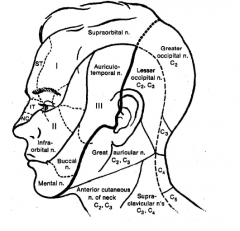
In the cervical region , the dorsal and ventral roots travel just posterior to the vertebral artery. The formed spinal nerve is just posterolateral to the vertebral artery, and the ventral and dorsal rami are found lateral to the vertebral artery. The dorsal ramus skirts the superior articular process of its same level, traveling posteriorly to the zygapophyseal joints, deep back muscles, and skin of the back. If present, the dorsal ramus of the C1 spinal ramus will be found wedged between the posterior arch of the atlas and the vertebral artery as it courses in the groove for the vertebral artery (Schaeffer, 1953 ). The first and second cervical nerves travel anterior to the vertebral artery, whereas the remaining cervical spinal nerves travel posterior to this vessel.
What are the two ganglia that connect to each other?
Each spinal cord segment has two ventral roots that connect by a white ramus to a spinal sympathetic ganglion. These ganglia communicate with each other up and down the spinal cord, forming two sympathetic chains, one on each side of the vertebral column. Only those pairs of spinal sympathetic ganglia from T1 to L2 receive inputs via the white rami. Those above T1 receive inputs from fibers from the thoracic segments that climb the sympathetic chain. Those ganglia below L2 receive inputs from preganglionic fibers that descend the chain from the lower thoracic and lumbar ganglia (see Figure 4.9.3 ). Each pair of spinal sympathetic ganglia send out an efferent branch to effector organs. These fibers travel from the ganglia to the spinal nerve by a gray ramus communicans. It is gray because the axons it contains are unmyelinated postganglionic fibers. Thus, only some of the sympathetic ganglia have white rami, but all have gray rami.
What is the dorsal root?
Each dorsal root (also named the posterior root, radix posterior, radix dorsalis or radix sensoria) is attached to the dorsolateral sulcus of the spinal cord by a series of rootlets arranged in a line – the dorsal root entry zone (DREZ). Dorsal roots are larger than the ventral roots, with thicker and more numerous fibers. Primary afferent fibers of the dorsal roots are either myelinated or unmyelinated. It is estimated that there are 7,100 dorsal root fibers in the L3 dorsal root in the mouse; 2,650 of these are myelinated and 4,550 unmyelinated ( Biscoe et al., 1982 ). The dorsal roots contain sensory fibers from the skin, subcutaneous and deep tissues, and viscera.
What are the connections between the sympathetic nervous system and the spinal cord?
Each segment of the spinal cord corresponds to a pair of sympathetic ganglia (paravertebral ganglia), one on each side of the column. These communicate up and down by interganglionic branches, so the set of ganglia resembles beads on a string and is called the sympathetic chain. The preganglionic cells originating in the cord synapse in the ganglia with postganglionic cells. These postganglionic cells send efferents to the skin, sweat glands, and blood vessels of the body, as shown at the left. The efferent output to the viscera, shown to the right, can occur via postganglionic cells in the paravertebral ganglia or via prevertebral ganglia. The prevertebral ganglia include the celiac, superior mesenteric, and inferior mesenteric ganglia. They receive inputs from preganglionic fibers that travel through the chain. All of the visceral organs receive sympathetic efferents as shown. Although the targets of efferent output are shown separately here, in reality both sympathetic chains give rise to both types of efferents. The adrenal medulla receives preganglionic input directly; it acts like a ganglion.
What is the peripheral nervous system?
The peripheral nervous system is mainly derived from the neural crest. This is a population of cells that detaches from the lateral margins of the neural plate during neurulation (see Fig. 2.2 ). Neural crest cells that come to lie dorsolateral to the neural tube ultimately become the primary sensory neurons of the dorsal root ganglia. These neurons are initially bipolar but their central and peripheral processes fuse at a common T-shaped extension of the cell body to form a single continuous axon. For this reason they are described as pseudounipolar (Greek: pseudo-, false). The central processes of the dorsal root ganglion cells innervate the alar (sensory) plate of the neural tube, whereas the peripheral processes enter the spinal nerves at each segmental level (see Fig. 2.4B ).
Where does the phrenic nerve originate?
The phrenic nerve is formed from the ventral roots of C3–C5; its primary component arises from the C4 anterior primary ramus. The three roots join at the lateral border of the anterior scalene muscle, and the phrenic nerve passes inferiorly along the anterior surface of this muscle, posterior to the sternomastoid and omohyoid muscles, and into the chest. It travels close to the internal mammary artery, the root of the lung, and the pericardium. It communicates with the sympathetic chain, the accessory nerve, and the hypoglossal nerve. 6
Which system is a system of efferent nerves that connect the CNS with internal organs?
The autonomic nervous system is a system of efferent nerves that connect the CNS with internal organs, but unlike the motoneurones that connect directly with skeletal muscles, the pathway between the spinal cord and a visceral organ is interrupted by a synapse within an autonomic ganglion.
Why is the ventral horn larger than the thoracic horn?
The ventral horn consists of the cell bodies of motoneurones, and the size of the ventral horn in the lumbar region is greater than in the thoracic region because of the large motor innervation of muscles in the lower limb. Similarly in the cervical cord, many motoneurones are used to control the upper limbs, hand and fingers, so the cervical ventral horn is large.
What are the dorsal columns?
The dorsal columns contain the axon collaterals (branches) of sensory neurones which synapse on neurones in the medulla. The information carried by these axons is concerned with touch and vibration.
What is the structure of the spinal cord?
The spinal cord has a cylindrical segmented structure, each segment being derived from the somites of the embryo. A spinal nerve arises from each side of every segment and these segmental nerves leave the vertebral canal through the inter-vertebral foraminae.
Where are the autonomic neurones found?
The cell bodies of these autonomicneurones are found in the lateral horn of the grey matter of the 1st thoracic to 2nd lumbar, and sacral segments 2-4 (see below).
Where does the spinal cord end?
The spinal cord ends at the second lumbar segment of the vertebral column, below which the lower lumbar, sacral and coccygeal nerves travel through the subarachnoid space to reach the appropriate intervertebral foraminae where they leave the spinal canal.
Which part of the spinal cord is protected by the vertebral column?
The spinal cord is protected by the vertebral column, to which the outermost of the meninges, the dura mater, is attached.
What is the ventral root?
Anatomical terminology. In anatomy and neurology, the ventral root, motor root or anterior root is the efferent motor root of a spinal nerve . At its distal end, the ventral root joins with the dorsal root to form a mixed spinal nerve.
What is the root of a spinal nerve?
Ventral root of spinal nerve. In anatomy and neurology, the ventral root, motor root or anterior root is the efferent motor root of a spinal nerve . At its distal end, the ventral root joins with the dorsal root to form a mixed spinal nerve.
What is the medulla spinalis?
Medulla spinalis. A spinal nerve with its anterior and posterior. The motor tract. Diagrammatic transverse section of the medulla spinalis and its membranes. A portion of the spinal cord, showing its right lateral surface. The dura is opened and arranged to show the nerve roots.
What nerve sends out cones looking for target cells until the person stops growing?
C. As a person matures, the axonal projections of a spinal nerve send out growth cones looking for target cells until the person stops growing.
What nerve was damaged close to the knee?
D. The vagal nerve was damaged close to the knee.