What is the superficial fascia of the scalp?
The superficial fascia in the scalp is a firm, fibro-fatty layer, intimately adherent to the skin, and to the Occipitofrontalis and its aponeurosis; behind, it is continuous with the superficial fascia at the back of the neck; laterally, it is prolonged into the temporal region, where it is looser in texture.
What are the fascia and muscles of the head?
The Fasciæ and Muscles of the Head. a. The Muscles of the Scalp. The superficial fascia in the cranial region is a firm, dense, fibro-fatty layer, intimately adherent to the integument, and to the Epicranius and its tendinous aponeurosis; it is continuous, behind, with the superficial fascia at the back of the neck; and, laterally,...
What is the anatomy of the scalp?
The scalp refers to the layers of skin and subcutaneous tissue that cover the bones of cranial vault. In this article, we shall look at the anatomy of the scalp – its layers, neurovascular supply, and any clinical correlations. The scalp consists of five layers. The first three layers are tightly bound together, and move as a unit.
What is the subgaleal fascia?
The subgaleal fascia is the layer usually referred to as the "loose areolar layer" or the "subaponeurotic plane". This layer cleaves readily, allowing the skin, subcutaneous tissue Figure 6 1 Layers of the scalp above the superior temporal line (top insert) and below the superior temporal line (right inset).

Does the scalp have fascia?
Also referred to as the superficial fascia, the connective tissue of the scalp is a fibrofatty layer. This layer forms the bridge between the skin and the epicranial aponeurosis by connecting the two together. The tissue is also innervated with blood vessels and nerve endings.
How many fascias are in the head?
It consists of three fascial layers (or sheaths), which are: The investing layer of deep cervical fascia.
What tissue is on the scalp?
There are five layers to the scalp: the skin, connective tissue layer, galea aponeurotica, loose areolar connective tissue, and the pericranium.
Are there muscle on the scalp?
Two muscles are attached to the galea, in front, the frontalis, and behind, the occipitalis. The occipitalis muscle arises from here on the occipital bone, above the superior nuchal line.
Where is the fascia located?
Fascia is a thin casing of connective tissue that surrounds and holds every organ, blood vessel, bone, nerve fiber and muscle in place. The tissue does more than provide internal structure; fascia has nerves that make it almost as sensitive as skin.
Where is the deep fascia located?
Deep fascia surrounds bones, muscles, nerves, and blood vessels. It is commonly has a more fibrous consistency and rich in hyaluronan as compared to the other subtypes. Deep fascia tends to be highly vascularized and contain well developed lymphatic channels.
What is the layer of gunk on my scalp?
Sebum. The scalp produces a natural, waxy oil called sebum from glands beneath the skin. Some people produce more of this oil than others. Sebum plays an important role in protecting your skin from infection and helping keep it moist.
Why does my scalp ache?
Migraines, tension headaches, and autoimmune disorders like psoriasis can all cause the scalp to become inflamed, irritated, and painful. Sunburns, rashes, wounds, and insect bites also commonly cause scalp tenderness.
How do you massage your scalp?
How to massage your scalpUse the fingertips of both hands to apply light to medium pressure to your scalp, moving in small circles.Work your way across your scalp to cover all areas.Try to massage your scalp using your fingertips for at least 5 minutes at a time, several times a day.
Are there muscles on the top of your skull?
In other words, there is a muscle on the forehead (frontalis) and one on the back of the head (occipitalis), but there is no muscle across the top of the head. Instead, the two bellies are connected by a broad tendon called the epicranial aponeurosis, or galea aponeurosis (galea = “apple”).
How do you loosen your scalp muscles?
How to Release Tension From Your Head, Hair and ScalpStop Using Hair Elastics. Scrunchies are softer and gentler on your hair. ... Sleep With Your Hair Down. Just like us, our hair also needs its beauty sleep. ... Wear Relaxed Hairstyles. ... Try a Little Aromatherapy. ... Give Yourself a Head Massage.
What causes tight scalp muscles?
Tension headaches occur when neck and scalp muscles become tense or contract. The muscle contractions can be a response to stress, depression, head injury, or anxiety. They may occur at any age, but are most common in adults and older teens. It is slightly more common in women and tends to run in families.
Is there fascia in the face?
Fascia Elongates There are three layers of connective tissue that makes up the framework of the face + body. The deeper layer connects the skin to muscle and fat and forms the “girdle” of facial muscles— Superficial, Deep and Subserous. As the fascia dries out, it begins to fray or stiffen.
What is fascia of the neck?
The structures found in the neck are surrounded by a layer of subcutaneous tissue called the superficial fascia, while there are also layers of deep cervical fascia which distribute the structures in the neck into different compartments.
What is a fascia in anatomy?
Fascia is a sheath of stringy connective tissue that surrounds every part of your body. It provides support to your muscles, tendons, ligaments, tissues, organs, nerves, joints and bones.
What is the fascia on a roof?
The fascia is the attractive board along the side of the overhang and the roof that helps your roof appear finished. Your gutter sits atop the facia board. The fascia is also known as a “transition trim” between the home and the roofline. The fascia supports the shingles and helps to keep moisture out.
What are the layers of the scalp?
Applied Anatomy.-The scalp consists of five layers, viz. the skin, subcutaneous tissue, Occipitofrontalia and its aponeurosis, subaponeurotic areolar tissue, and pericra nium (fig. 580). But from a surgical standpoint it is better to regard the first three of these as a single layer, since they are all intimately united, and when torn off in an accident, or turned down as a flap in a surgical operation, remain firmly connected to each other. In consequence of the dense character of the subcutaneous tissue, the amount of swelling which occurs as the result of inflammation is slight; and a wound which does not involve the Occipitofrontalis or its aponeurosis does not gape. The blood-vessels which lie in this tissue do not contract when wounded, and therefore the hemorrhage from scalp wounds is often very considerable. It can, however, always be arrested by pressure -a matter of great importance, as it is often very difficult or impossible to pick up with forceps a wounded vessel in the scalp owing to the retraction of its cut ends.
What is the occipital musculofibrous layer?
The Occipitofrontalis (Epicranius) is a broad, musculofibrous layer which covers the top of the skull, from the nuchal lines to the eyebrows. It consists of four bellies–two occipital and two frontal-connected by an intervening aponeurosis, termed the epicranial aponeurosis (galea aponeurotica).
What is the difference between the frontal belly and the occipital belly?
Each Frontal belly (fig. 579) is thin, of a quadrilateral form, and intimately adherent to the superficial fascia. It is broader than the Occipital belly and its fibers are longer and paler in color. It has no bony attachments. Its medial fibers are continuous with those of the Procerus; its intermediate fibers blend with the Corrugator and Orbicularis oculi; and its lateral fibers are also blended with the latter muscle over the zygomatic process of the frontal bone. From these attachments the fibers are directed upwards, and join the epicranial aponeurosis in front of the coronal suture. The medial margins of the Frontal bellies are joined together for some distance above the root of the nose; but between the Occipital bellies there is a considerable, though variable, interval, occupied by an extension of the epicranial aponeurosis.
Which fascia is the cranial region?
The superficial fascia in the cranial region is a firm, dense, fibro-fatty layer, intimately adherent to the integument, and to the Epicranius and its tendinous aponeurosis; it is continuous, behind, with the superficial fascia at the back of the neck; and, laterally, is continued over the temporal fascia. It contains between its layers the ...
Which muscle moves the scalp backwards?
The Occipitales draw the scalp backward. By bringing alternately into action the Frontales and Occipitales the entire scalp may be moved forward and backward. In the ordinary action of the muscles, the eyebrows are elevated, and at the same time the aponeurosis is fixed by the Occipitales, thus giving to the face the expression of surprise;
Where are the medial margins of the frontales joined?
The medial margins of the Frontales are joined together for some distance above the root of the nose; but between the Occipitales there is a considerable, though variable, interval, occupied by the galea aponeurotica.
Where does the occipitalis originate?
The Occipitalis, thin and quadrilateral in form, arises by tendinous fibers from the lateral two-thirds of the superior nuchal line of the occipital bone, and from the mastoid part of the temporal. It ends in the galea aponeurotica.
Which layer of the scalp is closely connected to the integument?
It is closely connected to the integument by the firm, dense, fibro-fatty layer which forms the superficial fascia of the scalp: it is attached to the pericranium by loose cellular tissue, which allows the aponeurosis, carrying with it the integument to move through a considerable distance. Variations.
Which nerve is responsible for the frontalis?
Nerves. —The Frontalis is supplied by the temporal branches of the facial nerve , and the Occipitalis by the posterior auricular branch of the same nerve. Actions. —The Frontales raise the eyebrows and the skin over the root of the nose, and at the same time draw the scalp forward, throwing the integument of the forehead into transverse wrinkles.
Where is the scalp located?
Covering the surface of your head, the scalp, extends from the top of your forehead across to the epicranial aponeurosis of the head. Laterally, it reaches down to the external auditory meatus and zygomatic arch (cheekbone of the skull ). The scalp consists of 5 distincts layers:
What is the connective tissue of the scalp?
Connective tissue. Also referred to as the superficial fascia, the connective tissue of the scalp is a fibrofatty layer. This layer forms the bridge between the skin and the epicranial aponeurosis by connecting the two together. The tissue is also innervated with blood vessels and nerve endings.
How are hair follicles formed?
They are tubular and are formed from multiple layers of epithelial cells. The base of the follicle bulges, forming a hair bulb which surrounds the hair papilla. The bulb is invaginated by connective tissue known as dermal papilla. The dermal papilla contains many tiny blood vessels and nerve projections. This becomes the hair papilla once it invaginates into the hair bulb. The 3 innermost layers of epithelial cells within the hair follicle keratinize to produce the hair shaft. The outer epithelial layers form the hair’s epithelial sheath. The mass of cells from which the hair shaft is produced is referred to as the hair matrix.
What is the thicker layer of connective tissue beneath the skin called?
There is also a thicker layer of connective tissue beneath known as the reticular layer. This extends to the subcutaneous layer (hypodermis), which is positioned above the fascia. Within the subcutaneous layer, the basal portion of of sweat glands can be found. There are many hair follicles in the skin of the scalp.
Which layer of the hair follicle contains cells that become keratinised to an extent?
The innermost layer of the hair follicle contains cells which become keratinised to an extent, forming the medulla. The medulla is the very core of the shaft of hair. Surrounding the medulla, there is a keratinised layer of cells called the cortex. The cortex makes up the main body of the hair. The third cell layer of the hair follicle is also keratinised, forming a cuticle which is thin but hard. The cuticle is made up of keratin plates. These overlap creating a structure that supposedly prevents the hair from becoming matted.
Where does the occipitofrontalis muscle insert?
It is a thin but tough layer of fibrous tendinous tissue and is the site at which the occipitofrontalis muscle inserts into the tissue of the scalp. The occipital belly gives rise to it, whereas the epicranial aponeurosis inserts into the frontal belly of the occipitofrontalis.
How does hair shape?
The histology of your hair can vary slightly depending your ethnicity. When a cross section of a hair is made, its shape differs depending on the characteristics of your hair determined by your race, and the genes you get from your parents. For example, the straight hair of many Asian people gives a perfectly round cross section. Meanwhile, the wavey hair of European people gives an oval shaped cross section, and the curly hair of black people has a kidney-shaped cross section. Hair follicles (the sheath of cells that surround the base of each hair) head tend to be long and straight, but curly hair is often produced from curved hair follicles. Despite the unique differences between hair around the world, the basic histology of hair is universal.
What is the layer of the scalp?
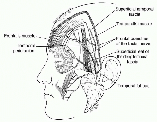
Figure 6 1 Layers of the scalp above the superior temporal line (top insert) and below the superior temporal line (right inset). Top inset : Skin, subcutaneous tissue, the musculoaponeurotic layer (galea in this illustration), the subgaleal layer of loose tissue, periosteum (pericranium), and bone of the skull. Right inset : Skin, subcutaneous tissues, the temporoparietal fascia (note temporal branch of VII N), the superficial layer of the temporalis fascia, a superficial pad of fat, the deep layer of temporalis fascia, the temporalis muscle above, the buccal fat pad below, skull.
Where is the subgaleal fascia located?
Anteriorly, the subgaleal fascia is continuous with the loose areolar layer deep to the orbicularis oculi muscles. Laterally, it is attached to the frontal process of the zygoma. This attachment continues along the superior surface of the zygomatic arch, above the external auditory meatus, and over the mastoid process. It terminates by fusing with the periosteum along the superior nuchal line.
What is the musculoaponeurotic layer?
The musculoaponeurotic layer, also inappropriately called the galea (which refers to aponeurosis only), consists of the paired frontalis (epicranius) and occipitalis muscle, the auricular muscles, plus a broad aponeurosis. The aponeurosis is the true galea and has two portions, an extensive intermediate aponeurosis between the frontalis and occipitalis muscles and a lateral extension into the temporoparietal region known as the temporoparietal fascia. Farther inferiorly, the temporoparietal fascia is continuous with the superficial musculoaponeurotic layer of the face (SMAS). The paired frontalis muscles originate from the galeal aponeurosis and insert into the dermis at the level of the eyebrows. An extension of the galea separates the two quadrilateral frontalis muscle in the middle of the forehead.
Which muscle is paired with the superficial musculoaponeurotic layer of the face?
Farther inferiorly, the temporoparietal fascia is continuous with the superficial musculoaponeurotic layer of the face (SMAS). The paired frontalis muscles originate from the galeal aponeurosis and insert into the dermis at the level of the eyebrows.
Which fascial plane allows free movement of the skin over the periosteum when the frontalis muscle is?
The loose tissue of the subgaleal fascia allows free movement of the skin over the periosteum when the frontalis muscle is contracted. Anatomic dissection have also revealed that the subgaleal frontalis muscle is contracted. Anatomic dissections have also revealed that the subgaleal fascia can be mobilized as an independent fascial layer. For the routine coronal approach to the fascial skeleton, however, this fascial layer is used only for its ease of cleavage.
Which layer of the skin cleaves readily?
The subgaleal fascia is the layer usually referred to as the "loose areolar layer" or the "subaponeurotic plane". This layer cleaves readily, allowing the skin, subcutaneous tissue. Figure 6 1 Layers of the scalp above the superior temporal line (top insert) and below the superior temporal line (right inset).
What is the mnemonic for the layers of the scalp?
The basic mnemonic for the layers of the scalp (Fig. 6-1) is : S = skin
What is the scalp?
The scalp is soft tissue and acts as a barrier to protect the cranial vault from physical trauma or infectious agents. Of the 5 layers of the scalp [ 1 ], the first three are firmly held together as a unit. The layers of the scalp can be remembered using the mnemonic SCALP:
What are the layers of the scalp?
The layers of the scalp can be remembered using the mnemonic SCALP: 1. Skin. The skin [ 2] contains several hair follicles and sebaceous glands. A gland is a group of cells that synthesize chemicals needed for bodily functions. Sebaceous glands secrete sebum or natural oil that lubricates the scalp.
How to keep your scalp healthy?
Tips To Keep Your Scalp Healthy. Exfoliate your scalp regularly to boost skin cell turno ver. It is important that dead skin cells shed to reveal a fresh layer of skin cells. Choose from exfoliating shampoos to hair scrubs to homemade scrubs. Massage your scalp to promote blood circulation.
How to get rid of scalp issues?
Most scalp issues arise from a dry scalp. Make sure you oil your hair, invest in deep conditioning treatments and stay away from hot showers that strip off the sebum on the scalp.
How to protect your scalp from the sun?
Protect your scalp from the harsh sun rays using a hat or a scarf. The skin on the scalp is prone to sun damage. You can also choose a shampoo containing sunscreen. Limit heat styling and chemical treatments as these can irritate your scalp over time. Opt for natural hairstyles whenever there is a chance.
What is the outer layer of the skull called?
Pericranium. Periosteum [ 6] is the outer layer of the skull bones (pericranium). It is a dense and irregular connective tissue that adheres to the calvarial bone of the skull. It has a vascular supply that supports the underlying calvarium.
What is the purpose of wrapping up your scalp?
Wrapping Up. Your scalp is a soft tissue protecting the inner layers of your head, besides growing hair from the follicles. Good hair care routines like cleansing, massaging and oiling ensure that there is an adequate supply of blood to the scalp. And, a healthy scalp is a precursor to healthy hair.
What is the scalp?
The scalp refers to the layers of skin and subcutaneous tissue that cover the bones of cranial vault. In this article, we shall look at the anatomy of the scalp – its layers, neurovascular supply, and any clinical correlations.
Which artery supplies the back of the scalp?
Occipital – supplies the back of the scalp. Anteriorly and superiorly, the scalp receives additional supply from two branches of the ophthalmic artery – the supraorbital and supratrochlear arteries. These vessels accompany the supraorbital and supratrochlear nerves respectively.
What is the thin connective tissue layer that separates the periosteum of the skull from the epicra?
Loose Areolar Connective Tissue – a thin connective tissue layer that separates the periosteum of the skull from the epicranial aponeurosis. It contains numerous blood vessels, including emissary veins which connect the veins of the scalp to the diploic veins and intracranial venous sinuses.
How many layers are there in the scalp?
Layers of the Scalp. The scalp consists of five layers. The first three layers are tightly bound together and move as a collective structure. The mnemonic ‘SCALP’ can be a useful way to remember the layers of the scalp: Skin, Dense Connective Tissue, Epicranial Aponeurosis, Loose Areolar Connective Tissue and Periosteum.
Why is my scalp bleeding?
The blood vessels within the layer are highly adherent to the connective tissue. This renders them unable to constrict fully if lacerated - and so the scalp can be a site of profuse bleeding.
Which veins connect the scalp to the diploic veins?
It contains numerous blood vessels, including emissary veins which connect the veins of the scalp to the diploic veins and intracranial venous sinuses.
What is the blood vessel that is adhered to the scalp?
The blood vessels to the scalp are adhered to dense connective tissue , preventing the vasoconstriction that normally occurs in response to damage. The blood supply to the scalp is made up of many anastomoses, which contribute to profuse bleeding. print Print this Article. star_border Rate this Article.